August 8, 2025
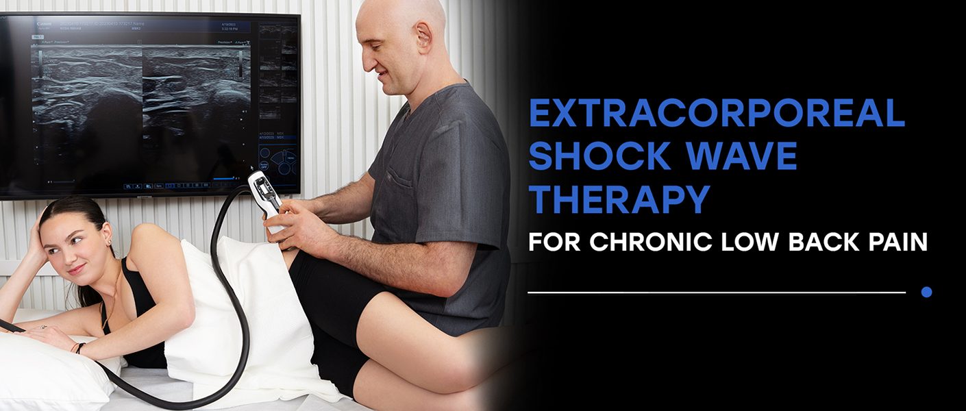 Chronic low back pain (LBP) is widely prevalent world-wide, and a leading cause of long-term disability. The United States, Australia, and Eastern Europe have the highest prevalence of LBP, with upwards of 225 cases per 100,000 population, according to a recent study published in Frontiers in Medicine. While trauma and overuse play a role, researchers are finding a strong correlation between LBP and body mass index (BMI). Despite LBP being an international health menace, medical doctors continue to treat pain symptoms while ignoring the underlying causes. But new research and advanced technologies are changing the game for back pain sufferers, providing innovative solutions for diagnosis, treatment and rehabilitation. Learn about the causes and types of low back pain, and how extracorporeal shockwave therapy can play a key role in restoring pain-free functional mobility for LBP patients.
The lumbopelvic region is the site of load transfer between the upper and lower body during any type of physical activity. To effectively mediate forces, the muscles of the core are activated to stabilize the region and protect the spine. Additional stability is provided by the fascia – a tough thin network of connective tissue that encases and connects muscles and organs, providing elastic tension and enabling tissue gliding.
But weakened and imbalanced muscles can fail to protect other structures, shifting force loads to fascia, tendons, ligaments and joints, and causing tissue damage and pain. Low back pain can arise from multiple causes, and patients often have more than one contributing factor.
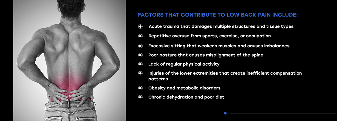
Factors that contribute to low back pain include:
With the exception of traumatic injuries, low back pain typically has a gradual onset. In many cases, pain arises from a bulging spinal disc that compresses a nerve, and such cases often self-resolve over a few days or weeks. LBP that persists for 3 months or longer and progressively worsens is considered a chronic condition.
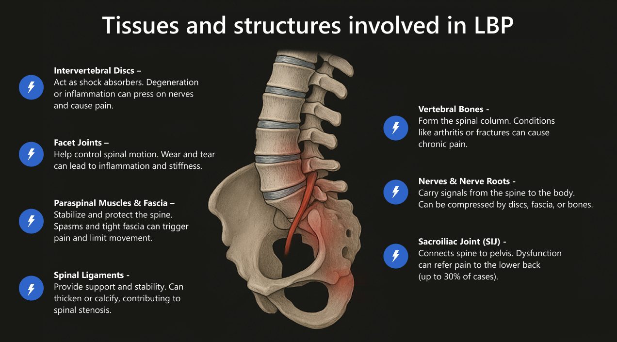
The lumbopelvic region houses a complex network of organs, muscles, joints, bones, nerves, blood vessels, connective tissue and fascia, and chronic LBP rarely arises from a single structure. For example, a bulging disc might irritate a nerve root while at the same time causing muscle spasms and placing stress on a facet joint.
Tissues and structures involved in LBP include:
The spinal column houses nerve roots that affect every part of the body, providing a conduit for messaging between the brain and peripheral tissues. In some chronic LBP cases, central sensitization can arise – a condition where pain perception is amplified in the central nervous system (CNS), even in the absence of ongoing tissue damage. Sensitization is often tied to psychological factors like stress or depression, and can prolong and exaggerate pain associated with chronic LBP.
Enthesopathy is a condition of the lower spine affecting the entheses—the attachment points of tendons, ligaments, or joint capsules to bones. It may involve inflammation, structural degeneration, or other changes that cause pain and reduced mobility. Enthesopathy is marked by pain or tenderness in the lower back at points where ligaments and tendons attach, along the vertebrae or near the iliac crest.
Radicular pain is caused by irritation or compression of a spinal nerve root, often from a herniated disc pressing against the nerve. Sciatica is a type of radicular pain caused by compression of the sciatic nerve that travels from the low back, all the way to the legs and feet. Spinal stenosis – a narrowing of the spinal canal – can also cause radicular pain as nerve roots are compressed within the narrowing space.
Inflammation in the low back caused by various types of arthritis can create non-mechanical LBP. Inflammatory low back pain is usually worse in the morning or after prolonged inactivity, causing stiffness that improves with movement.
Neuropathic LBP is caused by nerve damage, rather than direct structural issues. Post surgical nerve damage often appears after spinal procedures, caused by fibrosis or scar tissue. Nerve damage can also arise from diabetic neuropathy.
Pain felt in the lower back can sometimes originate from an internal organ or other non-spinal structure. Potential causes include kidney stones or infection, pelvic conditions, and rare but serious abdominal aortic aneurysm.
Pain with no identifiable structural or pathological cause accounts for about 85% of chronic LBP cases, leaving doctors baffled and patients frustrated. Potential causes are muscle tension, poor posture, stress, or minor biomechanical issues that are not visible with imaging.
Sadly, most medical doctors paint low back pain with a broad brush, relying on one-size-fits-all pain management strategies without differentiating between different types of pain. When pain persists or intensifies, MRI may be recommended to look for structural issues that can be surgically resolved. But MRI is costly and inconvenient for patients, and results are limited to static images that take days to process and analyze. In many cases, MRI fails to provide any insight at all into the cause of a patient’s LBP.
The diagnostic use of ultrasound for LBP is gaining traction in the broader medical community, especially for detecting SI-ligament or fascia dysfunction not visible on MRI.
A recent review by Bulgarian scientists supports adding high-resolution ultrasound to the clinician’s toolkit for functional and soft-tissue assessments of low back pain, noting that ultrasound is most valuable for assessing soft tissues vs. disc abnormalities.
Ultrasound imaging can be performed in-office, with portable and affordable equipment that takes up a fraction of the space of MRI. Unlike other imaging modalities, ultrasonography does not emit harmful radiation, and is considered safe for most patients. When administered by a skilled and experienced clinician, diagnostic ultrasound can provide invaluable information about the exact causes of low back pain, empowering doctors to go beyond pain management, to actually treating and healing the underlying mechanisms of LBP.
Conventional medical treatment of low back pain typically involves rest, ice and heat in the early stages, with the use of over-the-counter NSAIDs to treat pain and inflammation. When pain persists, anti-inflammatory drugs, opioids and antidepressants may be introduced, along with corticosteroid injections at the locus of pain.
Patients can suffer for years with flare-ups or unrelenting pain that requires increasing amounts of drugs to relieve symptoms. Surgery is usually reserved for structural issues like severe disc herniation and spinal stenosis, but doctors may turn to surgery as a last resort when other treatments fail. But back surgery poses high risks for the patient, with no guarantee of success.
Today, new advancements in research and technology are offering holistic alternatives to conventional LBP care that do not involve drugs or surgery, with the potential to reverse chronic low back pain and eliminate it for good. ESWT is an evidence-based, non-invasive technology that is revolutionizing LBP treatment. It uses high-frequency sound waves to trigger a cascade of biological reactions that promote tissue repair at the cellular level.
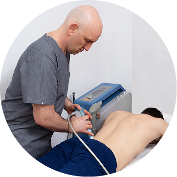
Chronic low back pain often involves more than one tissue type, and different tissues respond to specific types of shockwaves. Multimodal shockwave therapy uses a variety of shockwaves for optimal results. Advanced equipment allows clinicians to vary the depth and width of penetration, zoom in on specific tissues, and zoom out to cover larger areas. When applied under ultrasound guidance by an experienced clinician, ESWT provides an evidence-based methodology for fine-tuning LBP treatment.
Advantages of ESWT for chronic low back pain include:
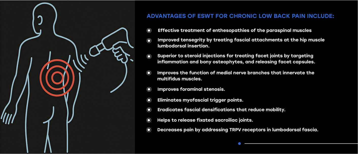
ESWT for LBP has been shown to improve functional mobility, with minimal side effects and long-term benefits. It addresses the underlying mechanisms of chronic low back pain, providing relief without drugs or surgery. ESWT can help to prepare tissues for additional treatments like physical therapy, aimed at restoring pain-free functional mobility.
A growing body of research is discovering innovative ways to combine ESWT with other methodologies for LBP diagnosis and treatment.
One study published in the Spine Journal explored the efficacy of shockwave therapy combined with low tension traction to regenerate and repair moderately and severely degenerated discs, and to explore its possible mechanisms of action. Since the intervertebral discs are avascular – meaning they have no blood supply – there is a limited flow of oxygen and nutrients to the discs, making it difficult to halt the degenerative process. In order to self-repair, discs are dependent on the exchange of nutrients and metabolites via molecular dispersion fluid from bony endplates.
The study combined ESWT with low tension traction to create a stable intervertebral environment for the repair and regeneration of degenerative discs. The researchers concluded that low energy ESWT combined with low tension traction provided a more stable intervertebral environment for the regeneration and repair of moderate and severe degenerative discs. This evidence holds great promise for a condition that was previously thought irreversible.
Pain generated from the lumbar facet joints account for up to one-third of LBP. Conventional treatment involves corticosteroid injections and medial branch radiofrequency neurotomy, both invasive therapies that carry the risk of complications, including infections and damage to the nerve roots or medial branch structures.
A Canadian study published in Neuroendocrinology Letters set out to explore the effectiveness of ESWT compared to both steroid injections and radiofrequency neurotomy. Results showed that ESWT had better long-term effects than steroid injections, and only slightly inferior efficacy compared to neurotomy. The shockwave group exhibited no adverse effects or complications, and patients reported significant long-term improvements in daily mobility.
A new 2025 randomized, sham-controlled study involved 128 patients with chronic lumbar facet syndrome. Patients were divided into two groups, with one group receiving focused ESWT, and the other receiving sham treatment. The treatment group showed significant reductions in pain intensity and disability at 6 and 12 months, and a follow-up with MRI showed resolution of bone marrow edema in nearly 60% of the treatment group, versus none in the control group.
Another study published in Biomechanics asserts that most back pain is caused by myofascial pain syndrome (MPS) – a chronic pain condition characterized by pain in the muscles and surrounding fascia. MPS typically involves the presence of trigger points—tight, sensitive spots in the muscle that can cause localized pain or refer pain to other areas of the body when pressed.
The researchers set out to demonstrate that ESWT can be used as a diagnostic tool for MPS when focused waves are directed at trigger points to provide a non-invasive mechanical stimulus. The objective was to produce referred pain patterns that implicate myofascial pain as the underlying cause of LBP. They concluded that focused ESWT provides ” a precise, reproducible, and non-invasive diagnostic approach for MPS in chronic LBP.”
In a 2022 study published in Neuromodulation, NYDNRehab clinical director Dr. Lev Kalika and his colleague, Dr. Bubnov set out to establish the efficacy of multimodal ESWT under ultrasound guidance, The aim of the study was to compare the effectiveness of US guidance for enhancing ESWT treatment of neuromusculoskeletal pathologies, muscle pain, and dysfunction.
In 2024, Dr. Kalika presented the results of a second study to the 26th World Congress of the International Society for Medical Shockwave Treatment. The study’s stated objective was to “investigate the impact of ultrasound guidance on the precision, direction, and depth of ESWT.” Results showed significant enhancement of subjective symptoms when precision adjustments were made based on ultrasound findings. They concluded that ultrasound guidance enhanced the functional application of ESWT, providing a more nuanced approach that rendered superior results.
Back pain sufferers often go from one doctor to the next, seeking long-term solutions to restore their functional mobility. However, conventional medicine is oriented to pain management, with surgery as a last resort when all else fails. At NYDNRehab, we take a more holistic approach to chronic low back pain, seeking to identify the underlying cause and eliminating pain at its source.
We use the highest-resolution ultrasound imaging to diagnose your condition and guide our procedures, for precise and effective treatment. Our high-grade multimodal shockwave equipment is second to none. When combined with Dr. Kalika’s 20+ years of clinical experience and professional expertise, ESWT at NYDNRehab provides low back pain sufferers with the most evidence-based and effective treatment options in NYC.
Che, Yan-Jun, et al. “Low energy extracorporeal shock wave therapy combined with low tension traction can better reshape the microenvironment in degenerated intervertebral disc regeneration and repair.” The Spine Journal 21.1 (2021): 160-177.
Low energy extracorporeal shock wave therapy combined with low tension traction can better reshape the microenvironment in degenerated intervertebral disc …
Ferdinandov, Dilyan, Dimo Yankov, and Martin Trandzhiev. “Common differential diagnosis of low back pain in contemporary medical practice: a narrative review.” Frontiers in Medicine 11 (2024): 1366514.
https://www.frontiersin.org/journals/medicine/articles/10.3389/fmed.2024.1366514/full
Kalika, Lev, and Rostyslav Bubnov. “PO236/# 817 TARGETED ULTRASOUND-GUIDED SHOCKWAVE THERAPY OF LOW BACK PAIN USING FOCUSED, DEFOCUSED AND RADIAL SHOCKWAVE: E-POSTER VIEWING.” Neuromodulation 25.7 (2022): S339.
https://www.neuromodulationjournal.org/article/S1094-7159(22)01183-7/abstract
Müller-Ehrenberg, Hannes, et al. “The Use and Benefits of Focused Shockwaves for the Diagnosis of Myofascial Pain Syndrome by Examining Myofascial Trigger Points in Low Back Pain.” Biomedicines 12.12 (2024): 2909.
https://www.mdpi.com/2227-9059/12/12/2909
Nedelka, Tomas, et al. “Mechano-transduction effect of shockwaves in the treatment of lumbar facet joint pain: comparative effectiveness evaluation of shockwave therapy, steroid injections and radiofrequency medial branch neurotomy.” Neuroendocrinol Lett 35.5 (2014): 393-397.
Mechano-transduction effect of shockwaves in the treatment of lumbar facet joint pain: comparative effectiveness evaluation of shockwave therapy, steroid …
Zhang, Chuan, et al. “The burden, trends, and projections of low back pain attributable to high body mass index globally: an analysis of the global burden of disease study from 1990 to 2021 and projections to 2050.” Frontiers in Medicine 11 (2024): 1469298.
The burden, trends, and projections of low back pain attributable to high body mass index globally: an analysis of the global burden of disease study from 1990 to …

Verified Expert Profiles
Dr. Lev Kalika is a world-recognized expert in musculoskeletal medicine. with 20+ years of clinical experience in diagnostic musculoskeletal ultrasonography, rehabilitative sports medicine and conservative orthopedics. In addition to operating his clinical practice in Manhattan, he regularly publishes peer-reviewed research on ultrasound-guided therapies and procedures. He serves as a peer reviewer for Springer Nature.
Dr. Kalika is an esteemed member of multiple professional organizations, including:
Below is a prime example of how ultrasound can take the guesswork out of diagnosis.

A bad physical therapy experience is one of the primary causes of unnecessary surgery
In this instance, an athlete was originally diagnosed with minor quadriceps muscle strain and was treated for four weeks, with unsatisfactory results. When he came to our clinic, the muscle was not healing, and the patients’ muscle tissue had already begun to atrophy.
Upon examination using MSUS, we discovered that he had a full muscle thickness tear that had been overlooked by his previous provider. To mitigate damage and promote healing, surgery should have been performed immediately after the injury occurred. Because of misdiagnosis and inappropriate treatment, the patient now has permanent damage that cannot be corrected.
The most important advantage of Ultrasound over MRI imaging is its ability to zero in on the symptomatic region and obtain imaging, with active participation and feedback from the patient. Using dynamic MSUS, we can see what happens when patients contract their muscles, something that cannot be done with MRI. From a diagnostic perspective, this interaction is invaluable.
Dynamic ultrasonography examination demonstrating
the full thickness tear and already occurring muscle atrophy
due to misdiagnosis and not referring the patient
to proper diagnostic workup
Demonstration of how very small muscle defect is made and revealed
to be a complete tear with muscle contraction
under diagnostic sonography (not possible with MRI)

Complete tear of rectus femoris
with large hematoma (blood)

Separation of muscle ends due to tear elicited
on dynamic sonography examination


