Extracorporeal shockwave therapy (ESWT) is a non-invasive therapeutic approach that falls into the category of regenerative medicine, used for treating injuries and pain syndromes. ESWT is a relatively new technology in the field of musculoskeletal medicine, having evolved over the past few decades to its current advanced state of efficacy.
A growing body of research supports the use of ESWT and other regenerative technologies for stimulating cellular progenesis, to promote and accelerate the healing of muscles, bones, nerves and connective tissues.
Scientists began exploring the potential use of shockwaves on human tissue in the 1960s and 70s, and by the mid 1980s, shock waves began to be used as a lithotripsy treatment to break up kidney stones and gallstones. This marked the advent of non-invasive technologies for in-vivo treatment of human tissues.
In its early stages, concerns that shockwaves to break up kidney stones could potentially harm the hip bones of patients receiving the treatment led to research on the impact of shock waves on bony tissue. Researchers soon discovered that shockwaves have an osteogenic effect on bone that can stimulate and accelerate the healing of fractures.
By the 1990s, research was being done to explore the effectiveness of shockwave therapy in the treatment of tendinopathies and bone disorders, including stress fractures, bone edema and avuncular necrosis. Studies expanded to encompass myofascial pain, cosmetic procedures, wound care, erectile dysfunction, management of spasticity, and a host of other orthopedic conditions.
Today, shockwave therapy has become the gold standard for regenerative medicine. It is often used to treat coronary disease and is used intraoperatively during open heart surgery. Its potential for treating neurodegenerative disorders like Parkinson’s disease and cerebral palsy is showing great promise.
ESWT uses a series of low-energy acoustic waves that are transmitted to the patient’s skin via a transducer, with a topical gel as a medium. It is completely non-invasive, and does not require anesthesia or pain medications.
Shockwaves work by triggering the body’s innate healing mechanisms to stimulate tissue repair and reduce pain. Many patients report significant pain reduction after a single treatment session.
Because ESWT triggers an inflammatory response, which is the body’s mechanism for healing, patients may experience temporary swelling and tenderness at the treatment site. This is a positive healing response, and it should not be treated with anti-inflammatory medications.
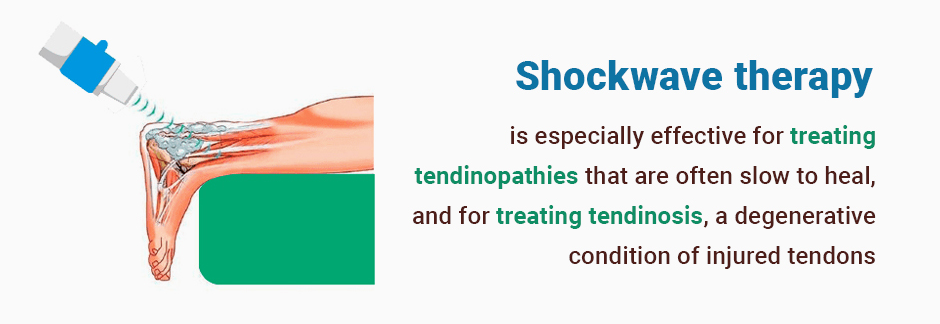
Shockwave therapy is especially effective for treating tendinopathies that are often slow to heal, and for treating tendinosis, a degenerative condition of injured tendons. It is also helpful for healing non-union bone fractures.
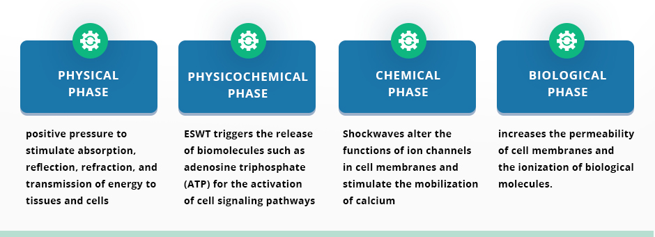
Until recently, the precise mechanisms of shockwave therapy were somewhat of a mystery, but it is now hypothesized that ESWT affects both cells and interstitial spaces in four distinct phases:
Physical Phase. In this phase, shockwaves create a positive pressure to stimulate absorption, reflection, refraction, and transmission of energy to tissues and cells.
Physicochemical Phase. In ESWT triggers the release of biomolecules such as adenosine triphosphate (ATP) for the activation of cell signaling pathways.
Chemical Phase. Shockwaves alter the functions of ion channels in cell membranes and stimulate the mobilization of calcium.
Biological Phase. ESWT modulates angiogenesis and facilitates the healing of bone and soft tissue wounds. Additionally, it appears that cavitation increases the permeability of cell membranes and the ionization of biological molecules.
The hypothesis of ESWT leading to tissue regeneration aligns with the principles of mechanotransduction. Mechanical load applied to the cytoskeleton stimulates cellular responses, leading to an increase in protein biosynthesis.
While the mechanisms of ESWT in alleviating pain are as yet unclear, there are two principal proposed hypotheses:
Shockwaves cause the degeneration of nerve fibers from small immunoreactive neurons, decreasing pro-inflammatory mediators.
Shockwaves trigger the release of endorphins and other analgesic molecules.
There are three types of shockwaves: focused, radial and defocused, and each has its advantages and disadvantages.
Focused shockwaves are the most regenerative of the three types, being 70% more effective than radial shockwaves, and 50% more effective than defocused shockwaves.
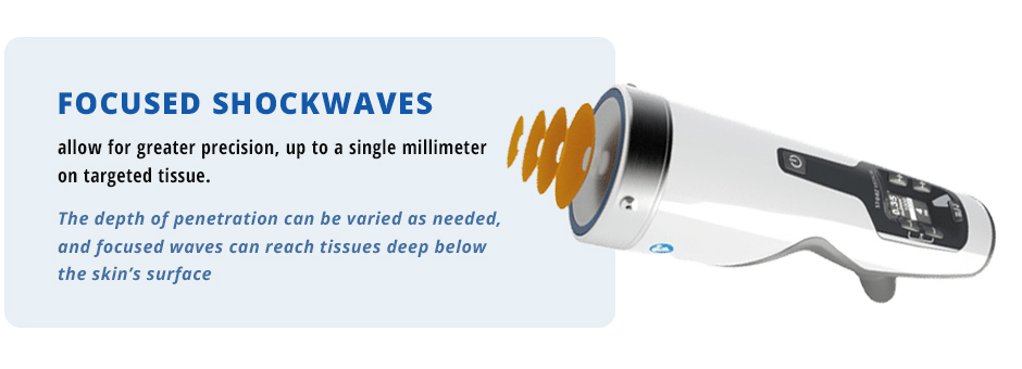
Focused shockwaves allow for greater precision, up to a single millimeter on targeted tissue. The depth of penetration can be varied as needed, and focused waves can reach tissues deep below the skin’s surface.
Despite its precision, focused shockwave therapy has a few disadvantages:
Focused shockwave needs to be performed under ultrasound guidance to ensure precise targeting, meaning the clinician must be trained in ultrasonography.
Focused shockwave clinicians need extensive training and experience for the procedure to be effective.
Focused shockwave therapy is more expensive than unfocused and radial shockwave.
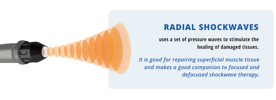
Radial shockwaves, also called extracorporeal pulse activation technology (EPAT), uses a set of pressure waves to stimulate the healing of damaged tissues. EPAT is not a true shockwave, but it does have a moderate regenerative effect on superficial tissues. It is good for repairing superficial muscle tissue and makes a good companion to focused and defocused shockwave therapy.
Defocused shockwaves are also called planar shockwaves or softwaves. They are more dispersed than focused shockwaves and do not penetrate as deeply. They are often used for cosmetic and dermatological procedures to remodel collagen within the dermis.
Defocused shockwaves are often used to treat cellulite. They can be useful in conditions that cover a large area, such as deep gluteal pain syndrome. Defocused shockwave therapy can be a good complement to focused shockwaves, but it can be expensive when used to get a regenerative effect.
The structures that make up the musculoskeletal system are interdependent, and problems rarely affect only a single structure. For example, non-traumatic tendon problems usually involve dysfunction at the myotendinous junction, myofascial pain and restriction, muscle stiffness, joint dysfunction and instability. Some of these changes originate at a location distant from the site of pain, or are caused by a combination of both.
Diagnostic ultrasonography coupled with a functional exam performed by an experienced practitioner are the recipe for success. In addition, using only a single type of shockwave is less likely to produce perfect and long-lasting outcomes. Combining different types of shockwaves will not only address the pain, but it will restore pain-free function. Guidance by ultrasonography helps the doctor to calculate the right amount of energy, frequency, and volume of pulses, and select the appropriate type of shockwaves used for different components of the injury.
Research shows that shockwave therapy is about 80-90 % effective, depending on the type of lesion being treated. However, most research is done using only a single type of shockwave. In our collective experience, ESWT performed holistically using different types of shockwaves can deliver 100% efficacy, often eliminating the need for other treatments or therapies.
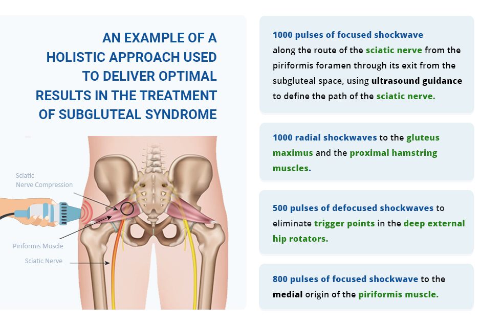
Following is an example of a holistic approach used to deliver optimal results in the treatment of subgluteal syndrome:
1000 pulses of focused shockwave along the route of the sciatic nerve from the piriformis foramen through its exit from the subgluteal space, using ultrasound guidance to define the path of the sciatic nerve.
800 pulses of focused shockwave to the medial origin of the piriformis muscle.
1000 radial shockwaves to the gluteus maximus and the proximal hamstring muscles.
500 pulses of defocused shockwaves to eliminate trigger points in the deep external hip rotators.
Shockwave therapy is the gold standard for regenerative therapies, yielding platinum results when performed by an experienced clinician.
Shockwave therapy is particularly effective in treating tendon disorders, including plantar fasciitis, shoulder tendinopathy, elbow tendinopathy, patellar tendinopathy and Achilles tendinopathy. In addition to its regenerative effects, ESWT stimulates the production of lubricin, a mucinous glycoprotein that facilitates tendon gliding.
One recent study of 384 patients with tendinopathies found that shockwave therapy significantly reduced pain and improved function and quality of life.
Myofascial pain syndrome occurs when areas of the myofascia, the thin tough sheath of tissue that surrounds and separates muscles and vital organs, become taught and develop tiny spasms in groups of muscle fibers, called trigger points, causing pain and dysfunction ESWT has been proven effective as a treatment for eliminating trigger points, relieving pain and restoring function of the myofascia.
Osteoarthritis involves the breakdown of joint cartilage and its underlying bone over time, resulting in joint pain and stiffness. While shockwave therapy does not work to stimulate the production of new cartilage, it is effective for relieving pain in the early stages of OA.
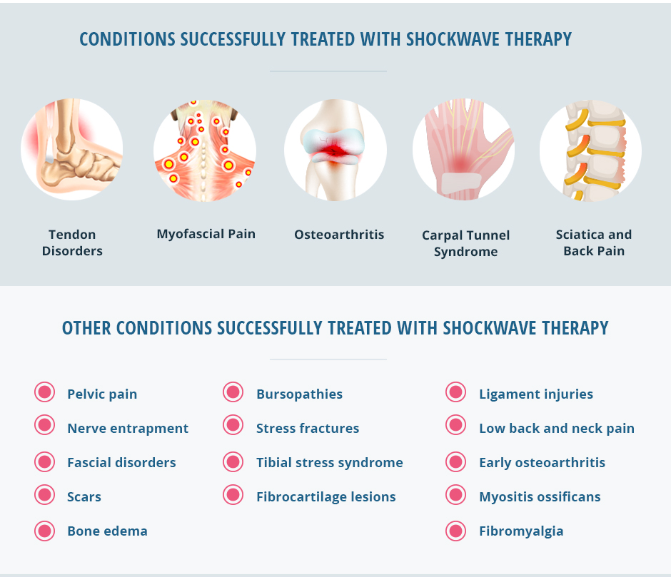
Research has shown ESWT to improve symptoms and functional outcomes in patients suffering from carpal tunnel syndrome. It is recommended that the underlying cause of neuropathy be addressed as part of the treatment protocol.
ESWT combining focused and radial shockwaves yield excellent results in treating sciatic nerve pain. It also has a positive effect on the facet joints of the spine, and is effective in relieving myofascial trigger points that are often a contributing cause of back pain.
Pelvic pain
Nerve entrapment
Fascial disorders
Scars
Bone edema
Bursopathies
Stress fractures
Tibial stress syndrome
Fibrocartilage lesions
Ligament injuries
Low back and neck pain
Early osteoarthritis
Myositis ossificans
Fibromyalgia
Effective shockwave treatment is largely dependent on the skill and experience of the clinician and the quality and precision of the technology used. Not all shockwave machines are equal in producing a regenerative effect, and inexpensive equipment can only deliver a minute fraction of a true shockwave.
At NYDNRehab, we use the latest and highest quality research-grade equipment to ensure that our patients get the full benefits of shockwave therapy. Our extensive experience and history of success with thousands of ESWT procedures make NYDNRehab the clinic of choice for shockwave therapy in NYC.
Dr. Lev Kalika has been treating patients with ESWT for over 15 years, and has performed over 2000 procedures. Dr. Kalika has been a member of the International Society of Medical Shockwave Therapy (ISMST) since 2008, and has published numerous research articles on the topics of ESWT, regenerative therapies, sports medicine, myofascial pain, ultrasound guided dry needling and rehabilitation.
Resourses
Dedes, Vasileios, et al. “Effectiveness and safety of shockwave therapy in tendinopathies.” Materia socio-medica 30.2 (2018): 131.
Zhang, Qing, et al. “Efficacy of extracorporeal shockwave therapy on pain and function in myofascial pain syndrome of the trapezius: a systematic review and meta-analysis.” Archives of physical medicine and rehabilitation 101.8 (2020): 1437-1446.

Verified Expert Profiles
Dr. Lev Kalika is a world-recognized expert in musculoskeletal medicine. with 20+ years of clinical experience in diagnostic musculoskeletal ultrasonography, rehabilitative sports medicine and conservative orthopedics. In addition to operating his clinical practice in Manhattan, he regularly publishes peer-reviewed research on ultrasound-guided therapies and procedures. He serves as a peer reviewer for Springer Nature.
Dr. Kalika is an esteemed member of multiple professional organizations, including:
Below is a prime example of how ultrasound can take the guesswork out of diagnosis.

A bad physical therapy experience is one of the primary causes of unnecessary surgery
In this instance, an athlete was originally diagnosed with minor quadriceps muscle strain and was treated for four weeks, with unsatisfactory results. When he came to our clinic, the muscle was not healing, and the patients’ muscle tissue had already begun to atrophy.
Upon examination using MSUS, we discovered that he had a full muscle thickness tear that had been overlooked by his previous provider. To mitigate damage and promote healing, surgery should have been performed immediately after the injury occurred. Because of misdiagnosis and inappropriate treatment, the patient now has permanent damage that cannot be corrected.
The most important advantage of Ultrasound over MRI imaging is its ability to zero in on the symptomatic region and obtain imaging, with active participation and feedback from the patient. Using dynamic MSUS, we can see what happens when patients contract their muscles, something that cannot be done with MRI. From a diagnostic perspective, this interaction is invaluable.
Dynamic ultrasonography examination demonstrating
the full thickness tear and already occurring muscle atrophy
due to misdiagnosis and not referring the patient
to proper diagnostic workup
Demonstration of how very small muscle defect is made and revealed
to be a complete tear with muscle contraction
under diagnostic sonography (not possible with MRI)

Complete tear of rectus femoris
with large hematoma (blood)

Separation of muscle ends due to tear elicited
on dynamic sonography examination


