August 14, 2023
In the center of each of your knees, there’s an anterior cruciate ligament (ACL). These ligaments are positioned diagonally. ACL injuries are fairly common, especially for athletes. To repair the damage, surgeons will sometimes graft tissues onto an ACL. That operation is an ACL reconstruction.
After such a reconstruction, it’s easy for the muscles around the knee, including the hamstrings and the quadriceps, to aid their patients.
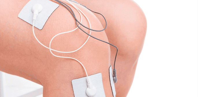
In 2012, the medical journal “Clinical Rehabilitation” published the results of a study that examined the effects of electro-stimulation on ACL reconstruction patients. This survey was set up as follows:
The results of this study were clear. The people who were administered electro-stimulation saw their swelling go down considerably after the first week. They experienced less pain as well. These three benefits were also noteworthy:
Granted, this research project was merely a pilot study. Among its deficiencies, it didn’t have a large group of subjects, and it didn’t involve a placebo. Nevertheless, it represents a starting point for scientific inquiry into electro-stimulation and ACL reconstruction recovery, and its conclusions are promising.
At this time, it’s almost certain that people who go through ACL reconstruction lose less muscle mass with electro-stimulation treatments than they would if they relied on exercise alone. Now, that’s not toning muscles and maintaining cardiovascular health. It’s just that, with the help of electro-stimulation, a patient will likely end up dealing with less pain, swelling, and atrophy.
With all of this information in mind, if you’re a docto their electro-stimulation sessions, which can be really soothing. Indeed, they can ease some of the anxiety that often accompanies injuries.
Then, once your patients realize how much faster they are recuperating, they’re sure to you and the electro-stimulation technology that you’ve adopted.

Verified Expert Profiles
Dr. Lev Kalika is a world-recognized expert in musculoskeletal medicine. with 20+ years of clinical experience in diagnostic musculoskeletal ultrasonography, rehabilitative sports medicine and conservative orthopedics. In addition to operating his clinical practice in Manhattan, he regularly publishes peer-reviewed research on ultrasound-guided therapies and procedures. He serves as a peer reviewer for Springer Nature.
Dr. Kalika is an esteemed member of multiple professional organizations, including:
Below is a prime example of how ultrasound can take the guesswork out of diagnosis.

A bad physical therapy experience is one of the primary causes of unnecessary surgery
In this instance, an athlete was originally diagnosed with minor quadriceps muscle strain and was treated for four weeks, with unsatisfactory results. When he came to our clinic, the muscle was not healing, and the patients’ muscle tissue had already begun to atrophy.
Upon examination using MSUS, we discovered that he had a full muscle thickness tear that had been overlooked by his previous provider. To mitigate damage and promote healing, surgery should have been performed immediately after the injury occurred. Because of misdiagnosis and inappropriate treatment, the patient now has permanent damage that cannot be corrected.
The most important advantage of Ultrasound over MRI imaging is its ability to zero in on the symptomatic region and obtain imaging, with active participation and feedback from the patient. Using dynamic MSUS, we can see what happens when patients contract their muscles, something that cannot be done with MRI. From a diagnostic perspective, this interaction is invaluable.
Dynamic ultrasonography examination demonstrating
the full thickness tear and already occurring muscle atrophy
due to misdiagnosis and not referring the patient
to proper diagnostic workup
Demonstration of how very small muscle defect is made and revealed
to be a complete tear with muscle contraction
under diagnostic sonography (not possible with MRI)

Complete tear of rectus femoris
with large hematoma (blood)

Separation of muscle ends due to tear elicited
on dynamic sonography examination


