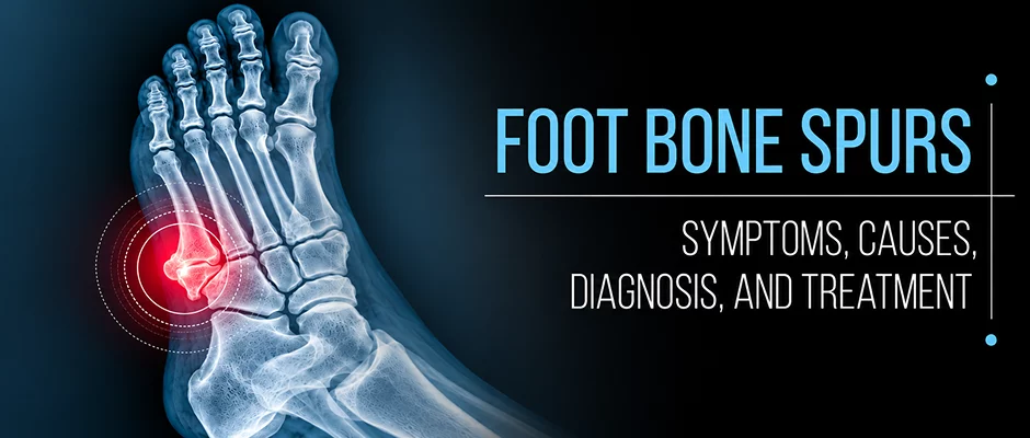
A bone spur in the foot sounds alarming, and on an X-ray it can look dramatic, but the spur itself is often not the reason your foot hurts. Understanding where these bony growths form, when they actually matter, and what genuinely helps can save you from unnecessary worry and the wrong treatment.

A bone spur, the common name for an osteophyte, is an abnormal bony outgrowth that develops along the edge of a bone, at a joint surface, or at the insertion point of a tendon or ligament. The term enthesophyte is used specifically when the growth occurs at a tendon or ligament attachment site, such as where the plantar fascia meets the heel bone.
These projections develop through a process of bone remodeling. In response to sustained mechanical loading, joint degeneration, or chronic soft-tissue tension, osteoblasts (bone-forming cells) deposit new bone material at structurally stressed sites. The result is typically a smooth, rounded bony prominence not a jagged spike, despite the word “spur.” Size varies considerably; most are small, though occasionally they become large enough to contact neighboring structures.
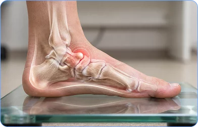
Bone spurs in the foot are a common radiographic finding. Their clinical significance ranges from completely irrelevant to functionally limiting, depending on location, size, and what surrounding tissues are involved. The presence of a spur on imaging should always be interpreted alongside a clinical examination, a principle that guides accurate diagnosis.
Osteophytes can form at several locations in the foot and ankle, each with its own clinical profile.
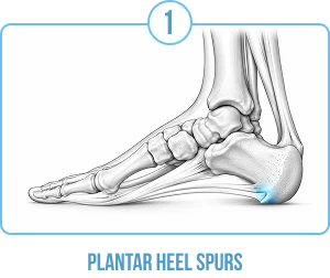
Plantar (Inferior) Heel, Calcaneal Enthesophytes
The most commonly discussed foot spur develops on the underside of the heel bone (calcaneus), at the attachment of the plantar fascia. These enthesophytes are strongly associated with plantar heel pain and plantar fasciitis, though the relationship between the spur and the pain is more nuanced than it may appear. The spur itself is frequently a secondary radiographic finding rather than the primary pain generator, a distinction explored in detail below.
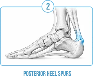
Posterior Heel, Achilles Insertion-Related Spurs
Bony changes can also develop at the posterior calcaneus where the Achilles tendon inserts. This is associated with insertional Achilles tendinopathy, a distinct condition from plantar fasciitis and may present with pain and tenderness at the back of the heel, often aggravated by the heel counter of a shoe.
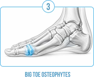
First Metatarsophalangeal (MTP) Joint, Big-Toe Osteophytes
Degenerative osteophytes at the first MTP joint are a hallmark of hallux rigidus, a condition involving progressive stiffness and pain at the big-toe joint. These spurs restrict joint motion and are driven by underlying cartilage degeneration, making them a fundamentally different entity from heel enthesophytes in terms of their cause, clinical behavior, and management.
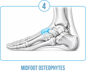
Midfoot, Tarsal Osteophytes
Osteophytes can develop across the midfoot joints in the setting of degenerative joint changes. A dorsal (top-of-foot) bony prominence, sometimes called a tarsal boss, may become symptomatic when footwear presses against it. This is one of several foot and ankle pain presentations that may be mistaken for a simpler soft-tissue problem until imaging is obtained.
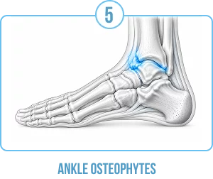
Ankle Joint
Osteophytes around the ankle joint are associated with post-traumatic arthritis, osteoarthritis, or chronic instability. Anterior ankle impingement, a condition common in athletes who repeatedly load the ankle in dorsiflexion, is one clinical context in which anterior ankle osteophytes cause symptoms.
Heel Spurs vs. Other Foot Osteophytes
It is important not to treat all foot bone spurs as a single condition. The underlying pathology, likely symptoms, natural history, and treatment approach differ substantially by location and context:
Applying statistics or treatment outcomes from studies on plantar heel spurs to other foot osteophytes or vice versa is clinically inappropriate. Each location requires its own evaluation.
This is one of the most important and most often misunderstood questions about foot osteophytes.
Most bone spurs are found incidentally, on imaging done for another reason. Many people have heel spurs on X-ray but no heel pain, and many people experience a great deal of heel pain but do not have a spur on X-ray. A bone spur seen on X-rays isn’t always the cause of symptoms.
If symptoms do develop, they are usually due to the spur’s interaction with the surrounding structures, a nerve, tendon, bursa or joint capsule, but not to the bony spur itself. Often the source of the pain is a soft tissue degeneration, fascial disease or joint disease associated with the bone lesion.
This distinction is clinically relevant. The clinical aim is not to remove the spur but to treat the underlying disease causing the symptoms in most cases. The bony prominence is usually still visible on X-rays after treatment.
When present, symptoms of a foot bone spur will vary depending on the surrounding structures and bones involved.
Plantar heel area:
Posterior heel (Achilles insertion):
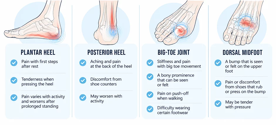
Dorsal midfoot:
General features that may accompany any symptomatic bone spur:
Not everyone who has foot bone spurs will have all of these symptoms. Sometimes a spur is discovered during evaluation of another condition and no treatment is necessary.
A bone spur is a growth that occurs in the foot as a result of prolonged mechanical stress, joint deterioration or chronic soft-tissue tension.
Degenerative joint disease (osteoarthritis):
Osteophytes are a known consequence of progressive loss of joint cartilage. Wear of cartilage exposes underlying bone to higher loading, and new bone forms along the margins of joints as part of the degeneration process.
Chronic tension at ligament and tendon attachment sites, especially where the plantar fascia attaches to the calcaneus, is related to enthesophyte formation.
Repetitive mechanical loading:
Activities and occupations that involve long periods of weight bearing, impact loading or repetitive joint motion may add cumulative stress at vulnerable sites.
Structural foot characteristics:
Flat feet (pes planus) or high arches (pes cavus) may change the way the foot is loaded, which can lead to increased stress at certain locations.
Previous injury:
Joint changes and alterations in joint mechanics after injury are related to the development of osteophytes.
Age:
Age is a factor, as the prevalence of foot osteophytes rises with age, which suggests that they are related to mechanical stress and cartilage changes over time.
Body weight:
Higher body weight has been linked to several foot conditions, including plantar heel pain.
Inflammatory arthropathies:
Some inflammatory diseases like ankylosing spondylitis and reactive arthritis can cause enthesophytes through different inflammatory processes than degenerative osteoarthritis.
Footwear:
Footwear that causes focal pressure and/or changes the mechanics of the foot can lead to focal stress at specific anatomical sites.





Most of these are associations that have been supported by epidemiological and clinical studies, but not proven as direct causes. Not everyone who has these risk factors will get symptomatic bone spurs.
Plantar fasciitis, which is actually a degenerative fasciopathy at the plantar fascia insertion on the heel, is one of the most prevalent causes of plantar heel pain. Researchers have noted that plantar calcaneal spurs and plantar fasciitis often occur together, and a calcaneal spur may represent a radiographic sign of chronic tension of the plantar fascia at that location.
But in most cases, the spur is not thought to be the cause of pain. Studies have shown that a significant proportion of people with radiographic spurs do not have pain in their heels while many patients with typical plantar fasciitis symptoms lack a spur on their X-ray. There is a varying relationship between spur size and symptom severity.
Current knowledge supports the idea that plantar fasciitis is a degenerative process of the plantar fascia itself, rather than acute inflammation, and that the presence of a heel spur (in the presence of other evidence) is a secondary radiographic finding, rather than the primary issue. Treatment is thus aimed at the underlying fascial pathology, load management and contributing biomechanical factors. See the NYDNRehab guide to plantar fasciitis for an in-depth look at the evaluation and treatment of this condition.
A heel spur cannot be dissolved, and a successful treatment of plantar fasciitis does not make it disappear. The spur is usually seen on X-rays long after the symptoms have gone away.
There are a number of conditions which can cause pain in the foot that may be confused with that of a bone spur. Correct diagnosis by a trained clinician is critical:
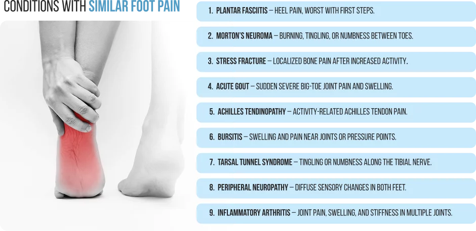
These conditions may coexist with bone spurs or may independently account for symptoms attributed to a spur. Clinicians must carefully correlate imaging findings with the full clinical picture.
The clinical history and physical examination are the first steps in diagnosis. A clinician will inquire about the timing, nature, and site of symptoms, the factors that make symptoms worse and better, activity level, footwear habits, and medical history.

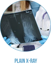
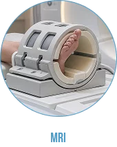
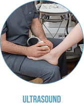
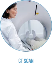
The interpretation of imaging findings should always take into account the clinical examination. A bone spur on X-ray does not mean a bone spur is the cause of symptoms; it is the job of the clinician to decide if the bone spur is clinically significant or a chance finding.
While most foot pain can be assessed on a non-urgent basis, some symptoms require immediate professional treatment. If you have any of the following symptoms, you should get medical help right away:
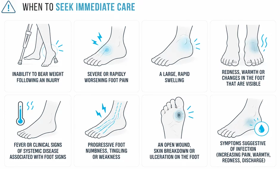
In people with diabetes, peripheral vascular disease or peripheral neuropathy, foot wounds or pressure sores need to be evaluated immediately, even if they don’t seem serious.
Many symptomatic foot bone spurs can be managed without surgery. Conservative treatment focuses on the underlying condition causing symptoms, rather than on removing the spur.
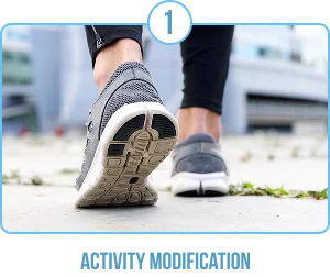
Avoiding or decreasing activities that trigger symptoms gives associated soft tissue irritation a chance to calm down. This is not to say no activity is allowed, but low impact activity without symptoms is usually fine during recovery.
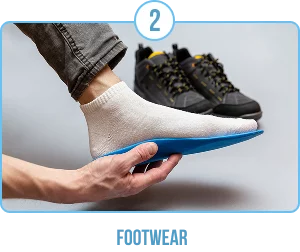
For many patients, footwear with sufficient cushioning, correct arch support and a roomy toe box can significantly decrease symptom burden. If you have an osteophyte on the middle of your foot (dorsal midfoot) or on your toes, it’s helpful to avoid shoes that put pressure on the osteophyte directly. Clinicians or specialists can provide individual advice depending on the foot structure and the site of the symptoms.
Physical therapy is a key element in the treatment of most foot osteophyte-related conditions and is usually structured. The program will vary based on the diagnosis.
There is evidence to support the following treatments for plantar heel pain and plantar fasciitis:
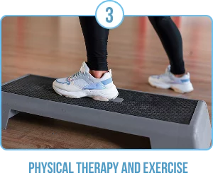
Rehabilitation with hallux rigidus and osteophytes of the first MTP emphasizes the preservation of available joint mobility, load management through the joint and strengthening of supporting musculature.
A structured loading program for the Achilles tendon is an essential part of the rehabilitation for insertional Achilles tendinopathy associated with posterior calcaneal changes and should be selected and progressed by a qualified clinician.
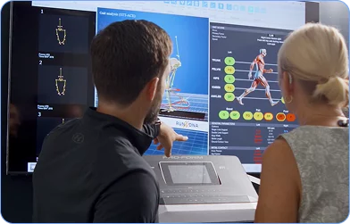
A physical therapist is able to evaluate movement patterns, determine the contributing biomechanical factors and create a program that is suitable for the patient’s condition. Dr. Lev Kalika at NYDNRehab uses advanced diagnostic imaging and customized motion analysis to determine which foot and ankle pain structure is causing pain and creates customized rehabilitation programs to address the patient’s pain.
Prefabricated or custom foot orthoses can redistribute plantar pressure and decrease the stress at areas of symptoms. There is generally supportive evidence for their use in plantar heel pain, although the quality and design of the studies differ and none of the orthosis types are universally superior. This will vary depending on the foot shape, the diagnosis and the patient. An experienced clinician knowledgeable about foot biomechanics can guide you in deciding whether or not an orthosis will be helpful and what kind.
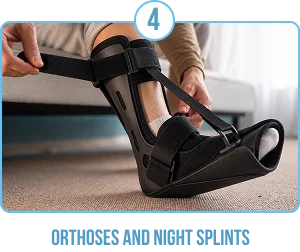
Night splints are used mainly in plantar fasciitis, using them to keep , to keep the foot dorsiflexed during the night. They are designed to keep the plantar fascia from tightening during sleep and lessen the morning pain experienced with the initial steps. There is moderate evidence for their use, and they may be especially helpful for patients with symptoms that are worst when they first get up after sleeping.
Some patients may be able to get some relief from pain from over-the-counter analgesics or nonsteroidal anti-inflammatory drugs (NSAIDs) for a short time while awaiting an appointment. These drugs treat pain and inflammation, but they will not change the underlying degeneration or the bony spur itself. Patients should take these medications as directed on the package, be aware of contraindications and should see a healthcare provider before taking these medicines regularly or if they have other health conditions.
If the conservative treatments do not relieve the pain, there are a few options for the procedure. Each is appropriate based on the specific diagnosis, the location of the symptoms and the individual clinical picture.

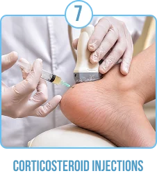
In some cases of foot osteophytes, like plantar fasciitis, corticosteroid injections can offer useful short-term pain relief. They are not specifically aimed at the spur itself, but at soft-tissue inflammation or irritation that is associated with it. There is evidence of their short-term efficacy in treating plantar heel pain, although this effect may diminish with time, and repeated injections can lead to the development of soft-tissue atrophy and, in the heel, plantar fascia rupture. The choice of whether or not to have an injection should be discussed carefully with a qualified clinician, taking into account the potential benefits, limitations and risks.
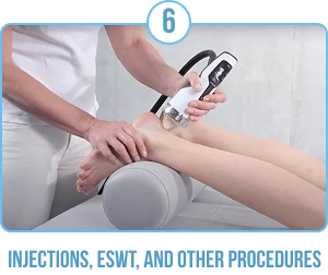
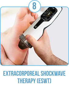
ESWT delivers acoustic energy to a targeted tissue area and has been studied primarily as a treatment for chronic plantar fasciitis and insertional Achilles tendinopathy not as a method of removing or dissolving a bone spur. There is generally good evidence for its use in chronic plantar heel pain that is unresponsive to other conservative treatments, although there are some differences in the studies making direct comparisons difficult. It is not a first-line treatment and should only be used after a good course of conservative treatment. The importance of its role in other osteophyte diseases of the foot is not as well established.
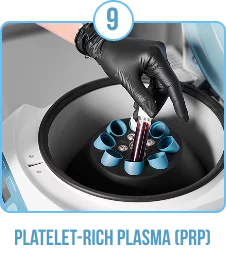
PRP injections are made by drawing a small amount of the patient’s blood, concentrating the platelets and injecting them into the target tissue to support healing. Studies of the use of PRP for plantar fasciitis and Achilles tendinopathy are still continuing. Some studies have revealed good results, others have revealed that it is not more effective than other treatments. The evidence is still mixed and condition-specific; PRP should not be used as a standard of care for all foot osteophyte related conditions. People who are thinking about getting PRP should talk to their treating doctor about the current evidence.
BMAC (Bone Marrow Aspirate Concentrate): an injection prepared from your own bone marrow. BMAC concentrates cells and other natural components from a small sample of your bone marrow. We may consider it for selected, more advanced joint or tendon problems when PRP alone may not be sufficient. Treatment combines precise ultrasound-guided injection with personalized rehabilitation.
MFAT (Micro-Fragmented Adipose Tissue): an injection prepared from your own fat tissue. MFAT uses a small amount of your fat, gently processed to preserve its natural cells and supporting tissue. We may consider it for selected, more advanced joint or soft-tissue problems when PRP alone may not be sufficient. Treatment combines precise ultrasound-guided injection with personalized rehabilitation.
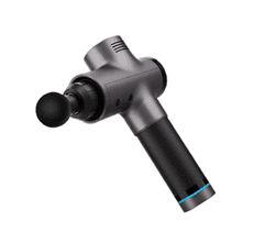
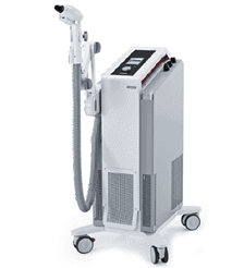
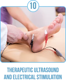
Some of these modalities may be used as supplements to a physical therapy program. Their use in particular for bone spur related conditions has not been well established, nor are they proven treatments for bone spur. They are generally only used in conjunction with other soft-tissue conditions, and as part of a comprehensive rehabilitation program
Surgical treatment for foot bone spurs is usually only considered after a proper course of conservative treatment has failed to provide adequate relief, and when symptoms are so severe that they interfere significantly with daily function or quality of life. The choice to undergo surgery is influenced by the diagnosis, the extent of structural abnormality, the functional consequences for the patient and the clinical and imaging evaluation.
The surgical treatment is very different depending on the location and the nature of the problem:
In some cases, minimally invasive surgical techniques are becoming available for some of these procedures, which may result in less soft tissue disruption and quicker recovery.
The recovery time after treatment for a symptomatic foot bone spur will depend greatly on the underlying diagnosis, the type of treatment, the location of the bone spur, and patient-specific factors. There is no universal recovery timeline that can be given for all conditions and procedures.
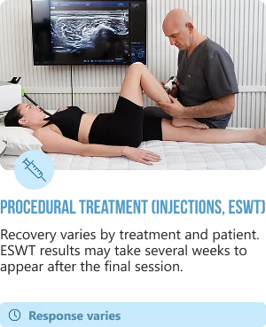

Patients should adhere to their surgeon’s individual postoperative timeline, not general ones. Physical therapy after surgery is usually an essential part of regaining strength, mobility and function and to help reduce the risk of recurrence.
Whatever the treatment method, it is crucial to be aware that the bony spur is usually still visible on imaging after symptoms have gone away. The success of treatment is assessed by how much the patient’s pain, function and quality of life improves, rather than by the absence of the radiographic finding.
The prevention of bone spur formation is not known to be reliable. Mechanical loading, joint degeneration, genetic factors and aging all play a role in the development of osteophytes, and all of these are factors that cannot be controlled. However, several general foot health strategies may help reduce mechanical stress on the foot, manage contributing factors, and potentially reduce the likelihood of symptomatic problems developing or worsening:






No. Many people with visible bone spurs in their feet have no symptoms. Bone spurs are often seen on X-rays done for another condition. The pain only happens when the spur rubs against or presses on other tissues, like nerves, tendons, bursae, or joint tissue. It is well documented that many spurs cause no pain.
A bone spur does not melt or fade away unless it is removed by surgery. The spur will not go away if the underlying cause is treated successfully, for example, if the symptoms of plantar fasciitis are resolved. Note that having a spur present on imaging does not necessarily imply that symptoms will be present. Many patients have lasting pain relief even though the spur is still present on x-rays.
Plantar fasciitis is a degenerative disorder of the plantar fascia at its attachment to the calcaneus that causes pain in the bottom of the foot, especially during the first steps in the morning. While a heel spur is a bony outgrowth at the same location, it is not the same condition as plantar fasciitis. Plantar fasciitis can occur without the presence of a spur, and a spur can be present without causing plantar fasciitis. The spur is usually regarded as a secondary finding and not as the primary pain generator.
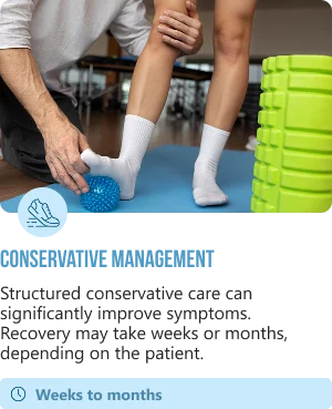
Recovery is dependent on the location, diagnosis, and treatment. With dedication, conservative treatment of plantar heel pain can yield improvement in weeks to months. Recoveries can differ depending on the procedure from several weeks for osteophyte removal, to six months or more for any procedure that requires tendon reattachment or joint fusion. A treating clinician can give a realistic timeframe in light of the particular situation.
No. Many patients with painful bone spurs in their feet improve with conservative treatment. Surgery is considered when symptoms are severe, functional limitation is severe, structural pathology is present and surgery is indicated, and reasonable amounts of conservative treatment have failed to yield adequate results. A decision is individual and should be made together with a qualified clinician.
Orthoses can redistribute plantar pressure and minimise mechanical stress at symptomatic areas. Evidence generally supports their use in plantar heel pain. Whether prefabricated or custom orthoses are appropriate and which type depends on the individual’s foot structure, diagnosis and clinical assessment. A clinician with a special interest in foot biomechanics can offer advice.
Request an appointment»

Our location: 11 West 25th Street 5th floor, New York, NY 10010
Dr. Lev Kalika is a world-recognized expert in musculoskeletal medicine. with 20+ years of clinical experience in diagnostic musculoskeletal ultrasonography, rehabilitative sports medicine and conservative orthopedics. In addition to operating his clinical practice in Manhattan, he regularly publishes peer-reviewed research on ultrasound-guided therapies and procedures. He serves as a peer reviewer for Springer Nature.
Dr. Kalika is an esteemed member of multiple professional organizations, including:

Below is a prime example of how ultrasound can take the guesswork out of diagnosis.

A bad physical therapy experience is one of the primary causes of unnecessary surgery

In this instance, an athlete was originally diagnosed with minor quadriceps muscle strain and was treated for four weeks, with unsatisfactory results. When he came to our clinic, the muscle was not healing, and the patients’ muscle tissue had already begun to atrophy.
Upon examination using MSUS, we discovered that he had a full muscle thickness tear that had been overlooked by his previous provider. To mitigate damage and promote healing, surgery should have been performed immediately after the injury occurred. Because of misdiagnosis and inappropriate treatment, the patient now has permanent damage that cannot be corrected.
The most important advantage of Ultrasound over MRI imaging is its ability to zero in on the symptomatic region and obtain imaging, with active participation and feedback from the patient. Using dynamic MSUS, we can see what happens when patients contract their muscles, something that cannot be done with MRI. From a diagnostic perspective, this interaction is invaluable.
Dynamic ultrasonography examination demonstrating
the full thickness tear and already occurring muscle atrophy
due to misdiagnosis and not referring the patient
to proper diagnostic workup
Demonstration of how very small muscle defect is made and revealed
to be a complete tear with muscle contraction
under diagnostic sonography (not possible with MRI)
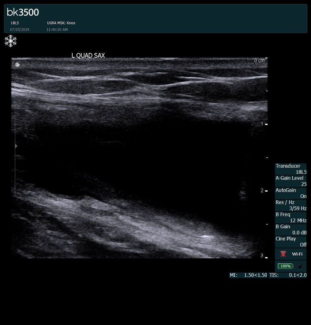
Complete tear of rectus femoris
with large hematoma (blood)
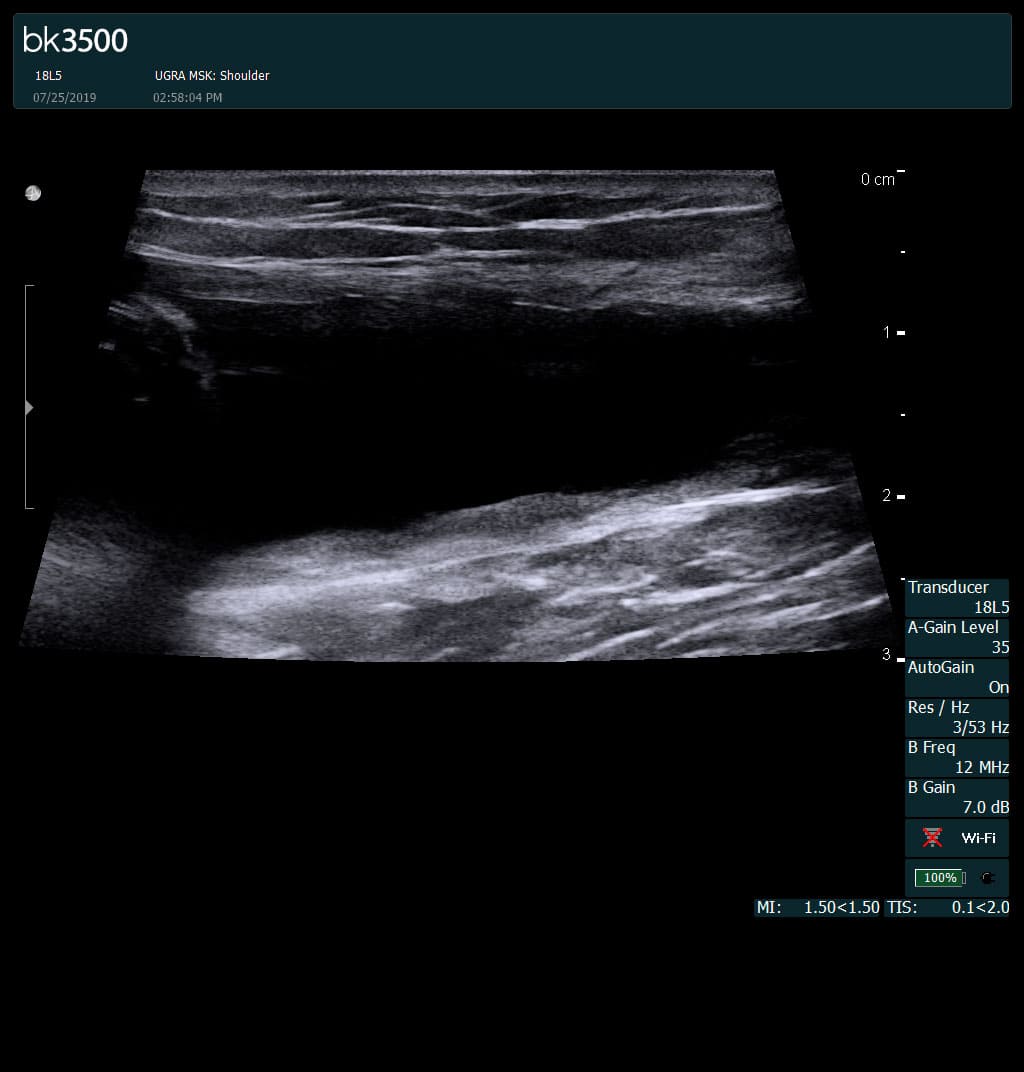
Separation of muscle ends due to tear elicited
on dynamic sonography examination

 Request Telehealth