HomeBlogHow Myofascial Release Therapy Helps Reduce or Eliminate Chronic Pain
How Myofascial Release Therapy Helps Reduce or Eliminate Chronic Pain
August 14, 2023
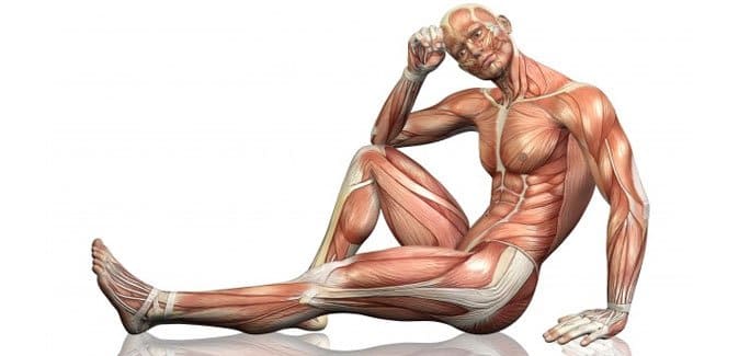
Just as your skin surrounds and supports your entire body, your fascia provides a second system of support, forming a web of tissue that encompasses your muscles, connective tissue, bones, nerves, blood vessels, and visceral organs, right down to the cellular level.
Healthy fascia is supple and elastic, providing support without restricting the underlying structures. However, when damaged, fascia can become rigid and inelastic, causing pressure and tension throughout your body. Fascial tension can be difficult to diagnose, as it does not appear on imaging scans like X-ray or MRI.
Tight fascia can restrict muscle function, creating movement deficiencies that can interfere with athletic performance and inhibit everyday activities. It can also become a source of chronic pain that keeps you from doing the things you love and reduces your overall quality of life. Myofascial release therapy (MFR) addresses the fascia surrounding your muscles and tendons, which can become overly tight for a variety of reasons, including disuse, overuse, or injury.
Conditions Treated with MFR Therapy
MFR therapy seeks toring function. Some conditions commonly treated with MFR therapy include:
Temporo-mandibular joint (TMJ) disorder
Carpal tunnel syndrome
Fibromyalgia
Migraine headaches
Shoulder pain
Hip pain
Back pain
General muscle and joint pain and dysfunction
Tight myofascial tissue can cause postural imbalances and joint misalignment by restricting movement in a particular area of your body, causing you to resolve.
Causes of Myofascial Pain Syndrome
There are two fundamental sources of myofascial pain. One stems from restricted movement of muscles and connective tissue that are bound by tight fascia. The other stems from damage to the affected tissue becomes inhibited, exacerbating the condition.
How MFR Therapy Works
During MFR therapy, the therapist gently massages the muscle fascia to stretch, pressure and tightness are released, and the tissue becomes pliable and elastic once again.
Because of the interconnectedness of bodily structures, the therapist may focus on a tight area that is nowhere near the area where the patient feels the most pain. MFR therapy addresses the entire network of muscles and fascia that may be causing your pain, reducing tension by releasing trigger points throughout your body.
MFR Therapy in NYC
If you suffer from headaches, TMJ syndrome, fibromyalgia or other chronic pain syndromes, the manual therapy specialists at NYDNRehab can help. We use advanced technologies and innovative therapies to its fullest.
Follow me:
About the Author
Dr. Lev Kalika is a world-recognized expert in musculoskeletal ultrasonography, with 20+ years of clinical experience in advanced rehabilitative medicine. In addition to operating his clinical practice in Manhattan, he regularly publishes peer-reviewed research on ultrasound-guided therapies and procedures.
Dr. Kalika is an esteemed member of the International Society for Medical Shockwave Treatment ((SMST), and the only clinician in New York certified by the ISMST to perform extracorporeal shockwave therapy. He is also an active member of the American Institute of Ultrasound in Medicine (AIUM), and has developed his own unique approach to dynamic functional and fascial ultrasonography.
In this instance, an athlete was originally diagnosed with minor quadriceps muscle strain and was treated for four weeks, with unsatisfactory results. When he came to our clinic, the muscle was not healing, and the patients’ muscle tissue had already begun to atrophy.
Upon examination using MSUS, we discovered that he had a full muscle thickness tear that had been overlooked by his previous provider. To mitigate damage and promote healing, surgery should have been performed immediately after the injury occurred. Because of misdiagnosis and inappropriate treatment, the patient now has permanent damage that cannot be corrected.
The most important advantage of Ultrasound over MRI imaging is its ability to zero in on the symptomatic region and obtain imaging, with active participation and feedback from the patient. Using dynamic MSUS, we can see what happens when patients contract their muscles, something that cannot be done with MRI. From a diagnostic perspective, this interaction is invaluable.
Dynamic ultrasonography examination demonstrating the full thickness tear and already occurring muscle atrophy due to misdiagnosis and not referring the patient to proper diagnostic workup
Demonstration of how very small muscle defect is made and revealed to be a complete tear with muscle contraction under diagnostic sonography (not possible with MRI)
Complete tear of rectus femoris with large hematoma (blood)
Separation of muscle ends due to tear elicited on dynamic sonography examination