September 22, 2025
Dorsalgia is a generalized clinical term used by doctors to describe back pain, regardless of its location or cause. It comes from the Latin root words: “dorsum,” meaning back, and “algia,” meaning pain. Despite its widespread prevalence, back pain is often described as “non-specific” due to the frequent absence of any identifiable mechanical issues.
Learn about different types and locations of back pain, and potential dorsalgia causes.
According to the World Health Organization (WHO), back pain is the leading cause of disability worldwide, with the highest number of cases occurring between the ages of 50-55. To understand the widespread prevalence of back pain, it is important to understand the relationship between the spinal column and the central nervous system.
Your spinal column provides a protective casing for the spinal cord, a key component of the central nervous system (CNS), made up of the brain and spinal cord. The spinal column is made up of bony vertebrae that provide a rigid structure for protecting and housing the spinal cord. Vertebrae are separated from one another by discs that absorb shock and provide a space where nerve roots exit the spinal cord and branch out into neurons, connecting the CNS to the peripheral nervous system that innervates the various parts of the body.
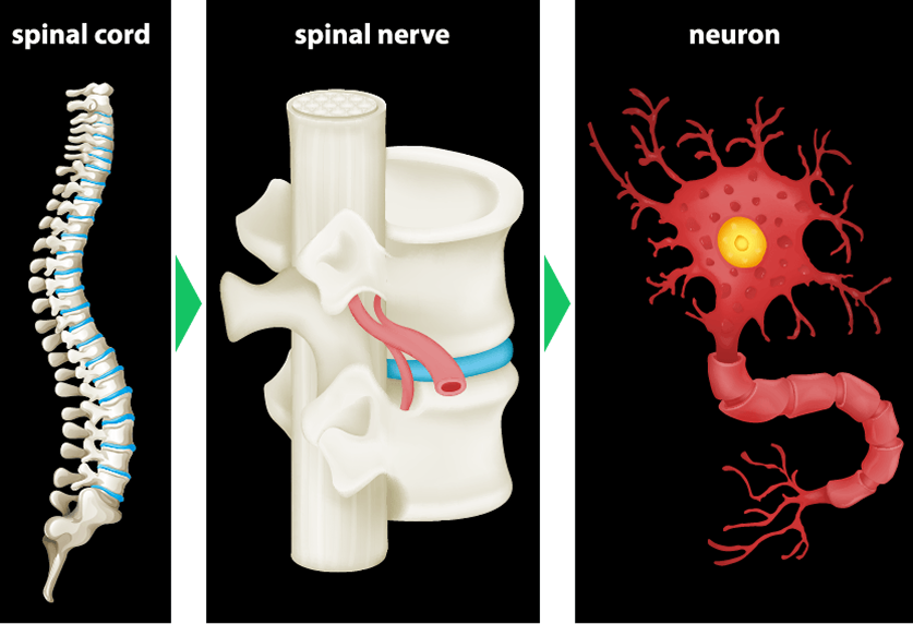
Thanks to the spinal cord, your nerves are able to seamlessly transmit millions of signals between your brain and the rest of your body. Peripheral sensory nerves send messages to your brain about the external environment, bodily functions, and the body’s position in space. At the same time, motor nerves send signals from the brain to the body, to govern metabolic and biological functions, and to produce movement.
Due to the spine’s unique architecture, nerve roots often become compressed, or “pinched” where they exit the spinal column, causing back pain. Nerves can also be compressed when the muscles and fascia that support the spine become weak, damaged or imbalanced.
Dorsalgia can be categorized by its location along the spinal column, or by its primary causes. Let’s begin with spinal architecture.
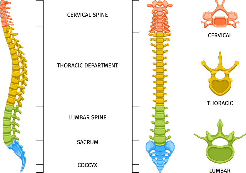
Nerves of the sacral spine govern reproductive and eliminatory functions, and pain in the coccyx – aka tailbone – is called coccydynia, often arising from a backward fall.
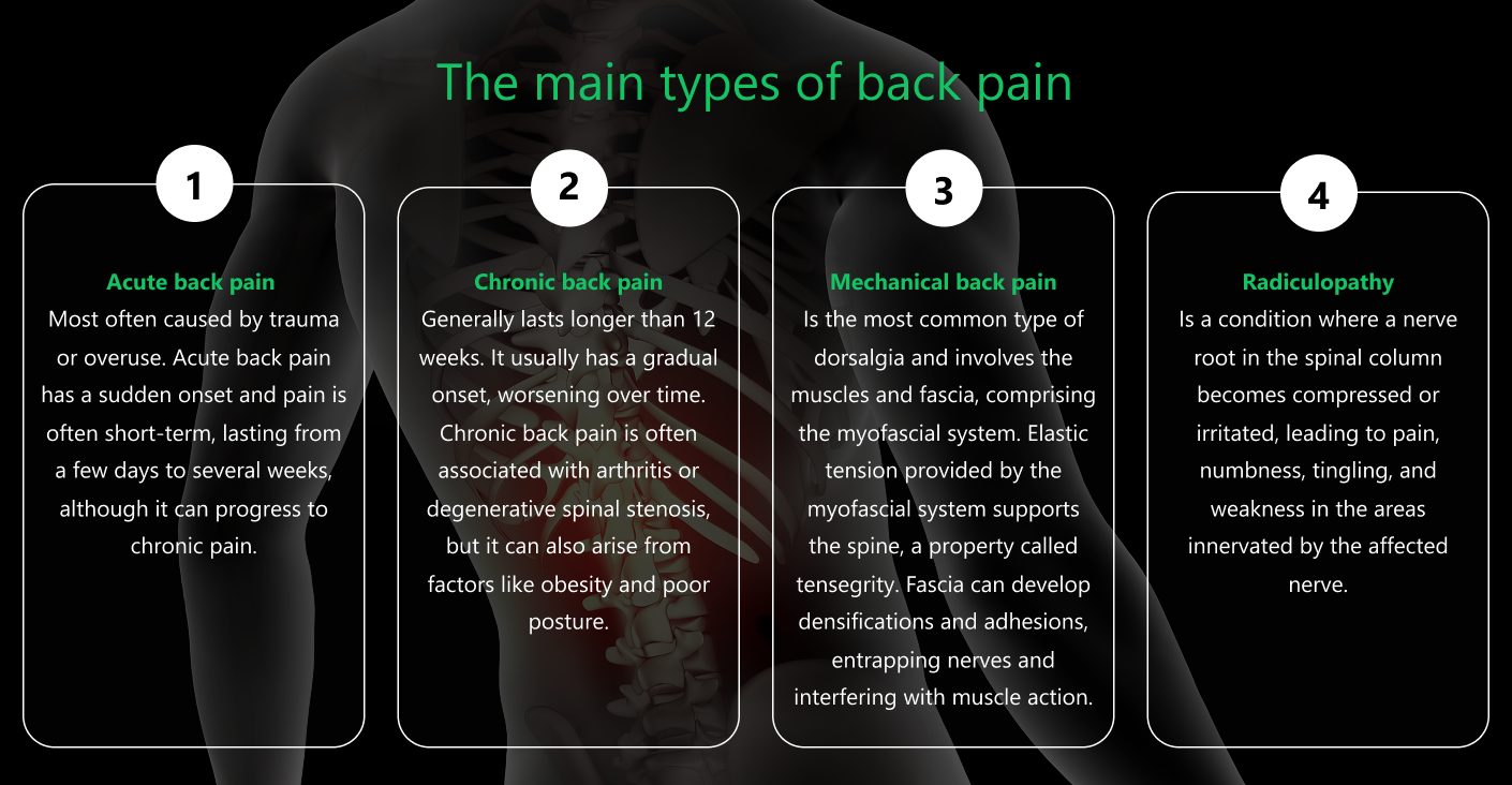
Another way to think about dorsalgia is in terms of its origins. Many factors can contribute to back pain, and more than one factor can be in play at the same time.
The main types of back pain include:
Dorsalgia symptoms can vary from one person to the next, depending on factors like physical activity level, posture, excessive sitting, and even metabolic health.
Common Symptoms of Dorsalgia include:
During your initial visit, your doctor will review your medical history and ask questions about the onset, duration, and intensity of pain. Your clinical exam may include assessments of posture, range of motion, and location of pain. Depending on the examination results, the doctor may use electromyography to test for nerve compression.
Medical doctors typically recommend rest, NSAID pain medications, muscle relaxants, and topical creams or ointments to treat dorsalgia symptoms. However, that approach may only mask your symptoms without addressing their underlying cause. Physical therapy and chiropractic care can help to improve posture, enhance spinal alignment, and promote mobility and stability.
Integrative therapy is a holistic approach to dorsalgia treatment that considers the whole patient, not just their symptoms. An integrative dorsalgia specialist may use a variety of alternative therapies to not only treat pain symptoms, but eliminate their contributing factors. Advanced holistic therapies are often combined, and used to prepare damaged tissues prior to physical therapy.
Advanced integrative therapies for dorsalgia pain include:

There are dozens of private clinics in Manhattan that offer back pain treatment, but most of them do not feature advanced technologies like ultrasonography and multimodal shockwave therapy. At NYDNRehab, our state-of-the-art clinic features the most advanced and evidence-based approaches available for dorsalgia diagnosis and treatment. Dr. Kalika’s experience and expertise make NYDNRehab the clinic of choice for dorsalgia treatment in NYC.

Verified Expert Profiles
Dr. Lev Kalika is a world-recognized expert in musculoskeletal medicine. with 20+ years of clinical experience in diagnostic musculoskeletal ultrasonography, rehabilitative sports medicine and conservative orthopedics. In addition to operating his clinical practice in Manhattan, he regularly publishes peer-reviewed research on ultrasound-guided therapies and procedures. He serves as a peer reviewer for Springer Nature.
Dr. Kalika is an esteemed member of multiple professional organizations, including:
Below is a prime example of how ultrasound can take the guesswork out of diagnosis.

A bad physical therapy experience is one of the primary causes of unnecessary surgery
In this instance, an athlete was originally diagnosed with minor quadriceps muscle strain and was treated for four weeks, with unsatisfactory results. When he came to our clinic, the muscle was not healing, and the patients’ muscle tissue had already begun to atrophy.
Upon examination using MSUS, we discovered that he had a full muscle thickness tear that had been overlooked by his previous provider. To mitigate damage and promote healing, surgery should have been performed immediately after the injury occurred. Because of misdiagnosis and inappropriate treatment, the patient now has permanent damage that cannot be corrected.
The most important advantage of Ultrasound over MRI imaging is its ability to zero in on the symptomatic region and obtain imaging, with active participation and feedback from the patient. Using dynamic MSUS, we can see what happens when patients contract their muscles, something that cannot be done with MRI. From a diagnostic perspective, this interaction is invaluable.
Dynamic ultrasonography examination demonstrating
the full thickness tear and already occurring muscle atrophy
due to misdiagnosis and not referring the patient
to proper diagnostic workup
Demonstration of how very small muscle defect is made and revealed
to be a complete tear with muscle contraction
under diagnostic sonography (not possible with MRI)

Complete tear of rectus femoris
with large hematoma (blood)

Separation of muscle ends due to tear elicited
on dynamic sonography examination


