August 14, 2023
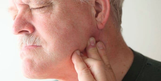
The temporomandibular joint, also known as the TMJ, joins your jaw to your skull. Try feeling it; it lies just in front of your ear hole. There are many causes for tmj disorder, some of which are poor posture, growth disorder, bad habits, inflammation, or injury.
The lower jaw bone, called the mandible, and the socket on the skull, called the temporal bone, make up the TMJ, or the temporomandibular joint. The articular disc, held in place by muscles and ligaments, lies between these two parts and causes the smooth opening and closing of the joint.
You may experience jaw joint pain or uncomfortable facial muscles, experience a clicking, grinding, or locking in the jaw, or suffer headaches and dizziness. If you have trouble opening and closing the mouth or feel pain when chewing hard food or yawning, you very likely have temporomandibular joint (TMJ) trouble.
Usually there is a combination of causes of tmj disorder: overactive jaw muscles, trauma, arthritis, neck stiffness or dysfunction, or even a sensitivity to stress-related pain.
Physiotherapy reduces pain and stiffness while restoring function and teaching management strategies. It is very thorough in giving tmj relief.
Massage the muscles that lie near the joint, using heat or ice for ten minutes to rest your phone between your shoulder and your ear, avoiding the joint.
Holding your neck stiffly, sitting or lying poorly, and holding your jaw in a back position can all hurt, as well as straining your neck. Simply be aware of how to keep your neck strong and mobile. Ask your physiotherapist for further information.
Ask your physiotherapist for further, more specific management techniques for tmj relief. The following are for starters.
Try standing or sitting upright. Draw your chin in until you give yourself a double chin, keeping your head straight on the whole while. Hold this position for at most 5 seconds, repeating it 8-10 times.
Lightly place your to relax the jaw muscles.
On to partially open and then fully open. Push it up with your finger. To keep the movement straight, practice in front of a mirror 6-8 times and 4-6 times daily.
Place the to keep the movement straight. Repeat this exercise 6-8 times 4-6 times per day.
Put the jaw into a neutral position, first partially open, then a knuckle width apart. Meanwhile, apply a gentle pressure with the thumb.

Verified Expert Profiles
Dr. Lev Kalika is a world-recognized expert in musculoskeletal medicine. with 20+ years of clinical experience in diagnostic musculoskeletal ultrasonography, rehabilitative sports medicine and conservative orthopedics. In addition to operating his clinical practice in Manhattan, he regularly publishes peer-reviewed research on ultrasound-guided therapies and procedures. He serves as a peer reviewer for Springer Nature.
Dr. Kalika is an esteemed member of multiple professional organizations, including:
Below is a prime example of how ultrasound can take the guesswork out of diagnosis.

A bad physical therapy experience is one of the primary causes of unnecessary surgery
In this instance, an athlete was originally diagnosed with minor quadriceps muscle strain and was treated for four weeks, with unsatisfactory results. When he came to our clinic, the muscle was not healing, and the patients’ muscle tissue had already begun to atrophy.
Upon examination using MSUS, we discovered that he had a full muscle thickness tear that had been overlooked by his previous provider. To mitigate damage and promote healing, surgery should have been performed immediately after the injury occurred. Because of misdiagnosis and inappropriate treatment, the patient now has permanent damage that cannot be corrected.
The most important advantage of Ultrasound over MRI imaging is its ability to zero in on the symptomatic region and obtain imaging, with active participation and feedback from the patient. Using dynamic MSUS, we can see what happens when patients contract their muscles, something that cannot be done with MRI. From a diagnostic perspective, this interaction is invaluable.
Dynamic ultrasonography examination demonstrating
the full thickness tear and already occurring muscle atrophy
due to misdiagnosis and not referring the patient
to proper diagnostic workup
Demonstration of how very small muscle defect is made and revealed
to be a complete tear with muscle contraction
under diagnostic sonography (not possible with MRI)

Complete tear of rectus femoris
with large hematoma (blood)

Separation of muscle ends due to tear elicited
on dynamic sonography examination


