August 14, 2023
Ankle sprains are common sports injuries that occur when the ankle rolls in a lateral motion, doing damage to more injuries down the road.
A suspected catalyst for CAI is peripheral nerve damage that occurs with a lateral ankle sprain. Inflammation and scar tissue at the injury site can reduce the space around a nerve structure, putting pressure on the nerve and interfering with optimal neural function. Remember that your nerves send and receive information to do, so a compressed or damaged nerve can undermine normal joint movement and cause instability.
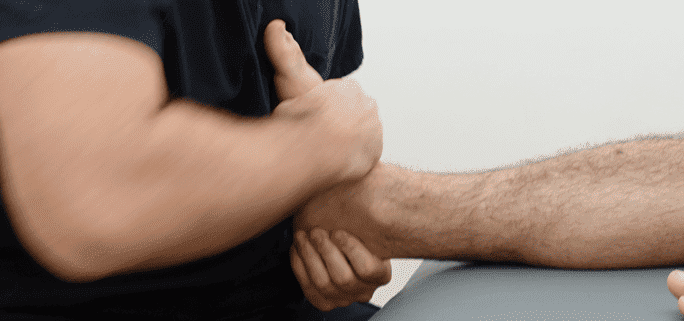
Manual nerve mobilization is a therapeutic technique applied tore optimal function and reduce foot and ankle pain. Nervous tissue is sensitive and must be mobilized gently. When done correctly, manual nerve mobilization can enhance the outcomes of traditional therapies by helping entrapped nerves function freely.
Traditional therapy for CAI includes strengthening and balance exercises, along with proprioceptive training. A 2016 study by Plaza-Manzano et al. sought to conventional therapies.
Fifty-six physically active subjects with CAI were randomly assigned tors over the course of four weeks. The second group of 28 received the same balance and strengthening programs, but also received manual therapy consisting of grade 3 joint mobilizations and peroneal nerve mobilizations.
Subjects were assessed before and at the end of the four week intervention, and again after one month of treatment. A battery of assessment instruments was used to assess pain, ankle stability, joint range of motion and strength. While both groups improved in all assessment categories, the researchers observed greater improvement in the group who received manual therapy.
The results of this study make a compelling case for the inclusion of manual peroneal nerve mobilization therapy in rehab proto improve ankle stability and relieve foot and ankle pain.

Verified Expert Profiles
Dr. Lev Kalika is a world-recognized expert in musculoskeletal medicine. with 20+ years of clinical experience in diagnostic musculoskeletal ultrasonography, rehabilitative sports medicine and conservative orthopedics. In addition to operating his clinical practice in Manhattan, he regularly publishes peer-reviewed research on ultrasound-guided therapies and procedures. He serves as a peer reviewer for Springer Nature.
Dr. Kalika is an esteemed member of multiple professional organizations, including:
Below is a prime example of how ultrasound can take the guesswork out of diagnosis.

A bad physical therapy experience is one of the primary causes of unnecessary surgery
In this instance, an athlete was originally diagnosed with minor quadriceps muscle strain and was treated for four weeks, with unsatisfactory results. When he came to our clinic, the muscle was not healing, and the patients’ muscle tissue had already begun to atrophy.
Upon examination using MSUS, we discovered that he had a full muscle thickness tear that had been overlooked by his previous provider. To mitigate damage and promote healing, surgery should have been performed immediately after the injury occurred. Because of misdiagnosis and inappropriate treatment, the patient now has permanent damage that cannot be corrected.
The most important advantage of Ultrasound over MRI imaging is its ability to zero in on the symptomatic region and obtain imaging, with active participation and feedback from the patient. Using dynamic MSUS, we can see what happens when patients contract their muscles, something that cannot be done with MRI. From a diagnostic perspective, this interaction is invaluable.
Dynamic ultrasonography examination demonstrating
the full thickness tear and already occurring muscle atrophy
due to misdiagnosis and not referring the patient
to proper diagnostic workup
Demonstration of how very small muscle defect is made and revealed
to be a complete tear with muscle contraction
under diagnostic sonography (not possible with MRI)

Complete tear of rectus femoris
with large hematoma (blood)

Separation of muscle ends due to tear elicited
on dynamic sonography examination


