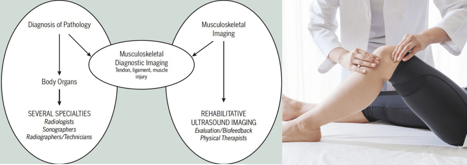
If you or a loved one has ever had a traumatic injury or stumped your doctor with regional pain from an unknown source, you have probably had an Xray or MRI. Those imaging methods are the most common types of diagnostic imaging for musculoskeletal pain, used to give your doctor a glimpse of what’s going on “under the hood.”
The problem with traditional imaging tools is that they give you still images, usually taken by a technician. Your doctor does not see the results until after they are processed, which can be within an hour or two for Xray, or within several days for MRI. In addition, both methods have inherent safety issues that make them inappropriate for some patients.
With still images, it is easy to overlook important clues about the source of pain, or miss hidden damage to bones, nerves and soft tissues that were not captured by the camera. Musculoskeletal diagnostic ultrasonography provides a safe, inexpensive and accurate alternative for detecting damage to the body’s structures and monitoring the healing process. Ultrasound technology is also showing promise as a tool for biomechanical analysis, helping athletes perform better with less risk of injury.
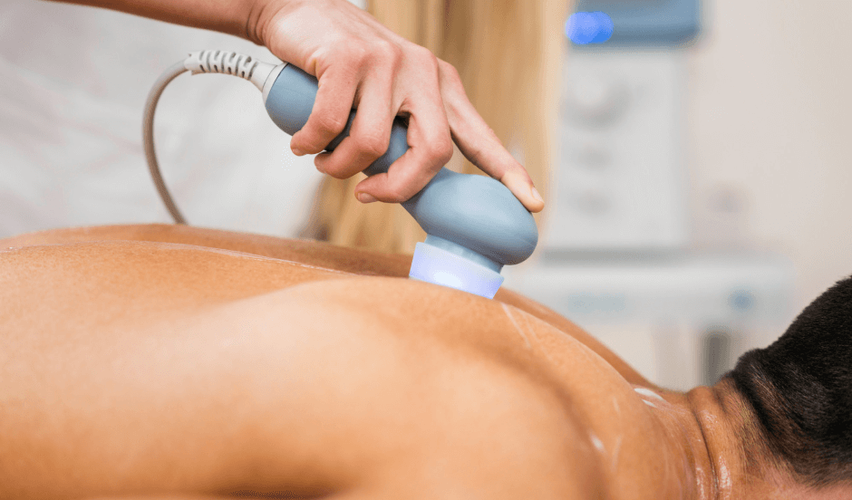
Ultrasound works by transmitting millions of high-frequency sound waves per second into the patient’s body, using a probe that can be moved and repositioned along the surface of the skin to gain a variety of views. When the sound waves hit a barrier, like an organ, bone, muscle, nerve or connective tissue, they echo back to the probe and are relayed to the ultrasound device. The reflected waves are calculated in terms of distance from the probe to the barrier, using the speed of sound and the time of each echo’s return. That information is then displayed as a two-dimensional image on the ultrasound screen, giving us a view of the body’s structures.

Accurate diagnosis immediately after an injury is key to getting the rehab ball rolling. If you are an athlete, early diagnosis means faster return to sport. Musculoskeletal diagnostic ultrasonography is fast, accurate and portable. An athletic injury can be examined right on the playing field, reducing the athlete’s risk of further injury and giving coaches a heads up if the athlete needs to be taken out of the game.
Instantaneous diagnosis in the rehab clinic means no delay in treatment. The patient can often be evaluated and treatment begun in the same session. During the diagnostic exam, the patient becomes an active participant in their own diagnosis, giving feedback and working with the clinician to reposition joints and limbs, so no damage goes undetected.
Your joints and muscles work interdependently to produce movement. Joint kinematics studies the position, angle, velocity, and acceleration of body segments in relation to one another. Joint kinematics is used during gait analysis to identify deficient motor patterns that may lead to injury and reduce performance. There are many tools for measuring joint kinematics, but they are often uncomfortable for the patient, which can detract from natural movement patterns.
A promising recent development is the use of a wireless sensor system using ultrasound technology. Only one wearable ultrasonic sensor is required, minimizing discomfort for the patient. The system measures joint angles, velocity and acceleration in terms of the speed of sound. The technology is inexpensive, accurate and less complex than other systems for measuring joint kinematics.
As your body heals, ultrasonography helps us accurately assess your rate and degree of recovery. Premature return to sport is one of the biggest causes of re-injury. MSUS imaging allows the athlete, coach and doctor to view damaged tissues in real time, with the athlete in motion.
Peak muscle torque is one marker for recovery that tells how much force a muscle or muscle group can produce. For example, measurements of knee extensor torque in the quadriceps may be used to evaluate ACL rehabilitation. Traditionally, isokinetic dynamometry has been the tool of choice for evaluating peak muscle torque, but it is unable to isolate the individual muscles of the quadriceps group.
Real-time ultrasound imaging enables us to view and measure the strength and function of each muscle with the knee in multiple positions, something that is not possible with isokinetic testing. We can use ultrasound to evaluate the muscle thickness of the individual muscles, to see which are healing, and which are underperforming. We can also compare muscle thickness of the injured and uninjured sides of he body, to get a baseline for recovery.
NYDNRehab is one of the foremost sports injury clinics in the United States. Our clinic features advanced technologies rarely found in a private clinical setting, including:
In addition to emergent technologies, we embrace innovative therapies to help you heal, rehabilitate and return to sport, stronger, faster and more agile, with lower risk of injury. Our clinical director, Dr. Lev Kalika, is a globally renowned expert in the field of rehabilitative musculoskeletal ultrasonography.
Resources
Ashnagar, Zinat, et al. “Ultrasound evaluation of the quadriceps muscles in pronated foot posture.” The Foot 38 (2019): 86-90.
Qi, Yongbin, et al. “Lower extremity joint angle tracking with wireless ultrasonic sensors during a squat exercise.” Sensors 15.5 (2015): 9610-9627.
Wadugodapitiya, Surangika, et al. “Ultrasound elastographic assessment of the stiffness of the anteromedial knee joint capsule at varying knee angles.” Bio-medical materials and engineering Preprint (2019): 1-12.

Verified Expert Profiles
Dr. Lev Kalika is a world-recognized expert in musculoskeletal medicine. with 20+ years of clinical experience in diagnostic musculoskeletal ultrasonography, rehabilitative sports medicine and conservative orthopedics. In addition to operating his clinical practice in Manhattan, he regularly publishes peer-reviewed research on ultrasound-guided therapies and procedures. He serves as a peer reviewer for Springer Nature.
Dr. Kalika is an esteemed member of multiple professional organizations, including:
Below is a prime example of how ultrasound can take the guesswork out of diagnosis.

A bad physical therapy experience is one of the primary causes of unnecessary surgery
In this instance, an athlete was originally diagnosed with minor quadriceps muscle strain and was treated for four weeks, with unsatisfactory results. When he came to our clinic, the muscle was not healing, and the patients’ muscle tissue had already begun to atrophy.
Upon examination using MSUS, we discovered that he had a full muscle thickness tear that had been overlooked by his previous provider. To mitigate damage and promote healing, surgery should have been performed immediately after the injury occurred. Because of misdiagnosis and inappropriate treatment, the patient now has permanent damage that cannot be corrected.
The most important advantage of Ultrasound over MRI imaging is its ability to zero in on the symptomatic region and obtain imaging, with active participation and feedback from the patient. Using dynamic MSUS, we can see what happens when patients contract their muscles, something that cannot be done with MRI. From a diagnostic perspective, this interaction is invaluable.
Dynamic ultrasonography examination demonstrating
the full thickness tear and already occurring muscle atrophy
due to misdiagnosis and not referring the patient
to proper diagnostic workup
Demonstration of how very small muscle defect is made and revealed
to be a complete tear with muscle contraction
under diagnostic sonography (not possible with MRI)

Complete tear of rectus femoris
with large hematoma (blood)

Separation of muscle ends due to tear elicited
on dynamic sonography examination


