May 3, 2024

 Many medical doctors try to help their patients manage OA pain with a combination of steroid injections and non-steroidal anti-inflammatory drugs (NSAIDs) until knee replacement surgery becomes inevitable. Ironically, those treatment approaches have been proven to worsen the symptoms of OA and accelerate cartilage erosion.
With nearly one-fifth of the world’s population suffering from OA and growing concerns about popular pain medications, there is a growing demand for holistic alternative therapies that treat OA pain and regenerate cartilage without drugs or surgery.
Learn how HA and PRP injections combined with shockwave therapy for knee osteoarthritis can help arrest cartilage erosion, stimulate cartilage synthesis, and restore pain-free joint function.
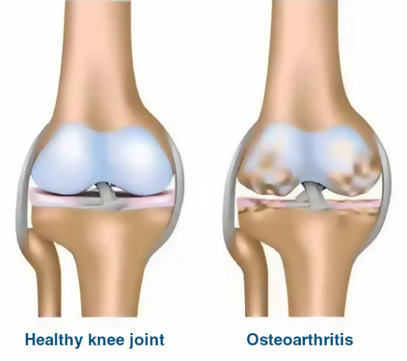
Cartilage is a special type of connective tissue that covers the ends of bones in diarthrodial joints — articulations where bones come together within a synovial joint capsule whose membrane secretes lubricating fluid. Cartilage lubricated by synovial fluid provides a smooth surface that minimizes bone friction during movement.
Types of diarthrodial joints include:
Diarthrodial joints of the ankles, knees, hips, elbows and shoulders are highly involved in load transfer during physical activity. The knees in particular are designed to withstand force loads many times the body mass of their owner. In active individuals, joint repetitive overload can sometimes damage joint cartilage, especially after an injury.
Articular cartilage is avascular, lacking blood vessels and nerves, which reduces its capacity for intrinsic repair. Many factors contribute to cartilage erosion associated with osteoarthritis:
Making positive lifestyle changes and addressing biomechanical factors can help minimize the rate and severity of cartilage erosion, while regenerative therapies can help stimulate cartilage synthesis.
A three-pronged approach of lifestyle behavior modification, physical therapy and cartilage regenerative therapy can dramatically reduce joint pain, encourage the regrowth of healthy cartilage, and help to restore optimal joint function.
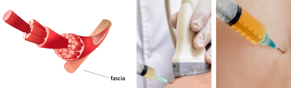
In its early stages. OA affects the soft tissues surrounding the joints, particularly the fascia. Fascia is a type of connective tissue that encases muscles and connects them to each other, and to other structures and organs throughout the body. In its healthy state, fascia is smooth and slippery, enabling muscles, organs, blood vessels and nerves to glide freely among other structures during movement. Fascia provides elastic tension within the body that protects its structural integrity – a property called tensegrity.
When joint injury occurs, multiple structures are affected, including fascia. When damaged, fascia becomes densified and sticky, losing its elastic properties and sometimes adhering to soft tissues and bony structures. When that happens, nerves and blood vessels can become compressed and entrapped, limiting muscle action and causing pain.
Because the knee is highly embedded with proprioceptors – mechanosensory neurons that inform your brain of your body’s position relative to gravity – damage to fascia surrounding the knee can suppress proprioception and severely reduce knee stability.
Restoring fascia to its healthy state helps to:
Damaged fascia in the early stages of OA can be treated with fascia manipulation therapy to elongate densified fibers and restore elasticity. In cases of fascia adhesions, ultrasound-guided hydrodissection may be used – a procedure where a jet of water or saline solution is injected to separate fascia from adjacent tissues, freeing up entrapped nerves and blood vessels.
Early treatment that includes fascia therapy can dramatically slow the progression of joint OA and prevent collagen erosion.
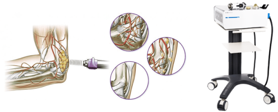
Extracorporeal shock wave therapy (ESWT) is a non-invasive procedure that delivers focused pulses of high-energy sound waves to the joint capsule and affected bone ends. ESWT promotes blood vessel formation and increases blood flow to the targeted tissues, for enhanced oxygen and nutrient delivery.
A growing body of scientific evidence shows ESWT to be effective in halting the progression of knee OA and promoting cartilage renewal:
ESWT is a recognized evidence-based approach for rebuilding cartilage in cases of degenerative knee OA. At the same time, ESWT reduces pain and inflammation, and improves joint function in patients with degenerative joint conditions.
When combined with other evidence-based approaches, ESWT offers an effective solution for treating degenerative joint conditions and reversing cartilage erosion.
Treatment approaches to musculoskeletal conditions are often multi-modal, and osteoarthritis is no exception. When combined with ESWT, injections of hyaluronic acid (HA) and platelet rich plasma (PRP) stimulate and provide support for the synthesis of new cartilage tissue.
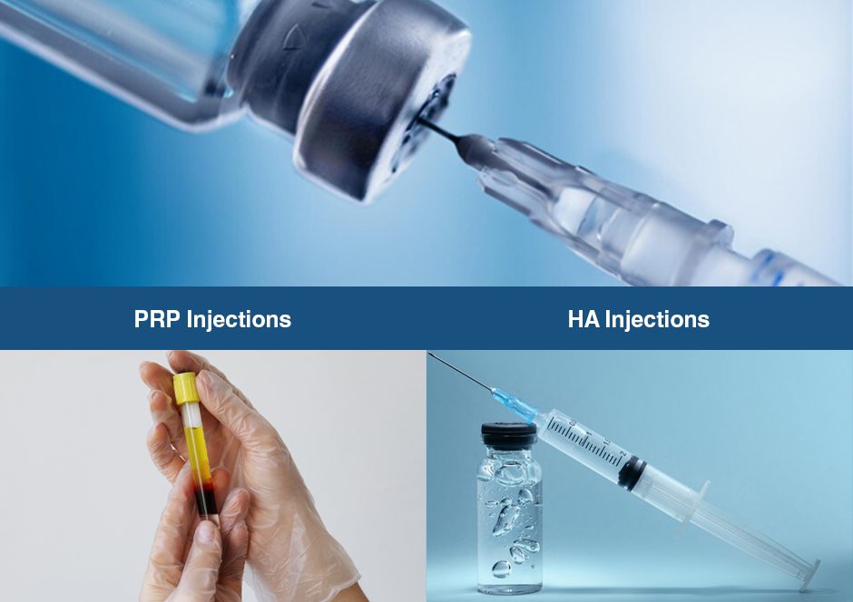
Platelet Rich Plasma (PRP) Injections
PRP therapy uses a sample of the patient’s own whole blood, spun in a centrifuge to extract a high concentration of platelets. Platelets are small cell fragments in human blood, manufactured in bone marrow. When PRP is injected into the joint capsule, it is exposed to collagen and releases biologically active factors that stimulate the synthesis of blood vessels and new cartilage cells.
Hyaluronic Acid (HA) Injections
Hyaluronic acid is a naturally occurring substance found in synovial fluid, fascia and soft connective tissues. Synovial fluid in osteoarthritic joints contains a lower concentration of HA than in healthy joints. When injected into the joint capsule, HA can improve the viscosity of synovial fluid, prevent cartilage degradation and promote its regeneration. At the same time, HA can help restore the slippery property of fascia that allows it to glide among other structures.
Safeguarding your joints against OA and cartilage erosion requires proactive measures involving positive lifestyle changes. Follow these guidelines to improve your overall health and reduce your risk of joint osteoarthritis and cartilage erosion:
Many clinics advertise shockwave therapy for knee osteoarthritis, but few have the advanced technologies and expertise needed to effectively target joint osteoarthritis. At NYDNRehab, our specialized equipment enables focused, defocused, linear and radial shockwave therapy, allowing us to target tissues with the correct type, frequency and depth of penetration.
Our shockwave and injection therapies are guided by ultrasound imaging, ensuring accurate delivery of ESWT, HA and PRP to the joint capsule. Our high-resolution ultrasound equipment is second to none when it comes to visualizing crystal-clear images of osteoarthritic joints.
You don’t have to live with debilitating osteoarthritis pain or take harmful drugs to treat it. Contact NYDNRehab today, and get holistic personalized shockwave therapy for knee osteoarthritis so you can enjoy being active again.

Verified Expert Profiles
Dr. Lev Kalika is a world-recognized expert in musculoskeletal medicine. with 20+ years of clinical experience in diagnostic musculoskeletal ultrasonography, rehabilitative sports medicine and conservative orthopedics. In addition to operating his clinical practice in Manhattan, he regularly publishes peer-reviewed research on ultrasound-guided therapies and procedures. He serves as a peer reviewer for Springer Nature.
Dr. Kalika is an esteemed member of multiple professional organizations, including:
Below is a prime example of how ultrasound can take the guesswork out of diagnosis.

A bad physical therapy experience is one of the primary causes of unnecessary surgery
In this instance, an athlete was originally diagnosed with minor quadriceps muscle strain and was treated for four weeks, with unsatisfactory results. When he came to our clinic, the muscle was not healing, and the patients’ muscle tissue had already begun to atrophy.
Upon examination using MSUS, we discovered that he had a full muscle thickness tear that had been overlooked by his previous provider. To mitigate damage and promote healing, surgery should have been performed immediately after the injury occurred. Because of misdiagnosis and inappropriate treatment, the patient now has permanent damage that cannot be corrected.
The most important advantage of Ultrasound over MRI imaging is its ability to zero in on the symptomatic region and obtain imaging, with active participation and feedback from the patient. Using dynamic MSUS, we can see what happens when patients contract their muscles, something that cannot be done with MRI. From a diagnostic perspective, this interaction is invaluable.
Dynamic ultrasonography examination demonstrating
the full thickness tear and already occurring muscle atrophy
due to misdiagnosis and not referring the patient
to proper diagnostic workup
Demonstration of how very small muscle defect is made and revealed
to be a complete tear with muscle contraction
under diagnostic sonography (not possible with MRI)

Complete tear of rectus femoris
with large hematoma (blood)

Separation of muscle ends due to tear elicited
on dynamic sonography examination


