After an injury, healing has to take place at the cellular level prior to restoring functional pain-free movement. The contents of every cell in your body is enclosed within a protective cell membrane made up of lipids. The membrane serves as a barrier to harmful substances outside the cell, but allows certain electrically charged elements like sodium, potassium, calcium, and magnesium to permeate the cell membrane under tightly controlled conditions.
Those elements, often referred to as electrolytes, carry a specific electrical charge that your cell can use to generate electricity for cellular processes, such as ATP production. In the cells of injured tissues, the flow of electrolytes becomes temporarily disrupted as damaged tissues heal, causing pain and interfering with movement. TECAR therapy dramatically speeds up tissue healing by normalizing electrical charges within damaged cells.
The flow of electrolytes across cell membranes is regulated by proteins called ion channels. When a cell is stimulated by a nervous impulse, positively charged ions are allowed to enter the cell, triggering electrical currents that become electrical pulses, called action potentials. In muscle cells, action potentials initiate the release of energy (ATP) within the cell to produce muscle contraction, which in turn produces movement.
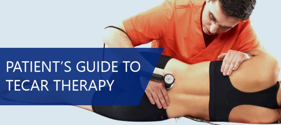
When cell membranes are damaged from trauma or overuse, the flow of ions across the cell membrane is disrupted, and the cell becomes dysfunctional. TECAR is an acronym for “transfer of energy capacitive and resistive therapy,” a new technology that helps to restore the ionic charge of damaged cells. TECAR therapy accelerates the healing process, for faster and more effective rehabilitation.
TECAR therapy requires a radiofrequency device, such as the INDIBA TC9 device, that generates an external electrical charge at the frequency of 448 kHz. When TECAR’s radiofrequency electromagnetic current (RFEC) is introduced to damaged tissues, TECAR therapy increases and stabilizes the exchange of ions in damaged cells, to accelerate healing.
TECAR therapy’s thermal effect helps to prepare damaged cells for fascia manipulation therapy, to release adhesions and trigger points that interfere with movement.
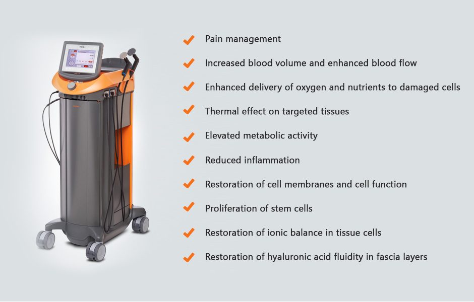
INDIBA therapy has multiple beneficial physiological effects, including:
Pain management
Increased blood volume and enhanced blood flow
Enhanced delivery of oxygen and nutrients to damaged cells
Thermal effect on targeted tissues
Elevated metabolic activity
Reduced inflammation
Restoration of cell membranes and cell function
Proliferation of stem cells
Restoration of ionic balance in tissue cells
Restoration of hyaluronic acid fluidity in fascia layers
INDIBA therapy is currently the most effective form of TECAR therapy. Just a few sessions combining INDIBA therapy with manual myofascial release therapy can achieve what months of physical therapy fail to accomplish.
INDIBA treatment is most effective when used prior to or in conjunction with other therapeutic approaches. We use INDIBA treatment for pain management in the early stages of injury, and to generate a thermal effect on tissues, to make them more pliable and responsive to manual therapies.
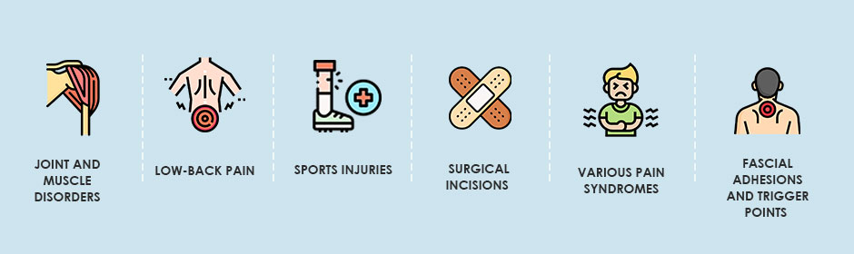
Conditions that benefit from INDIBA treatment include:
Joint and muscle disorders
Low-back pain
Sports injuries
Surgical incisions
Various pain syndromes
Fascial adhesions and trigger points
TECAR therapy is just one of several regenerative technologies we use at NYDNRehab to reduce pain and promote healing at the cellular level.
Physical therapy is an essential step to restoring functional movement after an illness or injury, but physical therapy is not always a stand-alone solution. Sometimes we need to address other issues before beginning physical therapy, to ensure the most successful outcomes.
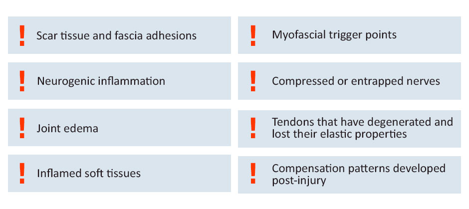
Issues that may need attention before beginning physical therapy include:
Scar tissue and fascia adhesions
Neurogenic inflammation
Joint edema
Inflamed soft tissues
Myofascial trigger points
Compressed or entrapped nerves
Tendons that have degenerated and lost their elastic properties
Compensation patterns developed post-injury
The clinic at NYDNRehab features a growing toolbox of regenerative technologies to prepare the body’s tissues for the rigors of physical therapy. Technologies like
TECAR therapy or shockwave therapy, ultrasound-guided needling procedures, and other new technologies can be game-changers that reduce pain and inflammation so you can begin to move freely. Regenerative technologies are designed to speed up the healing process and restore tissues to their normal healthy state.
Treating injured tissues with regenerative technologies like TECAR therapy prior to beginning physical therapy can dramatically shorten the duration and frequency of physical therapy sessions. Our goal is to restore functional movement as quickly as possible, without drugs or surgery. At NYDNRehab, we treat the whole patient, not just your symptoms.
1. Is TECAR therapy painful?
TECAR therapy is completely safe, painless and non-invasive. Most patients find INDIBA treatment to be relaxing, and many report feelings of general wellbeing and euphoria after INDIBA therapy.
2. Is INDIBA/TECAR therapy recommended for recovery after sports or exercise?
Yes, TECAR therapy is touted by athletes as a fast and effective way to reduce inflammation, promote healing of soft-tissue microtears, and minimize pain from delayed-onset muscle soreness (DOMS).
3. How many TECAR therapy sessions will I need for a sports injury?
It all depends on the location and severity of the injury, the types of tissues that are damaged, and other factors that are unique to the individual patient. TECAR therapy is usually used in conjunction with other therapeutic approaches. Our personalized approach to patient care ensures that you get the exact amount of INDIBA treatment necessary to heal from your injury.
4. How can you tell if INDIBA therapy is working?
In addition to patient feedback, we use the highest resolution diagnostic ultrasonography to monitor the progress of tissue healing. Our research-grade ultrasound equipment gives us the capacity to view microscopic changes via superb microvascular imaging (SMI), as well as sonoelastography, to test the elastic properties of tissues as they heal.
5. Is TECAR therapy used for cosmetic procedures?
While we don’t provide cosmetology services at NYDNRehab, radiofrequency therapy can be effective for improving cellular function anywhere in the body. You can find cosmetologists who provide an INDIBA facial, to improve the tone and texture of your facial skin at the cellular level. TECAR therapy has also been used successfully as an alternative to liposuction, to reduce localized fat stores in obese individuals.

Verified Expert Profiles
Dr. Lev Kalika is a world-recognized expert in musculoskeletal medicine. with 20+ years of clinical experience in diagnostic musculoskeletal ultrasonography, rehabilitative sports medicine and conservative orthopedics. In addition to operating his clinical practice in Manhattan, he regularly publishes peer-reviewed research on ultrasound-guided therapies and procedures. He serves as a peer reviewer for Springer Nature.
Dr. Kalika is an esteemed member of multiple professional organizations, including:
Below is a prime example of how ultrasound can take the guesswork out of diagnosis.

A bad physical therapy experience is one of the primary causes of unnecessary surgery
In this instance, an athlete was originally diagnosed with minor quadriceps muscle strain and was treated for four weeks, with unsatisfactory results. When he came to our clinic, the muscle was not healing, and the patients’ muscle tissue had already begun to atrophy.
Upon examination using MSUS, we discovered that he had a full muscle thickness tear that had been overlooked by his previous provider. To mitigate damage and promote healing, surgery should have been performed immediately after the injury occurred. Because of misdiagnosis and inappropriate treatment, the patient now has permanent damage that cannot be corrected.
The most important advantage of Ultrasound over MRI imaging is its ability to zero in on the symptomatic region and obtain imaging, with active participation and feedback from the patient. Using dynamic MSUS, we can see what happens when patients contract their muscles, something that cannot be done with MRI. From a diagnostic perspective, this interaction is invaluable.
Dynamic ultrasonography examination demonstrating
the full thickness tear and already occurring muscle atrophy
due to misdiagnosis and not referring the patient
to proper diagnostic workup
Demonstration of how very small muscle defect is made and revealed
to be a complete tear with muscle contraction
under diagnostic sonography (not possible with MRI)

Complete tear of rectus femoris
with large hematoma (blood)

Separation of muscle ends due to tear elicited
on dynamic sonography examination


