June 26, 2024
Likely as a result of continuous running and cutting while playing field hockey, a 25 year-old female experienced a plantar fascia tear. The tear was discovered via diagnostic imagining after the athlete had been suffering from painful plantar fasciitis for approximately six months.
After undergoing surgery in order to gait recovery and the neuromuscular training necessary for proprioception. The athlete began post-operative rehabilitation with the following basic goals:
The athlete received traditional rehabilitation therapy that included therapeutic movement, mobilization of joints, and NSAIDs.
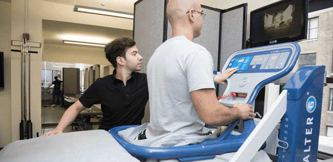
While receiving this therapy, she began the additional conditioning program that included workouts on the Anti-Gravity Treadmill. This four-phase program began in the fifth week after surgery and was scheduled for 10 weeks.
Full weight bearing began in phase one, along with beginning work on range of motion, gait re-education, pain management, and proprioception.
From weeks seven through nine, this phase continued the work from phase one and initiated a beginning cardiovascular training regimen. Additionally, phase two began to increase:
Phase three continued the work of phases one and two, including increases in volume and intensity.
This phase also decreased the angle of the surface on the Anti-Gravity Treadmill and initiated a full foot strike.
From weeks 13 through 15 after surgery, phase four maintained levels of volume and conditioning along with the same angle of surface from phase three. However, load was increased, and work to sports activity during this phase.
Feedback from the athlete was important to determine the speed of the treadmill workouts as well as the force of the impact the athlete would endure with each foot strike.
The rehabilitation program was successfully completed on schedule, and the athlete was given a green light by her physician to manage her gait and increase impact incrementally as pain levels allowed.
Four weeks after completion of the combined programs, the athlete returned to use the Anti-Gravity Treadmill in conjunction with her normal conditioning program.

Verified Expert Profiles
Dr. Lev Kalika is a world-recognized expert in musculoskeletal medicine. with 20+ years of clinical experience in diagnostic musculoskeletal ultrasonography, rehabilitative sports medicine and conservative orthopedics. In addition to operating his clinical practice in Manhattan, he regularly publishes peer-reviewed research on ultrasound-guided therapies and procedures. He serves as a peer reviewer for Springer Nature.
Dr. Kalika is an esteemed member of multiple professional organizations, including:
Below is a prime example of how ultrasound can take the guesswork out of diagnosis.

A bad physical therapy experience is one of the primary causes of unnecessary surgery
In this instance, an athlete was originally diagnosed with minor quadriceps muscle strain and was treated for four weeks, with unsatisfactory results. When he came to our clinic, the muscle was not healing, and the patients’ muscle tissue had already begun to atrophy.
Upon examination using MSUS, we discovered that he had a full muscle thickness tear that had been overlooked by his previous provider. To mitigate damage and promote healing, surgery should have been performed immediately after the injury occurred. Because of misdiagnosis and inappropriate treatment, the patient now has permanent damage that cannot be corrected.
The most important advantage of Ultrasound over MRI imaging is its ability to zero in on the symptomatic region and obtain imaging, with active participation and feedback from the patient. Using dynamic MSUS, we can see what happens when patients contract their muscles, something that cannot be done with MRI. From a diagnostic perspective, this interaction is invaluable.
Dynamic ultrasonography examination demonstrating
the full thickness tear and already occurring muscle atrophy
due to misdiagnosis and not referring the patient
to proper diagnostic workup
Demonstration of how very small muscle defect is made and revealed
to be a complete tear with muscle contraction
under diagnostic sonography (not possible with MRI)

Complete tear of rectus femoris
with large hematoma (blood)

Separation of muscle ends due to tear elicited
on dynamic sonography examination


