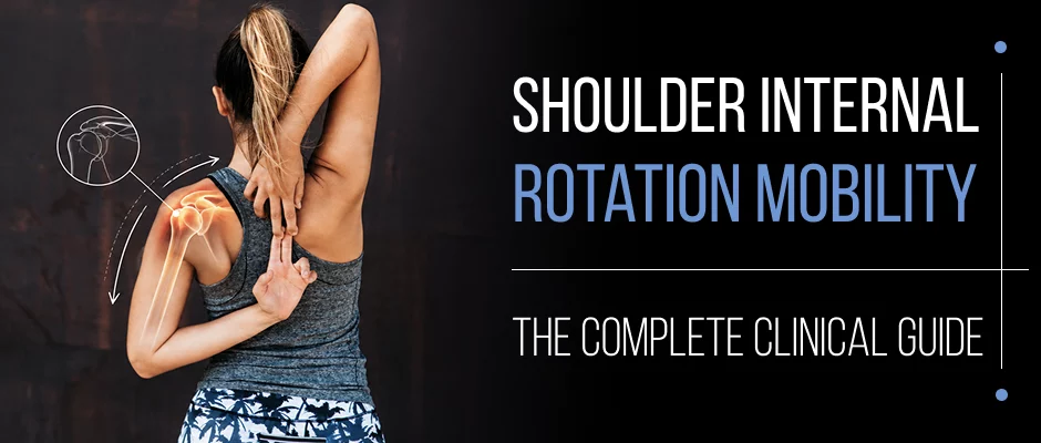
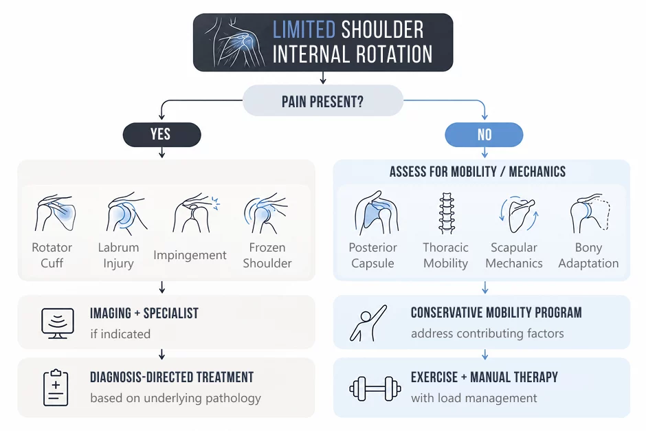
Reaching behind your back to fasten a seatbelt or scratch between your shoulder blades depends on one underrated movement: shoulder internal rotation. When it is limited, everything from throwing to sleeping can suffer, and the right fix depends entirely on why it is restricted.

Shoulder internal rotation is when your upper arm moves inwards toward your body, like when you reach behind you, put on a seat belt or tuck in your shirt.
This movement occurs at the glenohumeral joint and is mainly created by:
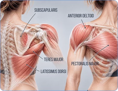
The clinical significance of internal rotation is that it is always accompanied by another movement. The scapula, thoracic spine and posterior capsule all have a direct effect on the amount of motion available and the smoothness, control, and pain-free nature of that motion.
At NYDNRehab, we emphasize understanding the reason behind the movement of a joint rather than just measuring how far it moves.
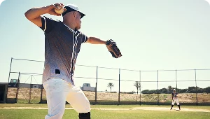
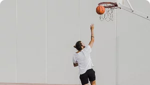
One of the most ignored factors that can cause shoulder pain and injury is restricted internal rotation, especially in overhead athletes and active adults.
Functional consequences include:
In 2003, Burkhart et al. found glenohumeral internal rotation deficit (GIRD) to be a significant factor in “dead arm” syndrome in pitchers. Later, Wilk et al. found that throwing athletes with GIRD ≥25° had a significantly higher risk of injuries.
In the non-athlete population, limited internal rotation can impact dressing, grooming, driving, and sleep, and is easily treated conservatively with targeted treatment when appropriately diagnosed.


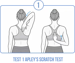
Try to touch the opposite shoulder blade with the back of your hand. Compare both sides. Limitation here is a result of internal rotation, adduction and extension.
Lying on your back with shoulder and elbow bent 90°. Turn the forearm towards the ground. If there is a side-to-side difference >20° then a clinical evaluation is warranted.
Just make a comparison of range on both sides. Non-throwing athletes, differences >20° are clinically significant.
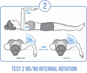
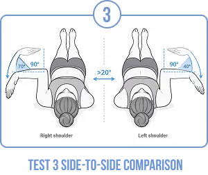
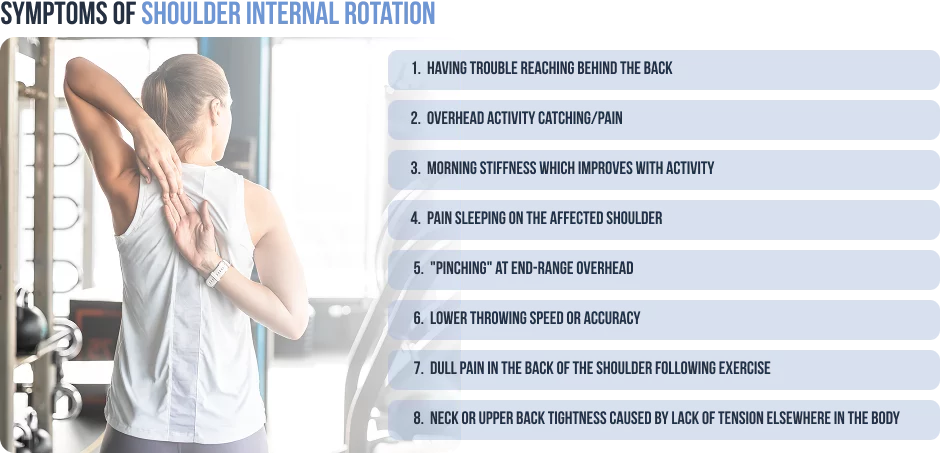
Posterior Capsule Tightness
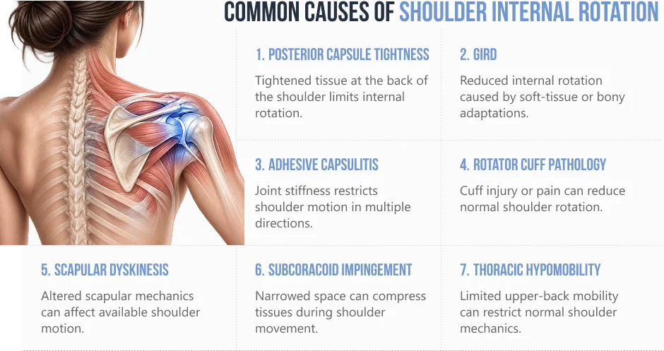
The most common cause in active adults and overhead athletes. The posterior capsule adapts by shortening and thickening in response to repetitive microtrauma, directly decreasing internal rotation.
GIRD (Glenohumeral Internal Rotation Deficit)
Most commonly observed in throwing athletes, a posterior capsule tightness and osseous (bony) humeral retroversion. The treatment is different if it’s soft-tissue or bony in origin and depends on the nature of the GIRD.
Adhesive Capsulitis
Global restriction (internal rotation, external rotation and abduction) is a hallmark of true frozen shoulder. More common in women over 40 and in people with diabetes.
Rotator Cuff Pathology
The most common injuries to the rotator cuff directly impair internal rotation, especially tears to the subscapularis. Any rotator cuff tear can cause pain that will also restrict motion. Injuries to subscapularis are often overlooked unless requested on imaging.
Scapular Dyskinesis
The resting position of the glenoid can be altered by the altered scapular mechanics, and can give a false impression of internal rotation restriction even when the glenohumeral joint is normal.
Subcoracoid impingement
The coracoid and humerous may be abnormally close together at birth.
When the interval is narrowed because of anatomy or abnormal kinematics, the lesser tuberosity and subscapularis tendon get squeezed between the coracoid.
Thoracic Hypomobility
Thoracic extension and rotation are decreased, which is very common in desk workers, and changes scapular movement and decreases available shoulder motion
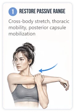
Cross-Body Stretch
Thoracic Extension Mobilisation (Foam Roller)
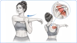

How: Cross arm at shoulder height. Gently over-press with the other hand.
Dosage: Hold 30 seconds × 3–5 reps, 2× daily.
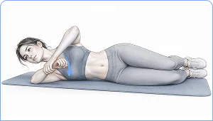
How: Lying on the affected side, elbows at 90°. Push the forearm down towards the floor.
Dosage: 30 seconds x 3-5 reps, 1x/day.
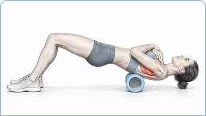
How: Position foam roller at T4-T8. Gently extend head over the roller. Pause 5-10 seconds between each segment.
Dosage: The number of extensions per segment is 3-5 1x/day.
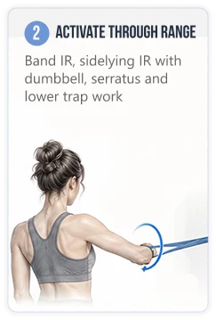
Band Internal Rotation at 90/90
Serratus Anterior Wall Slide
Y-Raise (Prone) Lower Trapezius
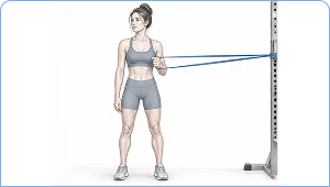
How: Band should be secured at shoulder level. Elbow at 90° and shoulder at 90°. Slowly turn your forearm inwards and then back out.
Dosage: 3 × 15 reps, 3× per week.
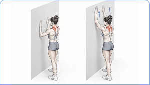
How: Facing a wall, forearms against wall. Move arms up, using pushing motion to move the wall. Lower slowly.
Dosage: 3 × 10 reps, 3–4× per week.
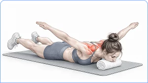
How: Lying on your stomach with your arms in Y position (135°) with your thumbs facing up. Raise arms while squeezing shoulder blades down. Lower slowly, holding for 2-3 seconds.
Dosage: 3 × 12–15 reps, 3× per week
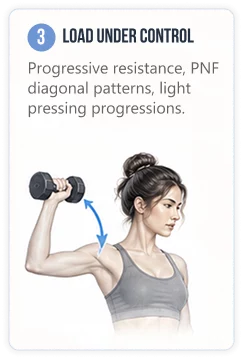
Mobility without motor control is instability. Once range is restored, the nervous system must actively own and control it under load.


For most patients who experience posterior capsule tightness, there is significant improvement in the first 4–8 weeks of regular, focused treatment. These are bony adaptations that can’t be altered through exercise; in these situations, the focus is on optimizing the total arc of motion, not restoring full symmetry, in throwers with humeral retroversion. Frozen shoulder can last for a longer period of 6-18 months.
This is dependent upon the diagnosis. It is generally safe, if done correctly, with moderate force, in cases of confirmed posterior capsule tightness without instability. Not recommended for patients with anterior instability, SLAP tears, or active impingement. The cross-body stretch is more well-tolerated by most patients and backed by similar evidence.
Yes, indirectly. If the shoulder cannot move freely, the body will move in the cervical spine, upper trapezius and thoracic area. This leads to chronic muscular overloading and pain patterns which seem to be from the neck but are actually caused by the lack of shoulder mechanics.
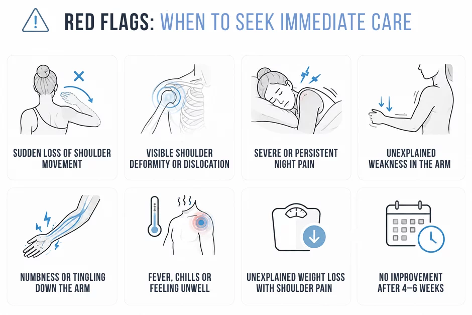
Not without guidance. Untreated or poorly treated rotator cuff tears may become worse if you stretch too vigorously. If you think you may have a tear, especially with a lot of weakness and limited motion, have your physician examine you and obtain appropriate imaging before starting a self-directed program.
Total arc of motion (TAM) is the total of internal and external rotation at 90° of abduction. TAM is approximately equal in both sides in healthy shoulders (usually 160–180°). A difference in TAM of over 10° from side to side is more clinically significant than a single-sided internal rotation deficit, especially for overhead athletes.
Yes, significantly. A rounded shoulder and forward head posture will decrease thoracic extension, change the resting position of the scapula, decrease the amount of up rotation that the scapula will have when elevating the arm and decrease how much glenohumeral internal rotation is available. Often, thoracic mobility work is a prerequisite to effective direct shoulder stretching.
Most cases of restrictive internal rotation can be treated conservatively with physical therapy, especially when it is caused by posterior capsule tightness, GIRD, scapular dyskinesis or thoracic hypomobility. Surgical referral is generally made when the structural lesion is diagnosed (labral tear, full thickness rotator cuff tear, glenohumeral OA) despite a good trial of conservative therapy or if there is a red flag symptom.
When the cause of a limitation of shoulder internal rotation is accurately diagnosed, most people will get much better. The problem is that external appearance of restricted motion is the same, and the cause of the restricted motion is quite different from person to person.
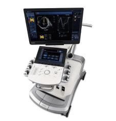
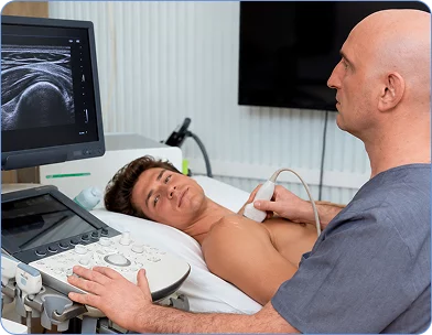
If you have been stretching regularly without any improvement, or if your symptoms persist despite all “right” stretching, it is almost certainly a diagnostic issue, and not an effort issue. The thorough evaluation of the shoulder, which involves clinical examination, motion analysis and, if applicable, real-time diagnostic ultrasound, can help determine the exact cause of your limited motion and help develop a treatment plan specifically geared to your presentation.
Request an appointment»

Our location: 11 West 25th Street 5th floor, New York, NY 10010
Dr. Lev Kalika is a world-recognized expert in musculoskeletal medicine. with 20+ years of clinical experience in diagnostic musculoskeletal ultrasonography, rehabilitative sports medicine and conservative orthopedics. In addition to operating his clinical practice in Manhattan, he regularly publishes peer-reviewed research on ultrasound-guided therapies and procedures. He serves as a peer reviewer for Springer Nature.
Dr. Kalika is an esteemed member of multiple professional organizations, including:

Below is a prime example of how ultrasound can take the guesswork out of diagnosis.

A bad physical therapy experience is one of the primary causes of unnecessary surgery

In this instance, an athlete was originally diagnosed with minor quadriceps muscle strain and was treated for four weeks, with unsatisfactory results. When he came to our clinic, the muscle was not healing, and the patients’ muscle tissue had already begun to atrophy.
Upon examination using MSUS, we discovered that he had a full muscle thickness tear that had been overlooked by his previous provider. To mitigate damage and promote healing, surgery should have been performed immediately after the injury occurred. Because of misdiagnosis and inappropriate treatment, the patient now has permanent damage that cannot be corrected.
The most important advantage of Ultrasound over MRI imaging is its ability to zero in on the symptomatic region and obtain imaging, with active participation and feedback from the patient. Using dynamic MSUS, we can see what happens when patients contract their muscles, something that cannot be done with MRI. From a diagnostic perspective, this interaction is invaluable.
Dynamic ultrasonography examination demonstrating
the full thickness tear and already occurring muscle atrophy
due to misdiagnosis and not referring the patient
to proper diagnostic workup
Demonstration of how very small muscle defect is made and revealed
to be a complete tear with muscle contraction
under diagnostic sonography (not possible with MRI)
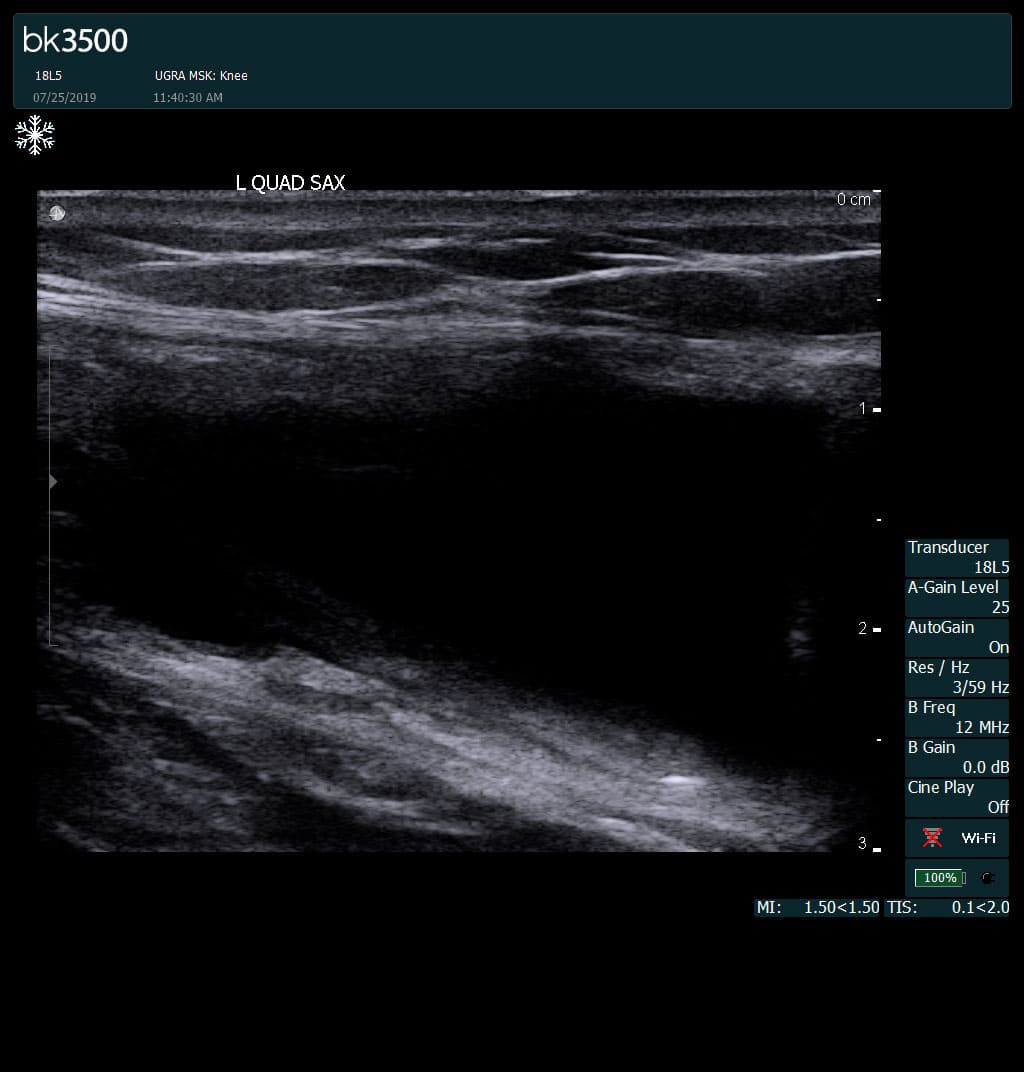
Complete tear of rectus femoris
with large hematoma (blood)
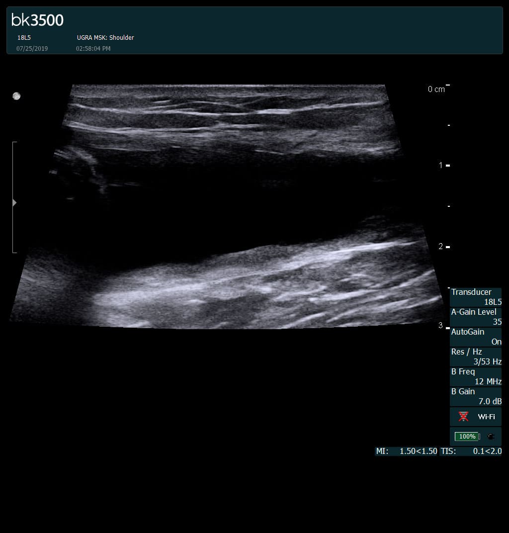
Separation of muscle ends due to tear elicited
on dynamic sonography examination

 Request Telehealth