- Home
- Who We Are
- What We Treat
- Hip
- Elbow and Arm
- Back Pain
- Low Back Pain
- Mid Back Pain
- Neck Pain
- Shoulder
- Wrist Pain
- Knee
- Foot and ankle
- Heel Pain
- Muscle Pain and Myofascial Pain Syndrome
- TMJ and Orofacial
- Womens Health
- Cervicogenic Headache
- Joint Hypermobility Syndrome
- Pelvic Pain
- Fibromyalgia
- Sciatica Pain (Sciatic Nerve)
- Shockwave Therapy for Lymphedema
- Carpal Tunnel Syndrome
- SI Joint Physical Therapy
- Golfers Elbow
- Holistic Podiatry Specialist
- Diagnosis
- Treatment
- Ultrasound Guided Procedures
- Physical Therapy
- Extracorporeal Shockwave Therapy ESWT
- Dynamic Neuromuscular Stabilization
- Fascia Manipulation Therapy
- Dry Needling
- Computer Assisted Rehabilitation Environment (C.A.R.E.N)
- High Energy Inductive Therapy (HEIT)
- Extracorporeal Magnetic Transduction Therapy
- Chiropractic
- Trigger Point Therapy
- Postural Reeducation and posture treatment
- Anatomy In Motion
- Acupuncture
- Tendon clinic
- Сupping therapy
- Gait Analysis LAB
- KINEO intelligent load and reactive neuromuscular training
- Pediatric Physical Therapy
- Neuromuscular therapy NYC
- Prolotherapy Specialist
- High Pressure Cryotherapy
- Blood flow restriction training
- INDIBA Tecar Treatment
- NESA Neuromodulation Therapy
- Sports Medicine
- Ultrasound Guided Procedures
- NYC Marathon
- 3D Running Gait Analysis
- Technology-Based Gait Analysis
- Computer Assisted Rehabilitation Environment (C.A.R.E.N)
- Dynamic Neuromuscular Stabilization
- Running Diagnosis
- Sports Injuries
- Running Services
- Return to Sports
- Sports Physical Therapy
- Sports Injury Prevention
- Golf Injury
- Anatomy in Motion
- Performance Lab
- 3D Motion Analysis
- Sonoelastography for Rehabilitation, Enhanced Performance and Injury Prevention
- Post-Exercise Recovery for Sports, Dance and Fitness
- The Most Comprehensive Assessment for Strength and Power is Driven by Technology
- Insurance
- Telehealth
- Contact Us


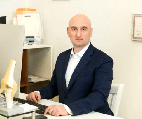
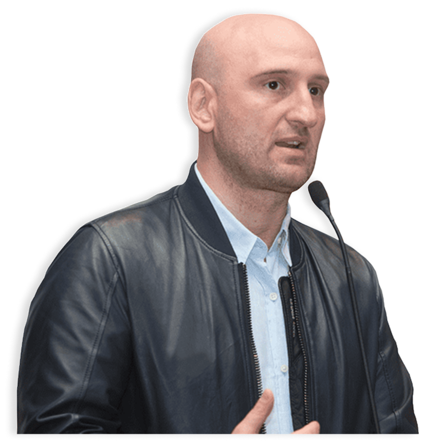
 Dr. Yuri Brosgol
 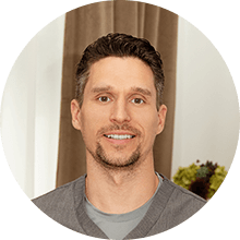 Dr. Michael Goynatsky
 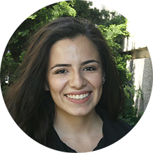 Dr. Daniela Escudero
  Dr. Michelle Agyakwah
  Dr. Tatyana Kapustina