Most people know from basic anatomy that tendons attach muscles to bones. This establishes a biomechanical lever system that causes the associated joint to rotate as the muscle shortens, thereby generating movement. Less understood is the important role of tendon tissue in providing elastic energy that enhances the velocity, force and quality of movement.
Plyometrics is a category of exercise that enhances tendon recoil during jumping, bounding and running activities. Regular plyometric training improves tendon elasticity, reducing muscle fatigue and contributing to force production, to enhance overall performance.

Tendons possess elastic properties that make them essential for generating and dissipating force. Tendon elasticity serves to reduce the workload of muscles and protect the body’s structures from impact forces. When you land from a jump, your tendons rapidly store energy as they elongate, releasing it more slowly as they recoil. The elastic energy released from tendons reduces the muscles’ workload and protects them from injury.
Tendons play three essential roles in human movement:
Without tendon elasticity, human movement would be slower, more energy-intensive and less efficient.
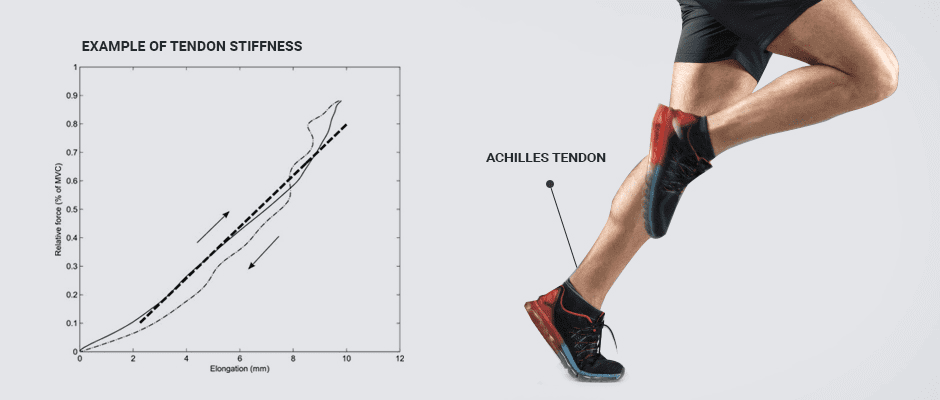
Depending on their location in your body and the role they play in movement, tendons have varying levels of stiffness. To understand this, think of rubber bands of varying thicknesses. A thin rubber band is quite compliant, stretching easily, but it releases a low amount of energy. A thick rubber band is stiffer and more difficult to stretch, but when released, it recoils with a large amount of force.
Your Achilles tendon is like a thick rubber band, able to elongate when subjected to the forces of gravity and muscle contraction and still maintain its form and function. When it recoils, it releases powerful elastic energy that is able to propel your body off the ground. You need both stiff and compliant tendons in different areas of your body, to provide stability and produce dynamic movement.
During the foot strike phase of the running gait cycle, the knee bends, the ankle dorsiflexes and the angle of the foot and lower leg at the ankle decreases. At toe-off, the ankle plantar flexes, the calf muscle shortens and the runner’s body propels upward. Since the calf muscle is responsible for plantar flexion, it seems logical to assume that its concentric contraction provides the force that causes upward propulsion. But that is not the case. The calf muscle’s role during running is to provide eccentric control of ankle dorsiflexion during the foot strike phase.
During dorsiflexion, the Achilles tendon is stretched like a rubber band, storing elastic energy. As the ankle is plantar flexed at toe-off, the Achilles tendon shortens, releasing elastic energy to propel the runner off the ground. If you had to rely on your calf muscles alone to do the work of running, you would quickly become fatigued, and your performance would suffer dramatically.
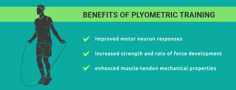
Plyometrics are jumping exercises that use your body weight, sometimes with added external loads, to improve your performance in jumping, sprinting, bounding and running activities. Plyometrics enhance the stretch-shortening cycle (SSC) of muscles and tendons, improving force production and the rate of tendon recoil.
Lower body plyometric training improves performance of the muscle-tendon complex in a number of ways:

Training is essential to athletic performance, but without nutritional support, it will only take you so far. Your tendons and other connective tissues are largely made up of collagen. Your cells build collagen from collagenase, protein enzymes that break down collagen in damaged tissue generate new healthy tissue.
Collagenase is supported and activated by certain nutrients, including:
Supplementing with Vitamin C and hydrolyzed collagen or gelatin can also boost collagen synthesis.
Tendons are continuous with muscle tissue, becoming denser and more collagenous as they approach their attachment to bone. They are less vascular than muscle, meaning they have a lower blood supply. When ruptured, their low vascularity makes tendons slow to heal due to a low supply of oxygen and nutrients.
Tendon ruptures require accurate diagnosis and careful management to restore their full function and elastic properties. Diagnostic ultrasound is an important tool for identifying tendon ruptures and evaluating their severity. It also provides important information about the healing process, helping us make appropriate decisions about treatment and loading during rehabilitation.
Extracorporeal shock wave therapy (ESWT) accelerates the healing process of tendons by stimulating neovascularization and increasing the flow of blood, oxygen and nutrients to the damaged tissues. When applied with ultrasound guidance, ESWT can accurately target injured tissues and stimulate cellular repair.
Tendons play an essential role in sports performance, and tendon injuries need to be managed with care. The sports medicine team at NYDNRehab uses the most advanced technologies and innovative therapies to restore tendon integrity and function.
Our clinic features the latest high-resolution ultrasound technology to diagnose and evaluate musculoskeletal injuries, and ESWT to promote healing and restore function. Our 3D gait analysis lab gives us quantitative data to identify motor deficiencies and compensation patterns that can contribute to tendon damage.
Our experienced sports physical therapists use plyometric training, Kineo smart loading, C.A.R.E.N virtual feedback, DNS and other advanced methodologies to rehabilitate injuries and optimize performance. Contact NYDNRehab today, and take your running and athletic performance to new heights, with less risk of injury.
Resources:
Arabatzi, Fotini, et al. “Effects of two plyometric protocols at different surfaces on mechanical properties of achilles tendon in children.” Asian Journal of Sports Medicine 9.1 (2018).
Konow, Nicolai, and Thomas J. Roberts. “The series elastic shock absorber: tendon elasticity modulates energy dissipation by muscle during burst deceleration.” Proceedings of the Royal Society B: Biological Sciences 282.1804 (2015): 20142800.
Sugisaki, Norihide, and Sadao Kurokawa. “Effect of lower-body plyometric training on athletic performance and muscle–tendon properties.” The Journal of Physical Fitness and Sports Medicine 3.2 (2014): 205-209.

Verified Expert Profiles
Dr. Lev Kalika is a world-recognized expert in musculoskeletal medicine. with 20+ years of clinical experience in diagnostic musculoskeletal ultrasonography, rehabilitative sports medicine and conservative orthopedics. In addition to operating his clinical practice in Manhattan, he regularly publishes peer-reviewed research on ultrasound-guided therapies and procedures. He serves as a peer reviewer for Springer Nature.
Dr. Kalika is an esteemed member of multiple professional organizations, including:
Below is a prime example of how ultrasound can take the guesswork out of diagnosis.

A bad physical therapy experience is one of the primary causes of unnecessary surgery
In this instance, an athlete was originally diagnosed with minor quadriceps muscle strain and was treated for four weeks, with unsatisfactory results. When he came to our clinic, the muscle was not healing, and the patients’ muscle tissue had already begun to atrophy.
Upon examination using MSUS, we discovered that he had a full muscle thickness tear that had been overlooked by his previous provider. To mitigate damage and promote healing, surgery should have been performed immediately after the injury occurred. Because of misdiagnosis and inappropriate treatment, the patient now has permanent damage that cannot be corrected.
The most important advantage of Ultrasound over MRI imaging is its ability to zero in on the symptomatic region and obtain imaging, with active participation and feedback from the patient. Using dynamic MSUS, we can see what happens when patients contract their muscles, something that cannot be done with MRI. From a diagnostic perspective, this interaction is invaluable.
Dynamic ultrasonography examination demonstrating
the full thickness tear and already occurring muscle atrophy
due to misdiagnosis and not referring the patient
to proper diagnostic workup
Demonstration of how very small muscle defect is made and revealed
to be a complete tear with muscle contraction
under diagnostic sonography (not possible with MRI)

Complete tear of rectus femoris
with large hematoma (blood)

Separation of muscle ends due to tear elicited
on dynamic sonography examination


