Achilles tendinitis and patellofemoral syndrome are common overuse injuries occurring in runners and other athletes that affect the Achilles and patellar tendons. Both can be painful and debilitating, and when left untreated, they can lead to degeneration of tendon tissue that causes irreversible damage.
Tendon elasticity has been getting significant attention recently as a critical factor for efficient and injury-free running. Elastic tendons are able to store energy and release it, contributing to force production and sparing muscles from overuse and energy depletion. In general, the stiffer the tendon, the greater its capacity for energy storage and release.
During training and rehab, it is important to keep in mind that tendons are designed for unique functionality, and undergo different loading patterns during different sports activities.
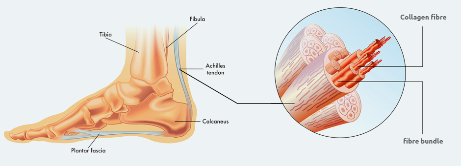
Tendons are tough connective tissues that are continuous with muscles and attach them to bones. Tendon tissue is made up of 70-80% collagen. The remainder is made up of proteoglycans and glycoproteins that include elastin, fibronectin, and tenascin-C. Glycoproteins give tendons elastic properties that allow them to return to their resting length after being stretched during loading. Tendons are encased in epitenon, a sheath of parallel fascicles that prevent them from adhering to neighboring tissues when shortening and lengthening.
To understand tendon elasticity, think of two rubber bands, one thin and one thick. The thin band stretches easily and returns to its original length without much force. When overstretched, a thin band with quickly lose its elastic properties. The thick band is difficult to stretch, and snaps back to its original length with tremendous force. Tendons are like the thick band. When stretched, they contribute significantly to force production by releasing elastic energy as they recoil to their original length.
Now, imagine that you stretch the thick rubber band repeatedly, elongating it to the point where its elastic fibers begin to fray. Eventually, it becomes less elastic and does not completely resume its pre-stretched form. The same thing can happen to tendons. When they are repeatedly overloaded with strain beyond their physiological limits, tendon stiffness is reduced, lowering elastic energy, causing failure and leading to irreversible plastic deformation.

During running, the soleus and gastrocnemius (calf) muscles work to plantar flex the foot, pointing the toes downward as the runner moves into the toe-off stage of the gait cycle. Simultaneously, the Achilles tendon is shortening, releasing elastic energy that contributes to force production to propel the runner’s body upward. The elastic stretch and recoil of the Achilles tendon contributes substantially to total energy storage and return by as much as 35%.
The patellar tendon is stiffer and thicker than the Achilles tendon, and it enhances knee mechanics during running by releasing elastic energy that helps the quadriceps muscles to extend the knee. As running speed increases, both knee flexion and contact time with the ground decrease, demanding a quick and strong release of elastic energy from the patellar tendon. Efficient running mechanics relies heavily on healthy elastic tendons, and injured or painful tendons should not be ignored.
Muscle and tendon loading patterns vary among athletes, depending on the sport. A 2017 study by Wiesinger et al. compared the patellar and Achilles tendon stiffness of water polo players, runners, ski jumpers and a control group.
The research team found that:
Another study by Cristi-Sanchez et al. (2019) found that elite soccer players have significantly stiffer patellar tendons compared to a control group, but found no significant difference in Achilles tendon stiffness. It is important for trainers and therapists to keep sport-specific differences in mind when training athletes for performance, and when rehabilitating tendon injuries.
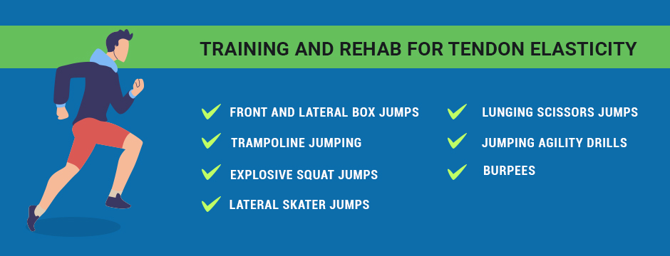
Plyometric (jumping) exercises are the best way to enhance tendon elasticity, but plyometric training needs to be tendon- and sport-specific. With that in mind, we would train a soccer player differently than we would train a runner.
Examples of plyometric exercisers include:
Plyometric training is essential, but it requires an intelligent approach that takes into account tissue stiffness, the tendons at stake, and sport-specific loading patterns. Done incorrectly, plyometric work can easily provoke symptoms.
At NYDNRehab, we utilize a comprehensive protocol for restoring tendon elasticity. Our methodology includes:
Resources:
Cristi-Sánchez, Iver, et al. “Patellar and Achilles Tendon Stiffness in Elite Soccer Players Assessed Using Myotonometric Measurements.” Sports health 11.2 (2019): 157-162.
Robi, Kelc, et al. “The physiology of sports injuries and repair processes.” Current issues in sports and exercise medicine (2013): 43-86.
Wiesinger, Hans-Peter, et al. “Sport-specific capacity to use elastic energy in the patellar and achilles tendons of elite athletes.” Frontiers in physiology 8 (2017): 132.

Verified Expert Profiles
Dr. Lev Kalika is a world-recognized expert in musculoskeletal medicine. with 20+ years of clinical experience in diagnostic musculoskeletal ultrasonography, rehabilitative sports medicine and conservative orthopedics. In addition to operating his clinical practice in Manhattan, he regularly publishes peer-reviewed research on ultrasound-guided therapies and procedures. He serves as a peer reviewer for Springer Nature.
Dr. Kalika is an esteemed member of multiple professional organizations, including:
Below is a prime example of how ultrasound can take the guesswork out of diagnosis.

A bad physical therapy experience is one of the primary causes of unnecessary surgery
In this instance, an athlete was originally diagnosed with minor quadriceps muscle strain and was treated for four weeks, with unsatisfactory results. When he came to our clinic, the muscle was not healing, and the patients’ muscle tissue had already begun to atrophy.
Upon examination using MSUS, we discovered that he had a full muscle thickness tear that had been overlooked by his previous provider. To mitigate damage and promote healing, surgery should have been performed immediately after the injury occurred. Because of misdiagnosis and inappropriate treatment, the patient now has permanent damage that cannot be corrected.
The most important advantage of Ultrasound over MRI imaging is its ability to zero in on the symptomatic region and obtain imaging, with active participation and feedback from the patient. Using dynamic MSUS, we can see what happens when patients contract their muscles, something that cannot be done with MRI. From a diagnostic perspective, this interaction is invaluable.
Dynamic ultrasonography examination demonstrating
the full thickness tear and already occurring muscle atrophy
due to misdiagnosis and not referring the patient
to proper diagnostic workup
Demonstration of how very small muscle defect is made and revealed
to be a complete tear with muscle contraction
under diagnostic sonography (not possible with MRI)

Complete tear of rectus femoris
with large hematoma (blood)

Separation of muscle ends due to tear elicited
on dynamic sonography examination


