August 14, 2023

Known as the Super 7, these seven tennis elbow exercises are collectively used as part of an overall treatment plan for physical injury to be done five times daily.
Every exercise in this regimen is designed to stretch, strengthen or relax a specific set of muscles. This treatment plan is ideal for the rehabilitation or prevention tennis elbow, making it beneficial for people who repeatedly lift or move at the elbow during work or while playing sports.
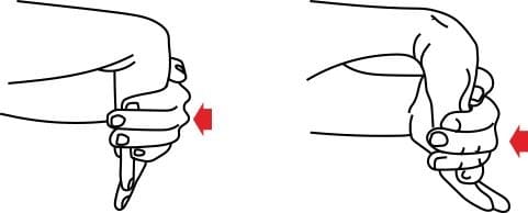
The first stretch loosens the wrist extensors, which are responsible for extending the wrist. During the first exercise, fully stretch the arm. While the arm is stretched, push the palm downward until the stretch is felt within the forearm.
The second stretch involves stretching the wrist flexors, which flex the wrist. Start by completely straightening the arm with the palms facing upward. Then, like exercise number one, push the palm downward.
It is important to overdo the exercises, which can exacerbate the injury.
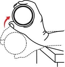
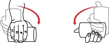
The first building exercise helps the wrist extensors. Start by making the palm point downward while holding the weighted object. Lift the wrist to pull the object back. Keep this lift steady for two seconds before lowering the weight.
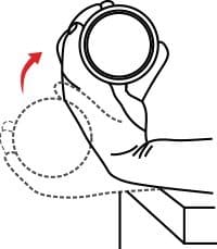
This exercise concentrates on the wrist flexors. Face the palm upward, and hold the weight. Lift the wrist, and keep this movement steady for two seconds before carefully lowering the wrist.
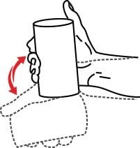
During this exercise, the wrist deviators are strengthened. Make a thumbs up while holding the weight. Then, move only the wrist upward and downward.
The final exercise focuses on the wrist supinato a maximum of 50 times.
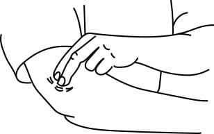
Last on the list is a massage. The sore area should be rubbed for five minutes by applying firm pressure with two fingers.
Sometimes, a certain exercise may worsen the pain. If this happens, immediately call a physical therapist or doctor.

Verified Expert Profiles
Dr. Lev Kalika is a world-recognized expert in musculoskeletal medicine. with 20+ years of clinical experience in diagnostic musculoskeletal ultrasonography, rehabilitative sports medicine and conservative orthopedics. In addition to operating his clinical practice in Manhattan, he regularly publishes peer-reviewed research on ultrasound-guided therapies and procedures. He serves as a peer reviewer for Springer Nature.
Dr. Kalika is an esteemed member of multiple professional organizations, including:
Below is a prime example of how ultrasound can take the guesswork out of diagnosis.

A bad physical therapy experience is one of the primary causes of unnecessary surgery
In this instance, an athlete was originally diagnosed with minor quadriceps muscle strain and was treated for four weeks, with unsatisfactory results. When he came to our clinic, the muscle was not healing, and the patients’ muscle tissue had already begun to atrophy.
Upon examination using MSUS, we discovered that he had a full muscle thickness tear that had been overlooked by his previous provider. To mitigate damage and promote healing, surgery should have been performed immediately after the injury occurred. Because of misdiagnosis and inappropriate treatment, the patient now has permanent damage that cannot be corrected.
The most important advantage of Ultrasound over MRI imaging is its ability to zero in on the symptomatic region and obtain imaging, with active participation and feedback from the patient. Using dynamic MSUS, we can see what happens when patients contract their muscles, something that cannot be done with MRI. From a diagnostic perspective, this interaction is invaluable.
Dynamic ultrasonography examination demonstrating
the full thickness tear and already occurring muscle atrophy
due to misdiagnosis and not referring the patient
to proper diagnostic workup
Demonstration of how very small muscle defect is made and revealed
to be a complete tear with muscle contraction
under diagnostic sonography (not possible with MRI)

Complete tear of rectus femoris
with large hematoma (blood)

Separation of muscle ends due to tear elicited
on dynamic sonography examination


