
A torn meniscus does not mean you will need surgery. Sometimes, the knee will lock, making it impossible to fully extend or bend the leg. This is more likely to require surgical intervention, as this type of meniscus tear is much more resistant to conservative care. Other types of meniscus tears respond well to physical therapy, with a 90 percent success rate.
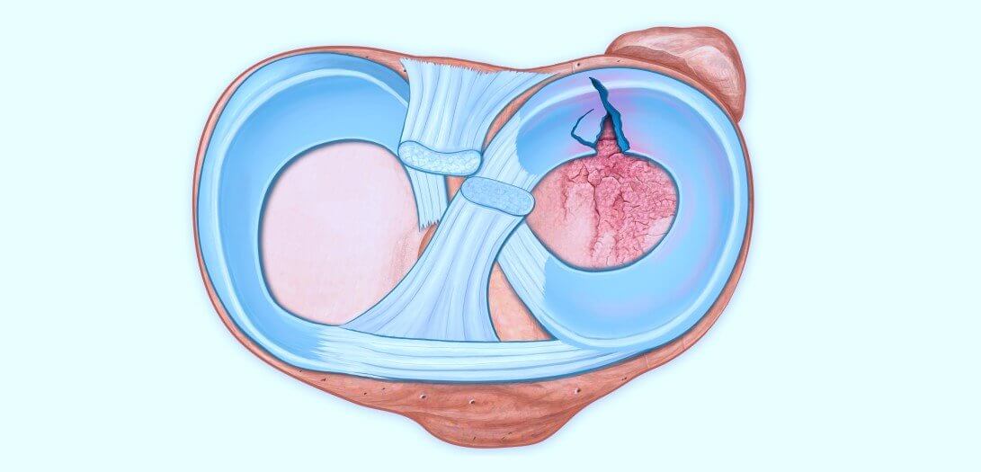
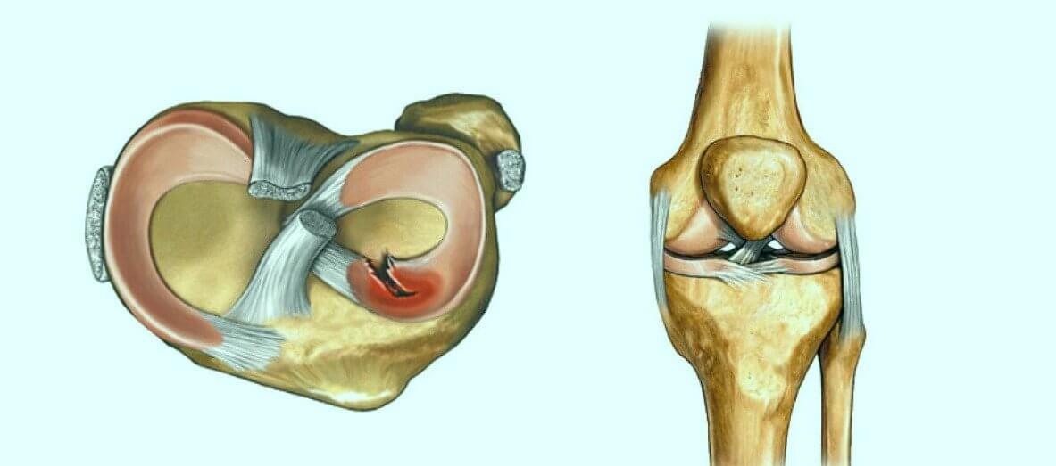
The knee has two large pieces of cartilage shaped like the letter C, located on top of the tibia bone. Their purpose is to protect the bones in the upper leg from hitting the bone in your lower leg.
Stress between the bones is absorbed by the meniscus when you are active, such as when walking, jumping, climbing or running. A meniscus is rubbery, but it can tear when rotating or twisting the knee with your foot in contact with the ground.
A tear of the meniscus is a rather common injury, particularly in people who play sports. About 40 percent of people over 65 also have them, and as many as half a million people suffer from them each year.
When you tear your meniscus, you may have a popping sound or sensation in your knee. Immediately afterward, you will feel pain on the inside or on outside of the knee. Swelling of the knee will be next, but it may take more than one day before it’s noticeable. People involved in sports may even be able to play for a couple of days afterwards. Once the swelling starts, the pain will likely be very noticeable, and it will be stiff at times.
One of the symptoms of a torn meniscus is the knee locking. It is also possible that your leg cannot be straightened, but it will hurt much more when the knee is twisted, or when squatting. There may also be a strong feeling that the knee is not going to support you, or that it is giving way.
In some situations with a meniscus tear, you will be able to carry on with most activities. If you use your knees in daily activities, you may be unable to continue to do them. Just going upstairs, or even walking, can cause serious pain. Participating in athletics is likely to cause the pain to start again, along with swelling.
In many cases, pain and swelling may subside in four to six weeks. However, if your knee cannot be straightened, you should see a doctor. You should also see a doctor if the knee locks and cannot be bent.
There are several activities that can cause a torn meniscus. The primary activity is sports, especially those where you make quick stops or sudden turns, as in basketball or football. There is an increase in the number of children with torn menisci because of the many children’s sports programs now available.
People who make sudden turns or deep squats, or who do heavy lifting are also subject to tears. After the age of 30, the meniscus usually begins to grow weaker and thinner, which is apt to be a common cause of tears in people of that age. At an older age, even simple movements may result in a tear, such as just getting up out of a chair.
Another torn meniscus cause is osteoarthritis. This common disease will weaken and further thin out the meniscus. If the joint did not already have osteoarthritis, it will likely develop if a piece of the meniscus moves around in the knee, causing more damage.
Once a tear of the meniscus is suspected, the doctor will first ask you what you were doing when it happened. The doctor will also likely move your knee and leg into different positions to help determine where the problem is and what might be causing it. Tenderness will also be watched for along the meniscus line.
It is also possible that an MRI might be needed, which will enable better viewing of the cartilage and describing specific damage. An x-ray or ultrasound will not reveal the whole span of the cartilage. However, since 90 percent of patients with meniscus tears respond to conservative care, diagnostic ultrasound combined with joint line tenderness becomes a diagnosis of exclusion, and the patient can proceed straight to physical therapy.
Some people find that a meniscus with a small tear is not that bothersome. Others find it difficult to carry on with their normal activities. The difference may be the location and type of tear.
If the individual takes it easy, it is likely that the pain and swelling will decrease. This may allow for many normal activities to continue. Problems are apt to be seen when bending, or when going up or down stairs.
The meniscus has a blood supply that extends to about one-third of the outermost edges. This can enable faster healing where the blood supply is most abundant. In the past, the inner part of the meniscus was thought to be incapable of self-healing, but this has proven false in more recent research. When a meniscus is repaired with sutures, even where there is a low blood supply, it will heal.
In the past, many doctors believed that part of the meniscus would not heal, and they simply cut away the torn part of the meniscus. Unfortunately, this means an increased likelihood of getting osteoarthritis in that joint.
It is possible to treat very small tears in the meniscus yourself, as they simply heal on their own with time. Treatment of torn meniscus involves several steps, but it will help provide some relief to your painful and swollen knee.
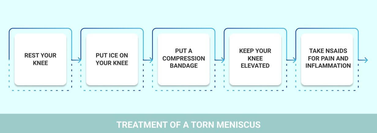
Rest Your Knee – Stay off of your knee as much as possible. You should not use it for any activity other than just walking. You will also probably find it helpful to use crutches to avoid putting much weight on it.
Put Ice on Your Knee – Ice will enable you to reduce any swelling, but do not keep ice on it for more than 20 minutes at a time. Use ice one time about every two to three hours, for a couple of days.
Put a Compression Bandage on Your Knee – Swelling can also be limited by using a compression type bandage. Keep Your Knee Elevated – When not walking, it will help to keep your knee elevated with a pillow.
Take NSAIDs for Pain and Inflammation – Some over-the-counter medications will help to reduce the inflammation. These include medicines such as Motrin and Aleve. Use sparingly.
After the swelling has gone down, you may start a quadriceps strengthening program in your gym.
This should be composed of:
Make sure to use a range of movement that is pain free, and that exercises do not hurt afterwards. Use three sets of ten reps, progressing to fifteen. Progress your weight gradually.
If this still fails, you may want to consult your chiropractor for an individualized guided session.

Verified Expert Profiles
Dr. Lev Kalika is a world-recognized expert in musculoskeletal medicine. with 20+ years of clinical experience in diagnostic musculoskeletal ultrasonography, rehabilitative sports medicine and conservative orthopedics. In addition to operating his clinical practice in Manhattan, he regularly publishes peer-reviewed research on ultrasound-guided therapies and procedures. He serves as a peer reviewer for Springer Nature.
Dr. Kalika is an esteemed member of multiple professional organizations, including:
Below is a prime example of how ultrasound can take the guesswork out of diagnosis.

A bad physical therapy experience is one of the primary causes of unnecessary surgery
In this instance, an athlete was originally diagnosed with minor quadriceps muscle strain and was treated for four weeks, with unsatisfactory results. When he came to our clinic, the muscle was not healing, and the patients’ muscle tissue had already begun to atrophy.
Upon examination using MSUS, we discovered that he had a full muscle thickness tear that had been overlooked by his previous provider. To mitigate damage and promote healing, surgery should have been performed immediately after the injury occurred. Because of misdiagnosis and inappropriate treatment, the patient now has permanent damage that cannot be corrected.
The most important advantage of Ultrasound over MRI imaging is its ability to zero in on the symptomatic region and obtain imaging, with active participation and feedback from the patient. Using dynamic MSUS, we can see what happens when patients contract their muscles, something that cannot be done with MRI. From a diagnostic perspective, this interaction is invaluable.
Dynamic ultrasonography examination demonstrating
the full thickness tear and already occurring muscle atrophy
due to misdiagnosis and not referring the patient
to proper diagnostic workup
Demonstration of how very small muscle defect is made and revealed
to be a complete tear with muscle contraction
under diagnostic sonography (not possible with MRI)

Complete tear of rectus femoris
with large hematoma (blood)

Separation of muscle ends due to tear elicited
on dynamic sonography examination


