Whether you are suffering from chronic pain or an acute injury, you want to recover as quickly as possible so you can get back to your normal daily activities. One exciting therapy that is helping thousands of patients to heal and restore full function is ESWT, or extracorporeal shock wave therapy. ESWT is a non-invasive treatment that has been shown to accelerate tissue repair and increase bone density.
ESWT was discovered by urologists using lithotripsy, a type of shock wave therapy, to break up and disperse patients’ kidney stones. They discovered that patients who had the kidney stone procedure also experienced increased bone density and new tissue growth as a result of lithotripsy treatment.
The benefits of shock wave therapy were soon explored for other applications. In a study sponsored by the Food and Drug Administration (FDA), patients suffering from chronic plantar fasciitis saw an average reduction in pain of 92% after only one ESWT treatment.
“Extracorporeal” simply means that the treatment is performed outside the body, without injections or incisions, eliminating the risks and expenses of invasive treatments that are often only marginally effective. Moreover, ESWT is an outpatient treatment that requires no lengthy recovery period or time off from work, and carries no risk of causing further damage.
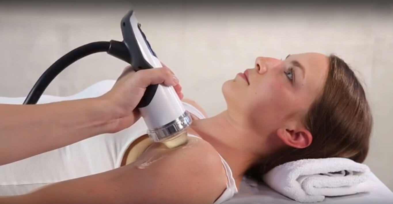
During an ESWT session, the patient is placed in a comfortable position, with the injured area resting on a soft water-filled membrane. A local anesthesia may be used to minimize discomfort. The technician will then use an ultrasound scan to view and target the damaged area. The area is then treated with shockwaves, which are intense, very short energy waves that travel faster than the speed of sound.
ESWT produces ultrasonic waves of energy that, when transmitted into tissue, have a profound effect on reducing pain and stimulating healing. ESWT has two levels of transmission, low energy and high energy. Low energy waves have an analgesic effect by disrupting cell membranes, reducing the patient’s pain.
When high energy waves comes in contact with damaged tissue, they cause a direct biological reaction by increasing blood flow to the area, and initiating a reparative response. Application of high energy waves stimulates the production of fibroblasts—cells in connective tissue that produce collagen and other fibers—and the growth of new healthy tissue.
Although its uses are varied, ESWT is particularly effective in treating injuries where connective tissue attaches to bone. Attachment points of ligaments and tendons in the shoulder, elbow, hip, knee, ankle and foot all respond well to ESWT.
Common conditions treated with ESWT include:
ESWT works especially well for chronic injuries that do not appear to be getting better after an extended period of time. The procedure seems to be able to jump-start the healing process, restoring it to an active phase of healing.
ESWT is one of the most effective non-invasive treatments approved by the FDA. Because the procedure is non-invasive, risks are minimal, and ESWT is frequently recommended as an alternative to surgery.
The trained specialists at NYDNRehab have performed thousands of ESWT procedures for connective tissues throughout the body, with exceptional results, We use real-time diagnostic ultrasound to target the site of injury, and follow up with comprehensive rehabilitation programs like eccentric strengthening exercises and correction and retraining of movement mechanics. Our goal is to restore you to full function, so you can return to the activities you love.

Verified Expert Profiles
Dr. Lev Kalika is a world-recognized expert in musculoskeletal medicine. with 20+ years of clinical experience in diagnostic musculoskeletal ultrasonography, rehabilitative sports medicine and conservative orthopedics. In addition to operating his clinical practice in Manhattan, he regularly publishes peer-reviewed research on ultrasound-guided therapies and procedures. He serves as a peer reviewer for Springer Nature.
Dr. Kalika is an esteemed member of multiple professional organizations, including:
Below is a prime example of how ultrasound can take the guesswork out of diagnosis.

A bad physical therapy experience is one of the primary causes of unnecessary surgery
In this instance, an athlete was originally diagnosed with minor quadriceps muscle strain and was treated for four weeks, with unsatisfactory results. When he came to our clinic, the muscle was not healing, and the patients’ muscle tissue had already begun to atrophy.
Upon examination using MSUS, we discovered that he had a full muscle thickness tear that had been overlooked by his previous provider. To mitigate damage and promote healing, surgery should have been performed immediately after the injury occurred. Because of misdiagnosis and inappropriate treatment, the patient now has permanent damage that cannot be corrected.
The most important advantage of Ultrasound over MRI imaging is its ability to zero in on the symptomatic region and obtain imaging, with active participation and feedback from the patient. Using dynamic MSUS, we can see what happens when patients contract their muscles, something that cannot be done with MRI. From a diagnostic perspective, this interaction is invaluable.
Dynamic ultrasonography examination demonstrating
the full thickness tear and already occurring muscle atrophy
due to misdiagnosis and not referring the patient
to proper diagnostic workup
Demonstration of how very small muscle defect is made and revealed
to be a complete tear with muscle contraction
under diagnostic sonography (not possible with MRI)

Complete tear of rectus femoris
with large hematoma (blood)

Separation of muscle ends due to tear elicited
on dynamic sonography examination


