Our patient was a 22 year-old male presenting with low back pain, scoliosis, and external hip and foot rotation.
The patient had previously sought orthopedic consultations and underwent MRI. The suspected diagnosis was ischio-femoral impingement, a condition associated with hamstring syndrome, to which his external hip and foot rotation were attributed. The rotations were corrected at the thoracic and upper lumbar level (L1-2), and the sacroiliac joint (SIJ) position was restored, but his treatment did not provide significant relief.
In addition to a general clinical exam, we conducted a comprehensive assessment that involved a battery of physical tests and functional neuromuscular ultrasonography. The patient’s health history was carefully reviewed to better understand his overall health profile, as well as any previous treatment he had received.
We determined that the patient’s foot and hip external rotation were related to his spinal and pelvic posture.
We took a holistic and multimodal approach to target the underlying causes of the patient’s pain and dysfunction.
As a result of our treatment protocol, the patient reported multiple positive outcomes:
Our patient’s complaints of back pain, scoliosis and external hip and foot rotation were resolved through a comprehensive assessment and a targeted multimodal treatment approach, resulting in significant improvements in the patient’s overall condition.

Verified Expert Profiles
Dr. Lev Kalika is a world-recognized expert in musculoskeletal medicine. with 20+ years of clinical experience in diagnostic musculoskeletal ultrasonography, rehabilitative sports medicine and conservative orthopedics. In addition to operating his clinical practice in Manhattan, he regularly publishes peer-reviewed research on ultrasound-guided therapies and procedures. He serves as a peer reviewer for Springer Nature.
Dr. Kalika is an esteemed member of multiple professional organizations, including:
Below is a prime example of how ultrasound can take the guesswork out of diagnosis.

A bad physical therapy experience is one of the primary causes of unnecessary surgery
In this instance, an athlete was originally diagnosed with minor quadriceps muscle strain and was treated for four weeks, with unsatisfactory results. When he came to our clinic, the muscle was not healing, and the patients’ muscle tissue had already begun to atrophy.
Upon examination using MSUS, we discovered that he had a full muscle thickness tear that had been overlooked by his previous provider. To mitigate damage and promote healing, surgery should have been performed immediately after the injury occurred. Because of misdiagnosis and inappropriate treatment, the patient now has permanent damage that cannot be corrected.
The most important advantage of Ultrasound over MRI imaging is its ability to zero in on the symptomatic region and obtain imaging, with active participation and feedback from the patient. Using dynamic MSUS, we can see what happens when patients contract their muscles, something that cannot be done with MRI. From a diagnostic perspective, this interaction is invaluable.
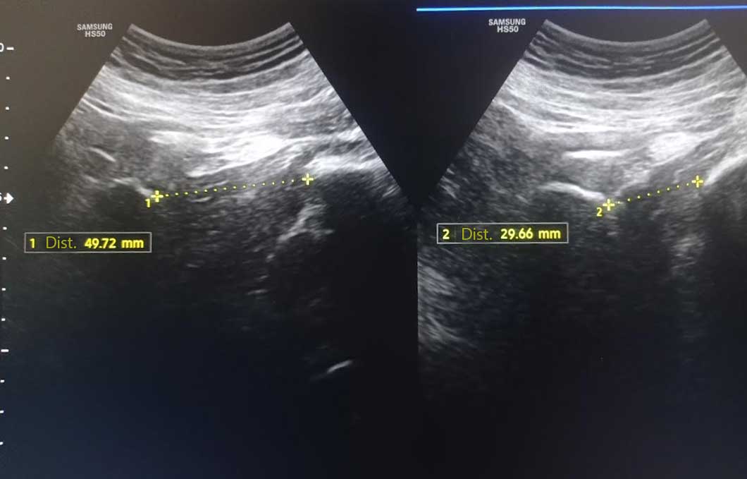
Dynamic ultrasonography examination demonstrating
the full thickness tear and already occurring muscle atrophy
due to misdiagnosis and not referring the patient
to proper diagnostic workup
Demonstration of how very small muscle defect is made and revealed
to be a complete tear with muscle contraction
under diagnostic sonography (not possible with MRI)

Complete tear of rectus femoris
with large hematoma (blood)

Separation of muscle ends due to tear elicited
on dynamic sonography examination


