Diagnostic examination performed by Dr. Lev Kalika, DC, RMSK, clinical director of NYDNRehab, registered in musculoskeletal sonography (RMSK, ARDMS/Inteleos) since 2012.

A 54-year-old recreational tennis player presented with persistent lateral elbow pain and an inability to return to tennis despite previous treatment. MRI had identified a high-grade tear of the common extensor tendon at the extensor carpi radialis brevis (ECRB) origin.
She was facing a decision: proceed with surgical repair, or attempt a further course of orthobiologic treatment.
For that decision, knowing that the tear was described as high grade was not enough. What mattered was how and where the extensor mechanism had structurally failed.
At NYDNRehab we performed a detailed multiplanar examination with high-resolution musculoskeletal ultrasound, tracing the common extensor complex from the lateral epicondyle into the ECRB muscle.
The injury proved more complex than a single tendon defect. Two distinct levels of structural failure were identified.
1. ECRB myoaponeurotic separation
The ECRB muscle was partially separated from the shared ECRB/EDC aponeurotic structure, with loss of the normal continuity between muscle and aponeurosis.
2. Deeper common extensor aponeurotic disruption
A second defect was identified through the deeper common extensor aponeurotic and tendinous fibers, closer to the attachment on the lateral epicondyle.
The underlying lateral collateral ligament complex appeared preserved. That distinction matters, because it separates a severe injury of the common extensor origin from an injury that also involves the ligamentous stabilizers of the lateral elbow, and the two carry different implications for stability and for treatment planning.
A report of a high-grade tear describes severity. It does not necessarily describe the architecture of the tear.
That distinction becomes particularly important when the choice is between surgery and orthobiologics. Tear size and morphology at the lateral elbow have been shown to track with symptoms and function, and larger intrasubstance tears are associated with a lower likelihood of responding to non-operative care. Severity alone, however, does not tell a clinician which tissue interfaces have failed.
The two modalities answer different questions. MRI is the established reference for grading the extent of a common extensor tendon tear. Ultrasound adds real-time, multiplanar and dynamic assessment of the same region, with side-to-side comparison in a single session. Used together, they describe both the size of the lesion and its structural organization. We have written more on that comparison in our article on ultrasound versus MRI for detecting tennis elbow tears.
If an orthobiologic approach is selected, this is not simply a matter of placing PRP into the ECRB tendon. The myoaponeurotic separation, the disrupted aponeurosis, and the residual tendon attachment are anatomically different targets, and reaching them requires ultrasound-guided delivery planned around the specific defects rather than around the general region of pain.
The findings are equally useful if the patient chooses surgery. A pre-operative map of where the extensor mechanism has failed, and which structures appear intact, gives the surgeon information that a severity grade alone does not provide.
Evidence at the lateral elbow supports keeping both routes open. A randomized trial found that ultrasound-guided tendon needling produced therapeutic results comparable to open-release surgery in chronic lateral epicondylosis, with an earlier return to work. Injection evidence is similarly nuanced: pooled randomized data show corticosteroid performing better in the first weeks, with platelet-rich plasma performing better beyond six months. None of this decides a single case. It does mean the decision deserves better anatomical information than a severity grade.
We use high-resolution musculoskeletal ultrasonography not only to identify pathology, but to map the anatomy of an injury before intervention. In practice that means tracing a structure along its full length, examining it in more than one plane, comparing it with the uninvolved side, and assessing behavior under movement and load rather than at rest alone.
This is the same diagnostic logic applied across our lateral elbow cases and our broader sports medicine practice: the treatment plan follows the anatomy, not the label.
For a patient deciding between surgical repair and orthobiologic treatment, that additional precision informs treatment planning and supports a more informed decision.
The question is not only how large the tear is. It is which structures have failed, and where.
If you have been told you have a high-grade tear of the common extensor tendon and are weighing surgery against a further course of conservative or orthobiologic care, a detailed diagnostic ultrasound examination can clarify what has actually failed. Contact NYDNRehab to schedule an evaluation at our Manhattan clinic.
Dr. Lev Kalika is a world-recognized expert in musculoskeletal medicine. with 20+ years of clinical experience in diagnostic musculoskeletal ultrasonography, rehabilitative sports medicine and conservative orthopedics. In addition to operating his clinical practice in Manhattan, he regularly publishes peer-reviewed research on ultrasound-guided therapies and procedures. He serves as a peer reviewer for Springer Nature.
Dr. Kalika is an esteemed member of multiple professional organizations, including:

Below is a prime example of how ultrasound can take the guesswork out of diagnosis.

A bad physical therapy experience is one of the primary causes of unnecessary surgery

In this instance, an athlete was originally diagnosed with minor quadriceps muscle strain and was treated for four weeks, with unsatisfactory results. When he came to our clinic, the muscle was not healing, and the patients’ muscle tissue had already begun to atrophy.
Upon examination using MSUS, we discovered that he had a full muscle thickness tear that had been overlooked by his previous provider. To mitigate damage and promote healing, surgery should have been performed immediately after the injury occurred. Because of misdiagnosis and inappropriate treatment, the patient now has permanent damage that cannot be corrected.
The most important advantage of Ultrasound over MRI imaging is its ability to zero in on the symptomatic region and obtain imaging, with active participation and feedback from the patient. Using dynamic MSUS, we can see what happens when patients contract their muscles, something that cannot be done with MRI. From a diagnostic perspective, this interaction is invaluable.
Dynamic ultrasonography examination demonstrating
the full thickness tear and already occurring muscle atrophy
due to misdiagnosis and not referring the patient
to proper diagnostic workup
Demonstration of how very small muscle defect is made and revealed
to be a complete tear with muscle contraction
under diagnostic sonography (not possible with MRI)
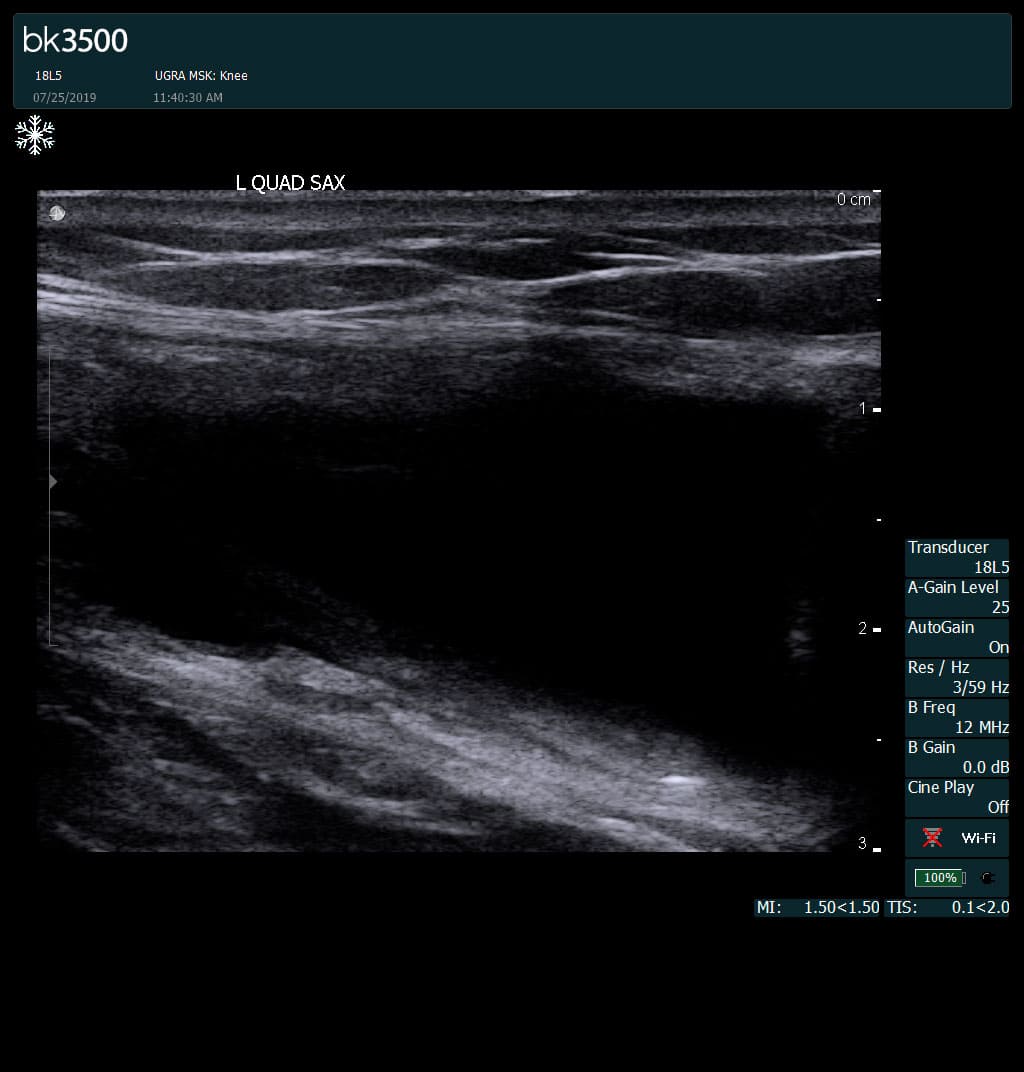
Complete tear of rectus femoris
with large hematoma (blood)
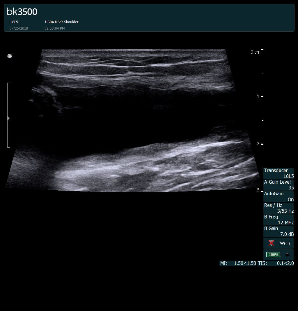
Separation of muscle ends due to tear elicited
on dynamic sonography examination


 Request Telehealth