January 2, 2025
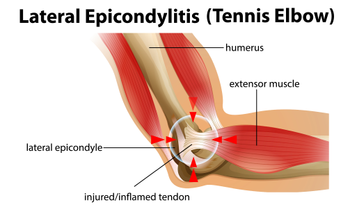
 Chronic lateral epicondylitis — aka tennis elbow – is a painful condition caused by repetitive overuse of the forearm wrist extensor muscles that attach to the epicondyle of the humerus bone, just above your elbow. Although tennis elbow can occur at any age, the condition is most commonly reported in adults aged 35-60. Despite its nickname, only about 20 percent of tennis players actually experience tennis elbow.
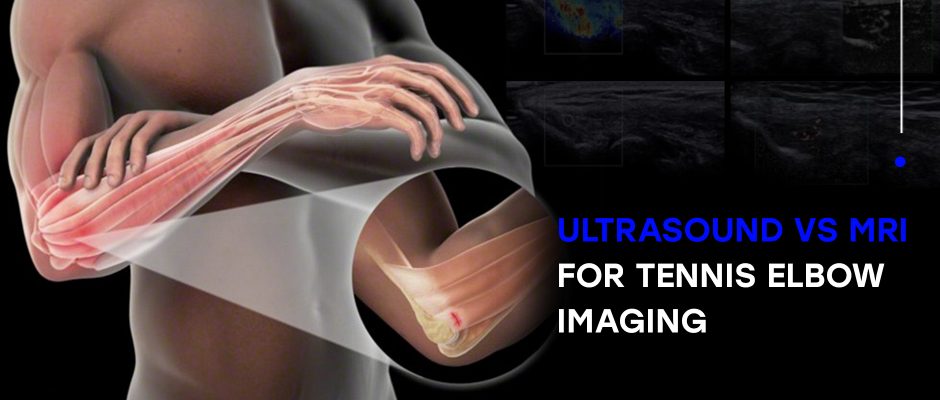
Learn about the causes and symptoms of chronic lateral epicondylitis, other conditions that mimic its symptoms, and why high resolution ultrasonography is far superior to MRI for diagnosing and treating elbow injuries.
In medical jargon, the suffix “itis” indicates an inflammatory condition, but in the case of lateral epicondylitis — as well as other tendon pathologies – this is usually a misnomer. In general, tendon tissue is avascular, meaning it has a limited blood supply, and microscopic evaluation of tendon tissue rarely shows signs of inflammation. In most cases, tendon injuries are marked by the degeneration of fibroblasts and disarray of collagen fibers.
While standard treatments for inflammation – such as ice, compression and NSAIDs – may help to soothe tennis elbow symptoms in the short run by desensitizing nerves, long-term relief requires a multifaceted approach aimed at realigning collagen fibers and rehabilitating the affected structures.
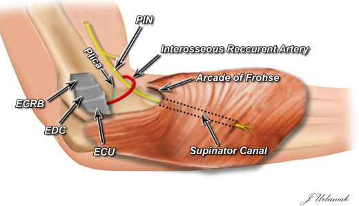
Early symptoms of tennis elbow begin as pain over the lateral elbow that intensifies during activity and abates during rest. Pain usually arises within 24-72 hours after repetitive wrist extension, with maximum tenderness upon palpation of the extensor carpi radialis brevis (ECRB), which is often the afflicted tendon. Even with reduced activity, tennis elbow symptoms can persist for months, with some cases taking over a year to resolve.

The intensity of tennis elbow symptoms often correlates with the severity of injury.
In conventional medicine, imaging is rarely used for diagnosis of tennis elbow. Instead, doctors rely on reported symptoms and results from the clinical exam. Only when treatments fail to alleviate persistent symptoms is MRI considered, to look for other causes such as osteophytes, degenerative joint disease, osteochondritis dissecans, or stress fractures, all of which share the same symptoms. However, in many cases, the limitations of MRI make it an impractical and ineffective diagnostic tool.
MRI’s shortcomings include:
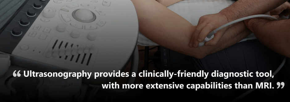
By comparison, ultrasonography provides a clinically-friendly diagnostic tool, with more extensive capabilities than MRI.
In addition, high resolution dynamic ultrasonography can distinguish tendinopathy from tendon ruptures. In most cases, patients with tendon tears also have partial ligament tears and/or ligament laxity, conditions that are difficult to visualize with MRI.
According to recent research, a unique imaging technique called stress ultrasonography can directly visualize the ulnar collateral ligament (UCL) – an elbow ligament that is frequently injured during repetitive overhead movements. Stress ultrasonography allows for assessment of ligament laxity, a distinct advantage over MRI.
A significant disadvantage of symptoms-based diagnosis is that other less common contributing factors may be overlooked. In fact, research indicates that, due to inappropriate handling of the condition, a substantial number of patients diagnosed with lateral epicondylitis do not experience positive therapeutic outcomes.
Lateral epicondylitis is only one of many conditions that affect the lateral elbow region and exhibit the same symptoms as tennis elbow. Failure to consider alternative causes of tennis elbow symptoms can prolong the patient’s suffering and potentially worsen the condition.
Diagnostic ultrasound enables the clinician to thoroughly examine the elbow and its surrounding tissues and structures. With dynamic ultrasound, doctors can assess how the various structures interact while in motion, compare the injured and uninjured elbows, and test for tissue laxity using sonoelastography.
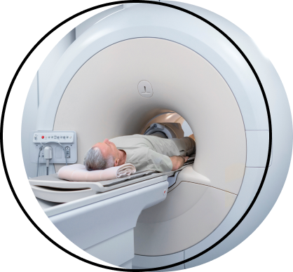
If you’ve ever had an MRI scan, you know what a costly and uncomfortable ordeal it can be. In many cases, doctors are unable to detect tissue damage based on MRI, making the procedure a total waste of time and money.
The clinic at NYDNRehab features the highest resolution ultrasound equipment currently available, with capabilities for sonoelastography and microvascular imaging. Our clinical director, Dr. Lev Kalika, is a recognized expert in diagnostic ultrasonography, credited with multiple scientific publications on the procedure.
If you suffer from chronic lateral epicondylitis or other chronic pain syndromes, we conduct same-day diagnostic ultrasound imaging in the comfort of our Midtown Manhattan clinic, for a fraction of the cost of MRI.
Contact NYDNRehab today to get a quick and accurate diagnosis, and a personalized treatment plan that gets fast results.

Verified Expert Profiles
Dr. Lev Kalika is a world-recognized expert in musculoskeletal medicine. with 20+ years of clinical experience in diagnostic musculoskeletal ultrasonography, rehabilitative sports medicine and conservative orthopedics. In addition to operating his clinical practice in Manhattan, he regularly publishes peer-reviewed research on ultrasound-guided therapies and procedures. He serves as a peer reviewer for Springer Nature.
Dr. Kalika is an esteemed member of multiple professional organizations, including:
Below is a prime example of how ultrasound can take the guesswork out of diagnosis.

A bad physical therapy experience is one of the primary causes of unnecessary surgery
In this instance, an athlete was originally diagnosed with minor quadriceps muscle strain and was treated for four weeks, with unsatisfactory results. When he came to our clinic, the muscle was not healing, and the patients’ muscle tissue had already begun to atrophy.
Upon examination using MSUS, we discovered that he had a full muscle thickness tear that had been overlooked by his previous provider. To mitigate damage and promote healing, surgery should have been performed immediately after the injury occurred. Because of misdiagnosis and inappropriate treatment, the patient now has permanent damage that cannot be corrected.
The most important advantage of Ultrasound over MRI imaging is its ability to zero in on the symptomatic region and obtain imaging, with active participation and feedback from the patient. Using dynamic MSUS, we can see what happens when patients contract their muscles, something that cannot be done with MRI. From a diagnostic perspective, this interaction is invaluable.
Dynamic ultrasonography examination demonstrating
the full thickness tear and already occurring muscle atrophy
due to misdiagnosis and not referring the patient
to proper diagnostic workup
Demonstration of how very small muscle defect is made and revealed
to be a complete tear with muscle contraction
under diagnostic sonography (not possible with MRI)

Complete tear of rectus femoris
with large hematoma (blood)

Separation of muscle ends due to tear elicited
on dynamic sonography examination


