Heel pain is a common ailment among on-the-go New Yorkers who walk and stand on concrete on a daily basis. You can only ignore heel pain for so long before it becomes debilitating, slowing you down and interfering with your daily activities.
Heel pain can stem from many causes, and accurate diagnosis is critical for successful treatment and quick recovery. If you are looking for a heel pain specialist in Manhattan NYC, NYDNRehab features the most advanced technologies and therapies for heel pain diagnosis and treatment in NYC.
or

Clinical director & DC RMSK
Verified Expert Profiles
Dr. Lev Kalika is a specialist in foot and ankle pain, and nerve disorders. He is a world recognized expert in diagnostic musculoskeletal ultrasonography, with multiple research articles to his credit. Heel pain has long been a topic of particular interest to Dr. Kalika, making him a premiere heel pain specialist in Manhattan, NYC.
One of the greatest failures in treating heel pain is misdiagnosis and inappropriate treatment. It is not unusual for practitioners to diagnose heel pain as plantar fasciitis, which is typically treated with steroid injections. Yet 30-40% of all heel pain comes from other causes, especially nerve pathology, which is frequently missed on MRI.
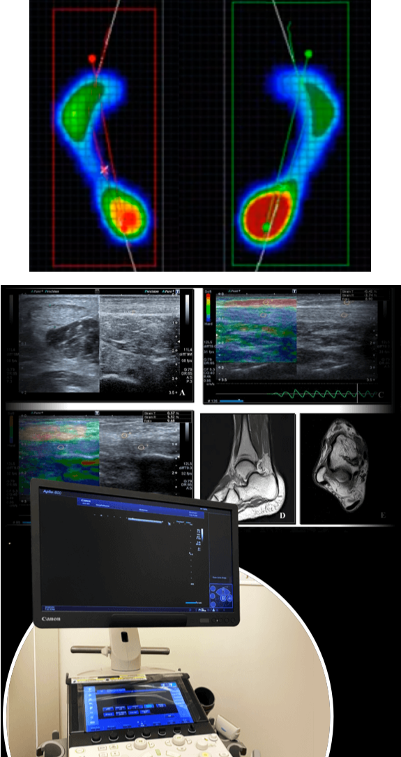
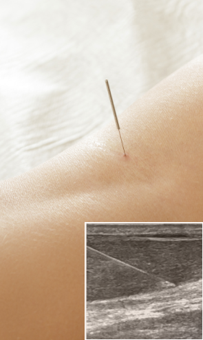
High resolution ultrasonography is rapidly replacing MRI as the imaging mode of choice for musculoskeletal pain and dysfunction. The NYDNRehab clinic features the highest resolution ultrasound equipment available, giving us crystal-clear images of muscles, tendons, ligaments, nerves and bones. Our research-grade ultrasound technology is rarely found in private clinics.
When coupled with Dr. Kalika’s experience and expertise, our cutting-edge technologies make NYDNRehab the clinic of choice for heel pain in NYC.
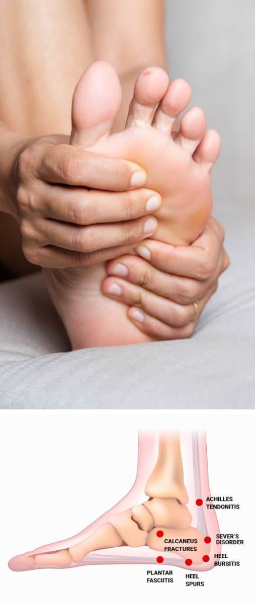
The foot/heel/ankle complex is made up of multiple interdependent structures that work together to propel you forward and upward. The foot’s intricate network of bones, muscles, connective tissues and nerves means that multiple issues can contribute to heel pain.
Plantar fasciitis
Caused by micro tearing of the resilient band of fascia that spans from your heel to your toes. Pain is most often felt on the bottom of the heel, and sometimes extends to the foot arch. The plantar fascia plays a key role in stabilizing the foot during walking and running. Its elastic properties contribute to energy production as it stretches, then shortens, propelling you forward and upward when walking, running or jumping. Plantar fasciitis is a painful condition that severely inhibits the function of your plantar fascia.
Achilles tendonitis and Achilles tendon ruptures.
The Achilles tendon attaches your calf muscles to your calcaneus, or heel bone. Like the plantar fascia, the elastic properties of the Achilles tendon contribute significantly to energy production during locomotion. If left untreated, an injured Achilles can degenerate over time, causing long-term pain and disability.
Heel bursitis
Heel bursitis is inflammation of the heel bursa pads, fluid-filled sacs that cushion soft tissues, preventing them from rubbing against rigid structures. Heel bursitis is felt deep within the heel, or sometimes at the back of the heel.
Calcaneus fractures
Calcaneus fractures are most often the result of trauma, such as a fall from a high elevation that crushes your heel bone upon landing. They can be caused by repetitive overuse during sports or exercise. In severe cases, surgery may be necessary to reconstruct the heel bone, followed by physical therapy to restore functional movement.
Heel spurs
Heel spurs are calcium deposits that arise from the heel bone. They rub against soft tissues, causing pain and inflammation. Heel spurs are common among running and jumping athletes. They are often misdiagnosed as plantar fasciitis, and vice versa.
Sever’s disorder
Sever’s disorder appears most frequently in children and teenage athletes. It occurs when the heel plate suffers micro traumas during a growth spurt, causing damage to the growth plate. The heel in growing children is more vulnerable to damage because the foot and heel grow faster than the muscles and connective tissues of the lower extremity, reaching their full size while other structures are still developing.
Repetitive overuse from sports and exercise
Trauma from falls and accidents
Poorly fitting or unsupportive footwear
Repetitive overuse from sports and exercise
rauma from falls and accidents
Poorly fitting or unsupportive footwear
The labyrinth of structures that make up the foot, heel and ankle can make it difficult to accurately diagnose heel pain. Misdiagnosis is common, resulting in ineffective treatment approaches that prolong the patient’s pain and suffering while wasting their time and draining their bank account.
At NYDNRehab, we leverage the most advanced technologies to ensure that we get to the underlying cause of your heel pain. Accurate diagnosis means we can quickly develop a treatment plan tailored to your personal needs.
Ultrasonography surpasses MRI and Xray for accurate diagnosis
We use the highest resolution diagnostic ultrasound to get crisp and clear images of the structures surrounding your heel. Ultrasound lets us view your heel in real time, with the patient in motion and providing feedback. It also lets us travel the length of large structures like bones, nerves and muscles, to pinpoint the exact area of injury or dysfunction.
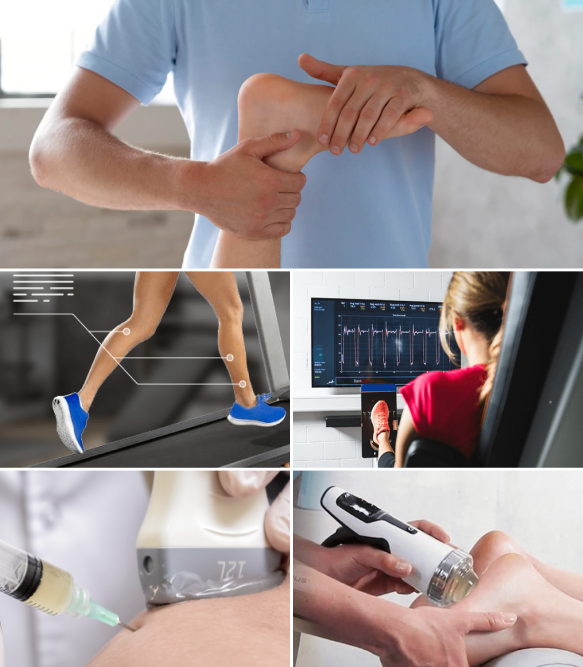
Technology-driven 3D gait analysis reveals mechanical deficits that underlie heel pain
We use the most advanced technologies for gait analysis to evaluate joint angles, muscle firing patterns, ground reaction forces, mass distribution and a myriad of other factors that impact gait. Our advanced equipment allows us to analyze gait in the transverse plane, a third dimension where most injuries occur. Our integrated gait analysis technologies take gait analysis to a whole new level, from observational to quantifiable, so we can work with you to restore healthy gait patterns based on hard data.
At NYDNRehab, we believe that what cannot be measured cannot be successfully treated. Our holistic and integrative approach to patient care keeps us constantly on the lookout for the latest non-surgical treatment options. Our personalized one-on-one treatment protocols mean that you will get the exact therapy you need for your unique condition.
The human body has its own innate healing mechanisms, but they sometimes need a nudge to activate and accelerate the healing process. Regenerative technologies expedite healing and recovery with minimal discomfort for the patient.
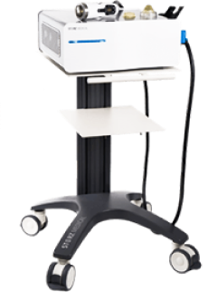
Focused Extracorporeal Shock Wave Therapy (ESWT) produces high-frequency sound waves to desensitize nerves and stimulate healing in damaged tendons, muscles and bones.
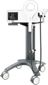
Extracorporeal Magnetic Transduction Therapy (EMTT) transmits high energy magnetic pulses that synchronize with the body’s own magnetic fields, triggering a regenerative response. EMTT waves can penetrate deep tissues, to target difficult-to-reach tendons, muscles, bones and nerves.
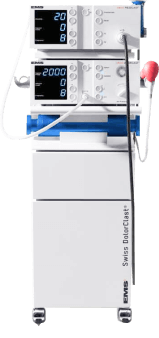
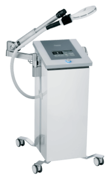
Extracorporeal Pulse Activation Technology (EPAT)
Extracorporeal Pulse Activation Technology (EPAT), also known as defocused shock wave therapy, uses acoustic pressure waves to enhance blood circulation, expediting delivery of oxygen and nutrients to damaged tissues and stimulating cellular metabolism.
High energy inductive therapy (HEIT) uses electromagnetic fields to penetrate cells, tissues, organs and bones, to reactivate the electrochemical function of cells and cell membranes. HEIT is used to stimulate healing of nerves, muscles and blood vessels.
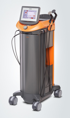
Our INDIBA Tecar therapy machine converts electrical current into a stable radio frequency current of 448 kHz, designed to increase and stabilize the exchange of ions in damaged cells, evoking a regenerative response that accelerates healing. INDIBA can be used to successfully treat joint and muscle disorders, low-back pain, sports injuries, surgical incisions and various pain syndromes. Another therapeutic effect of INDIBA is extreme and prolonged cellular hyperthermia. Due to this effect, INDIBA therapy combined with manual therapy and soft tissue tissue manipulation enables instantaneous release to occur, significantly shortening the number and duration of physical therapy sessions. What is normally accomplished in two months of physical therapy can be accomplished in 3-4 sessions with INDIBA.
Injection therapies use natural/neutral solutions that stimulate cellular repair by either nourishing or irritating the targeted cells. Guidance by ultrasound ensures that the injected substances hit their mark, for maximum effectiveness.
Dr. Kalika is currently a certified member of:
American Institute of Ultrasound Medicine

Active member of ISMST
International Society of Extra Corporeal Shockwave Therapy
Active member of GCMAS
Gait and Clinical Movement Analysis Society
Active member of NASS
North American Spine Society
Active member of IADMS
International Association of Dance Medicine and Science
Active member of Virtual Rehabilitation Society
Active member of ASRA
American Society of Regional Anesthesia and Pain Medicine
American Academy
Association of Orthopedic Medicine

Active member of Interventional Orthobiologics Foundation
Busy New Yorkers often put off seeing a heel pain doctor until the pain begins to interfere with their lifestyle. But postponing treatment can exacerbate your injury and lead to long-term disability. You don’t need to live with heel pain when the best heel pain doctor in New York is just around the corner. Contact NYDNRehab today, and get rid of your heel pain for good, so you can get back to your active New York lifestyle.
Clinical director & DC RMSK
 Dr. Yuri Brosgol
MD
Dr. Yuri Brosgol
MD
 Dr. Michael Goynatsky
DPT
Dr. Michael Goynatsky
DPT
 Dr. Daniela Escudero
DPT
Dr. Daniela Escudero
DPT
 Dr. Michelle Agyakwah
DC
Dr. Michelle Agyakwah
DC
 Dr. Tatyana Kapustina
L. Ac.
Dr. Tatyana Kapustina
L. Ac.

Verified Expert Profiles
Dr. Lev Kalika is a world-recognized expert in musculoskeletal medicine. with 20+ years of clinical experience in diagnostic musculoskeletal ultrasonography, rehabilitative sports medicine and conservative orthopedics. In addition to operating his clinical practice in Manhattan, he regularly publishes peer-reviewed research on ultrasound-guided therapies and procedures. He serves as a peer reviewer for Springer Nature.
Dr. Kalika is an esteemed member of multiple professional organizations, including:
Below is a prime example of how ultrasound can take the guesswork out of diagnosis.

A bad physical therapy experience is one of the primary causes of unnecessary surgery
In this instance, an athlete was originally diagnosed with minor quadriceps muscle strain and was treated for four weeks, with unsatisfactory results. When he came to our clinic, the muscle was not healing, and the patients’ muscle tissue had already begun to atrophy.
Upon examination using MSUS, we discovered that he had a full muscle thickness tear that had been overlooked by his previous provider. To mitigate damage and promote healing, surgery should have been performed immediately after the injury occurred. Because of misdiagnosis and inappropriate treatment, the patient now has permanent damage that cannot be corrected.
The most important advantage of Ultrasound over MRI imaging is its ability to zero in on the symptomatic region and obtain imaging, with active participation and feedback from the patient. Using dynamic MSUS, we can see what happens when patients contract their muscles, something that cannot be done with MRI. From a diagnostic perspective, this interaction is invaluable.
Dynamic ultrasonography examination demonstrating
the full thickness tear and already occurring muscle atrophy
due to misdiagnosis and not referring the patient
to proper diagnostic workup
Demonstration of how very small muscle defect is made and revealed
to be a complete tear with muscle contraction
under diagnostic sonography (not possible with MRI)

Complete tear of rectus femoris
with large hematoma (blood)

Separation of muscle ends due to tear elicited
on dynamic sonography examination