Hamstring Tendinopathy

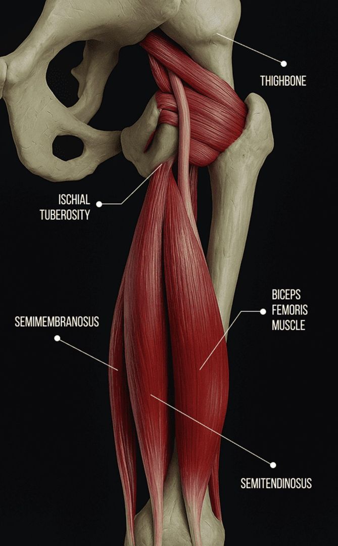
The three hamstring muscles – the semitendinosus, semimembranosus, and biceps femoris – are located at the back of the leg, acting at two joints to facilitate hip extension and knee flexion. During the gait cycle, the hamstrings help to stabilize the hip and knee joints while decelerating the lower leg in the swing phase. The hamstrings and the quadriceps work synergistically during physical activity to mediate force loads, generate forward and upward propulsion, and promote stability.

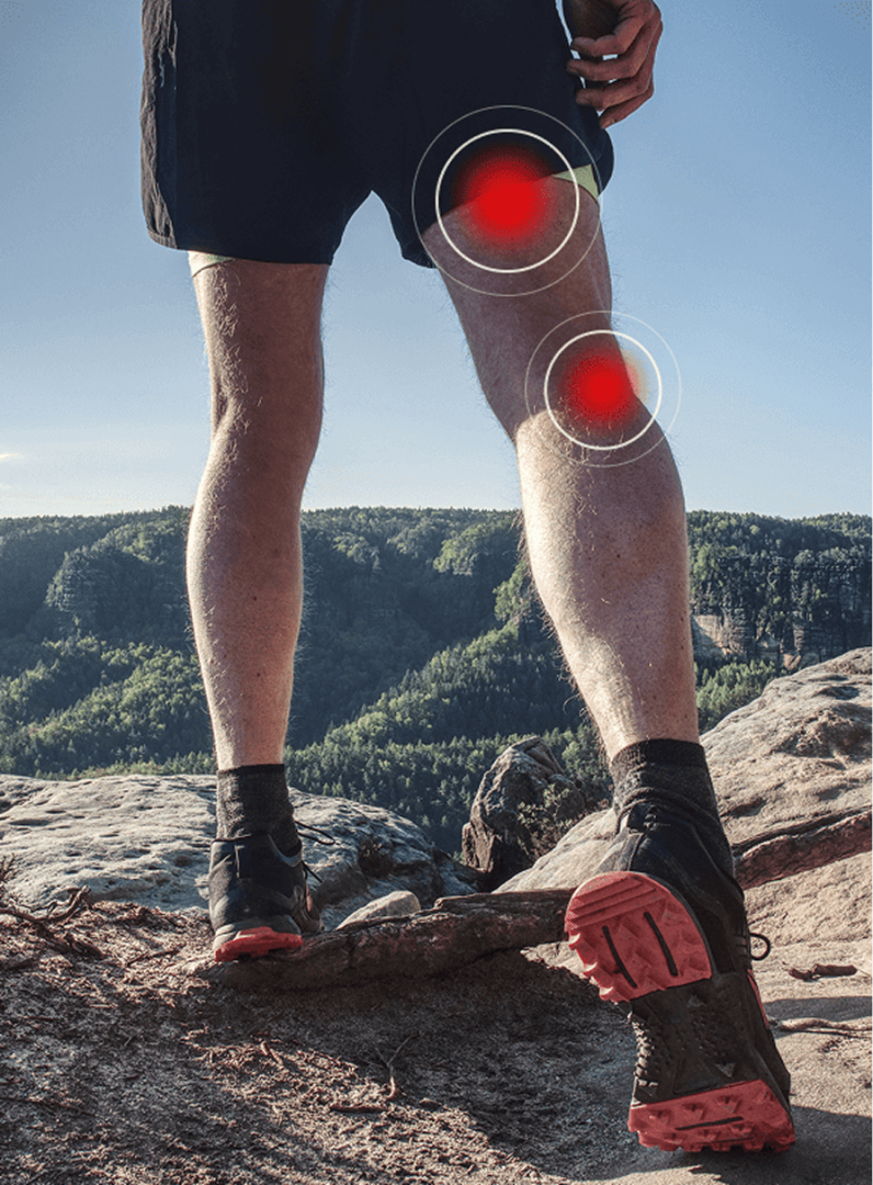
The hamstring muscles are attached to bones by tough, collagenous tendons, where propulsive forces and ground reaction forces converge to produce powerful movement. Hamstring tendon injuries are common in sprinters and runners, and in sports like football and basketball that require explosive bursts of power. Unless fully rehabilitated, injured hamstring tendons can degenerate over time, inhibiting gait and reducing mobility.

Contact us »

We’re located on 25th street in Manhattan NYC.
Open Monday-Friday, 10am-8pm.

Dr. Kalika and Dr. Brosgol are Leading the Charge in Advanced Injury Rehab

Dr. Lev Kalika
Dr. Lev Kalika

Clinical director & DC RMSK

Dr. Lev Kalika, DC clinical director of NYDNRehab, is an internationally recognized expert in diagnostic and musculoskeletal ultrasonography, with multiple research publications to his credit. Dr. Kalika has studied with some of the world’s most prestigious experts in diagnostic, fascia, and nerve ultrasonography, and has presented his research at multiple international conferences. Over the past 25 years, Dr. Kalika has had 100% success in treating hamstring tendinopathies, using physical rehabilitation, shockwave therapy and orthobiologics.

“My 20+ years of success in treating tendon injuries comes from a deep understanding of tendon pathologies, and from the ability to visualize each individual tendon. No two tendons are alike in their anatomy, biomechanics and function. Most tendons are superficial structures, and are much better visualized by high resolution ultrasonography versus MRI. A distinct advantage of diagnostic ultrasonography over MRI is its ability to visualize muscles and tendons in motion.” – Dr. Lev Kalika

Dr. Kalika is an active member of the American Institute of Ultrasound in Medicine (AIUM), and has developed his own unique approach to Dynamic Functional and Fascial Ultrasonography.

Dr. Yuri Brosgol

Orthobiologic specialist

Dr. Yuri Brosgol, MD is a neurologist with 20+ years of experience in treating pediatric and adult myofascial pain. Dr. Brosgol learned fascial hydro release methodology directly from Dr. Carla Stecco, the world’s leading specialist in fascial science. Dr. Brosgol has become a pioneer in the use of orthobiologic solutions, blazing the trail for transformative advancements in the treatment of musculoskeletal injuries.

Together, Dr. Kalika and Dr. Brosgol are combining their expertise to revolutionize the way joint and tendon injuries are treated. Dr. Kalika’s successful track record of diagnosing and rehabilitating tendon injuries, combined with Dr. Brosgol’s expertise in treating myofascial pain, makes NYDNRehab the clinic of choice for hamstring tendon rehab in NYC.

Why Choose NYDNRehab for Hamstring Injury Rehab?

When it comes to rehabilitating sports injuries, athletes are not willing to settle for mediocre treatment that only targets symptoms – they want fast and effective recovery that puts them back in play at pre-injury performance levels, with minimal risk of reinjury. Many conventional sports therapy clinics rely on cookie-cutter treatment protocols and antiquated recovery timelines that increase injury risk.

At NYDNRehab, we treat the whole athlete, not just your symptoms. Hamstring injuries typically cause compensation patterns that disrupt biomechanics along the entire kinetic chain, and full-spectrum tendinopathy rehab entails more than tissue healing. As holistic practitioners, we identify all factors that stand in the way of complete recovery, and address them in a progressive sequence that culminates in successful return-to-play.

Our clinic features some of the most advanced technologies and cutting-edge therapies currently available, backed by years of experience and patient satisfaction. Our personalized one-on-one approach to injury rehab ensures that you get the most appropriate therapy for your condition, so you can safely return to sports with confidence.

Hamstring Tendon Architecture and Function

The hamstring tendons are robust structures that connect the biceps femoris (long and short heads), semitendinosus, and semimembranosus muscles to bones of the pelvis, femur. and lower kinetic chain.

Each muscle and its tendinous attachments have their own unique architecture:
  • The biceps femoris muscle has a long head that originates at the ischial tuberosity of the pelvis, and a short head that originates about midway along the femur. The tendon inserts at the head of the fibula, at the lateral knee.
  • The semitendinosus muscle originates at the ischial tuberosity, with a long, cord-like tendon that inserts at the proximal tibia.
  • The semimembranosus muscle also originates at the ischial tuberosity, with a broader, membranous tendon inserting at the tibia and adjacent structures.
  • The three hamstring muscles share a common proximal tendon at the ischial tuberosity, and proximal tendinopathy is a frequent hamstring injury.
The hamstring tendons are mostly made of dense, fibrous collagen fibers with high tensile strength. They also contain elastin that allows them to stretch, and they are surrounded by a paratenon – a sheath of connective tissue that enables frictionless gliding among surrounding tissues. Irritation of the paratenon can lead to pain and other symptoms, known as paratenonitis Hamstring tendon collagen fibers are organized in parallel bundles, aligned along the direction of force production to optimize strength. The muscle-tendon junction (MTJ) is the transition zone between muscle fibers and their tendons, and the tendon-to-bone junction is where force generated from muscle contraction is transmitted from tendon to bone, enabling movement and providing joint stability.

Functions of the hamstring tendons include:

Knee flexion, where tendons pull against the tibia and fibula to bend the knee during running, jumping and kicking.

Hip extension, where the proximal tendons assist the gluteal muscles in straightening the hip when rising from a squat or sprinting.

Dynamic coordination with the quadriceps muscles at the front of the thigh to control and knee movement.

Knee stabilization during deceleration or landing, counteracting the forward movement of the tibia.

Pelvic stabilization, helping to maintain lower limb alignment.

Force transmission, transferring muscle contractile forces to bones to generate powerful movement while absorbing and distributing mechanical stress.

The proximal hamstring tendon is prone to strains caused by tensile overload, or tears due to high eccentric loads, where the muscle lengthens under tension during intense physical activity. It is also prone to compressive overload during repetitive hip flexion.

Due to their dense collagenous structure and limited blood supply, hamstring tendons can be slow to heal. Regenerative therapies and orthobiologics are used to stimulate tendon tissue neogenesis and realign collagen fibers that are in disarray due to injury.

Hamstring Tendinopathy
Symptoms and Risk Factors

Symptoms

  • Pain at the back of the hip where the hamstring tendons attach.
  • Buttock pain
  • Pain that increases with certain activities like bending forward, sitting, and running
  • Muscle stiffness in the hamstrings, particularly after periods of inactivity
  • Feelings of hamstring muscle weakness or instability

Risk Factors

  • Training errors, such as sudden increases in training volume or intensity
  • Worn and unsupportive footwear
  • Insufficient warmup before intense activity
  • Poor static and dynamic posture
  • Previous lower extremity injuries
  • Frequent use of corticosteroids, antibiotics and statin drugs
  • Over-40 age demographic
  • Female sex
  • Weak or tight hamstrings and quadriceps
  • Prolonged sitting
  • Training on hard surfaces

Successful Treatment Begins with Accurate Diagnosis

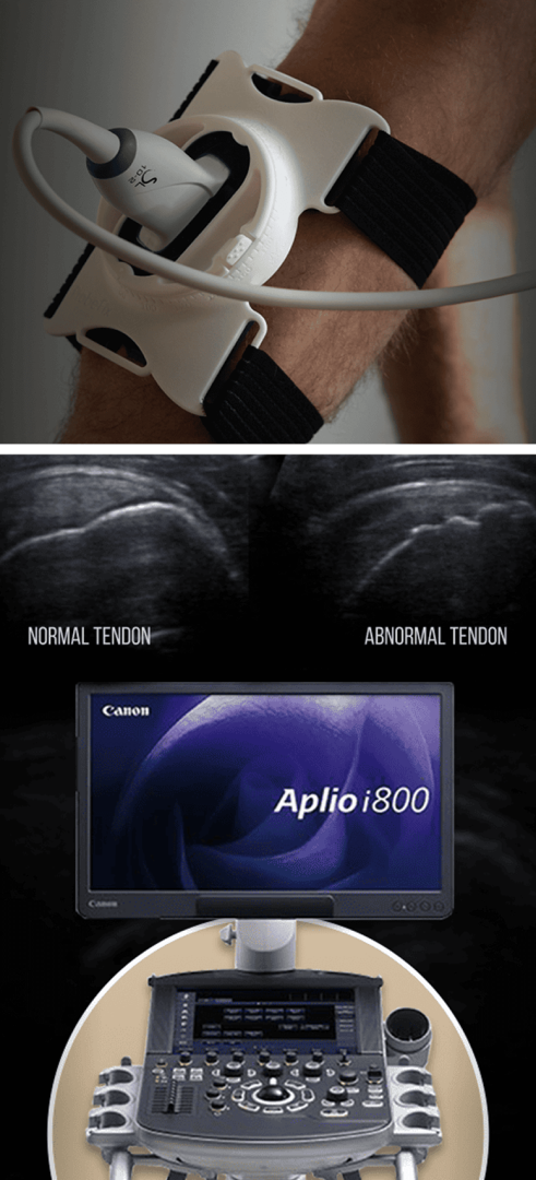
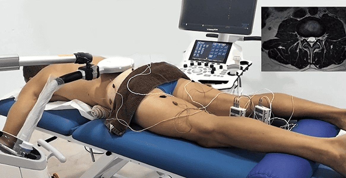
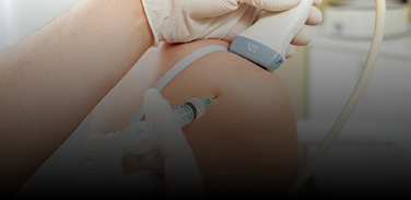
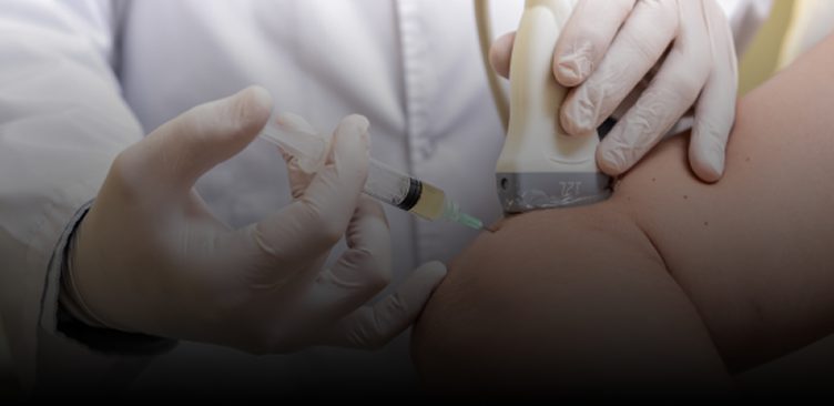
NYDNRehab features research-grade ultrasound equipment with the highest resolution available in New York City. Our equipment gives us capabilities for sonoelastography to test for tendon stiffness, and superior microvascular imaging (SMI) to assess inflammation and detect vascular neogenesis. Sonelastography and SMI are the latest advancements in tendon imaging, and are not possible with other radiological modalities.

NYDNRehab is among the first sports medicine clinics to feature dynamic ultrasonography using the USONO ProbeFix device. ProbeFix attaches directly to the athlete, allowing us to visualize dynamic real-time images of damaged tissues during sport-specific actions. ProbeFix can even be synced with motion capture cameras to produce 3D images of muscles, fascia, bones and joints during physical activity. This game-changing technology gives us a huge advantage for diagnosing tendon injuries and restoring optimal sport-specific biomechanics.

Ultrasound imaging has multiple advantages over MRI:

  • Much higher resolution
  • On-site imaging and interpretation – no waiting for lab results
  • Capabilities for dynamic assessment in real time
  • Ability to visualize fascia adhesions related to tendon degeneration
  • Capabilities for superior microvascular imaging (SMI) to assess the inflammatory component of tendinopathies
  • Capabilities for sonoelastography to assess tendon stiffness
  • Ability to compare the injured and non-injured limb in the same session
  • Ability to track patient progress via dynamic imaging during treatment
1

Running Diagnostics

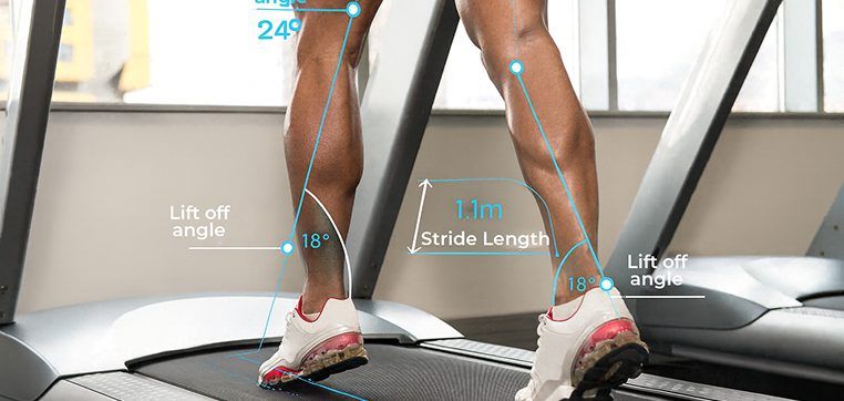
Our state-of-the-art 3D running analysis lab enables us to identify biomechanical and neuromuscular deficiencies that are invisible to the naked eye, in all three planes of motion. Most running injuries occur in the transverse (rotational) plane, which cannot be analyzed in two dimensions. 3D gait analysis empowers us to identify critical errors in running gait, and to distinguish between injuries and compensation strategies.

2

Gait analysis enables us to:

  • Measure and quantify functional limitations, impaired movement and disability
  • Identify movement deviations that are undetectable during the clinical examination
  • Distinguish compensation patterns from their root cause, so that treatment interventions target the whole injury, and not just the symptoms
Data from gait analysis equips us to design effective treatment strategies that address the source of tendinopathy, retraining the athlete to run more efficiently, prolong their running career, and prevent future injuries.

3

ForceFrame Max muscle testing for strength and symmetry

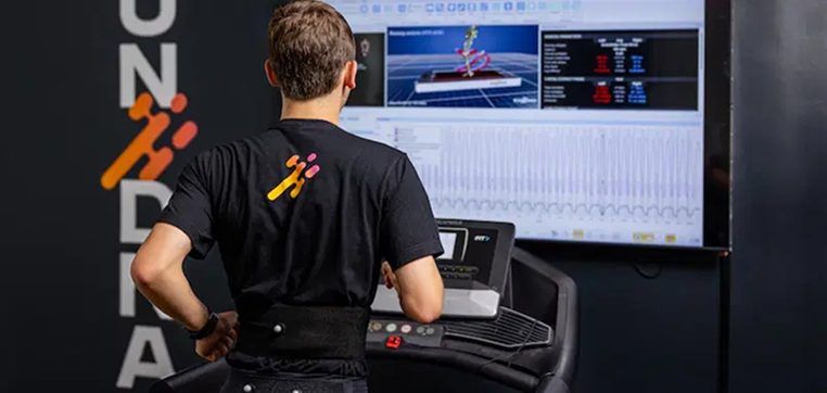
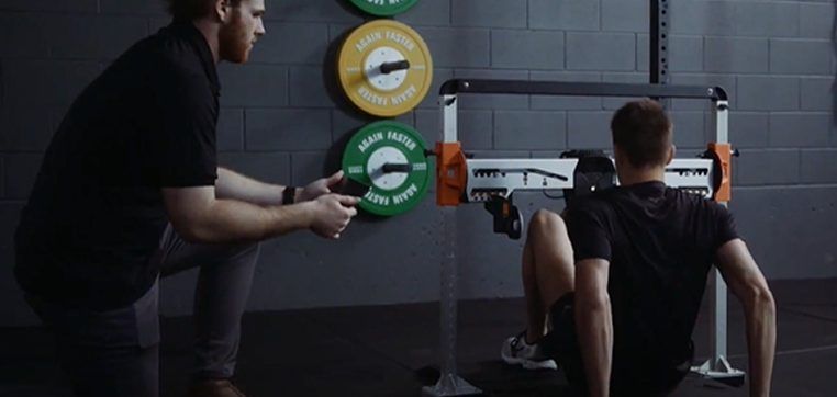
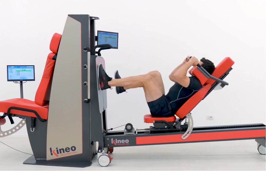
Most people – especially physically active people – develop muscle imbalances and compensation patterns over time that reduce movement efficiency. ForceFrame lets us accurately test individual muscle groups on both sides of the body for strength and symmetry, helping us to identify and correct muscle imbalances, asymmetrical muscle strength, inefficient muscle firing patterns, and compensation patterns developed from past injuries.

Link Between Hamstring Tendinopathy
and Myofascial Dysfunction

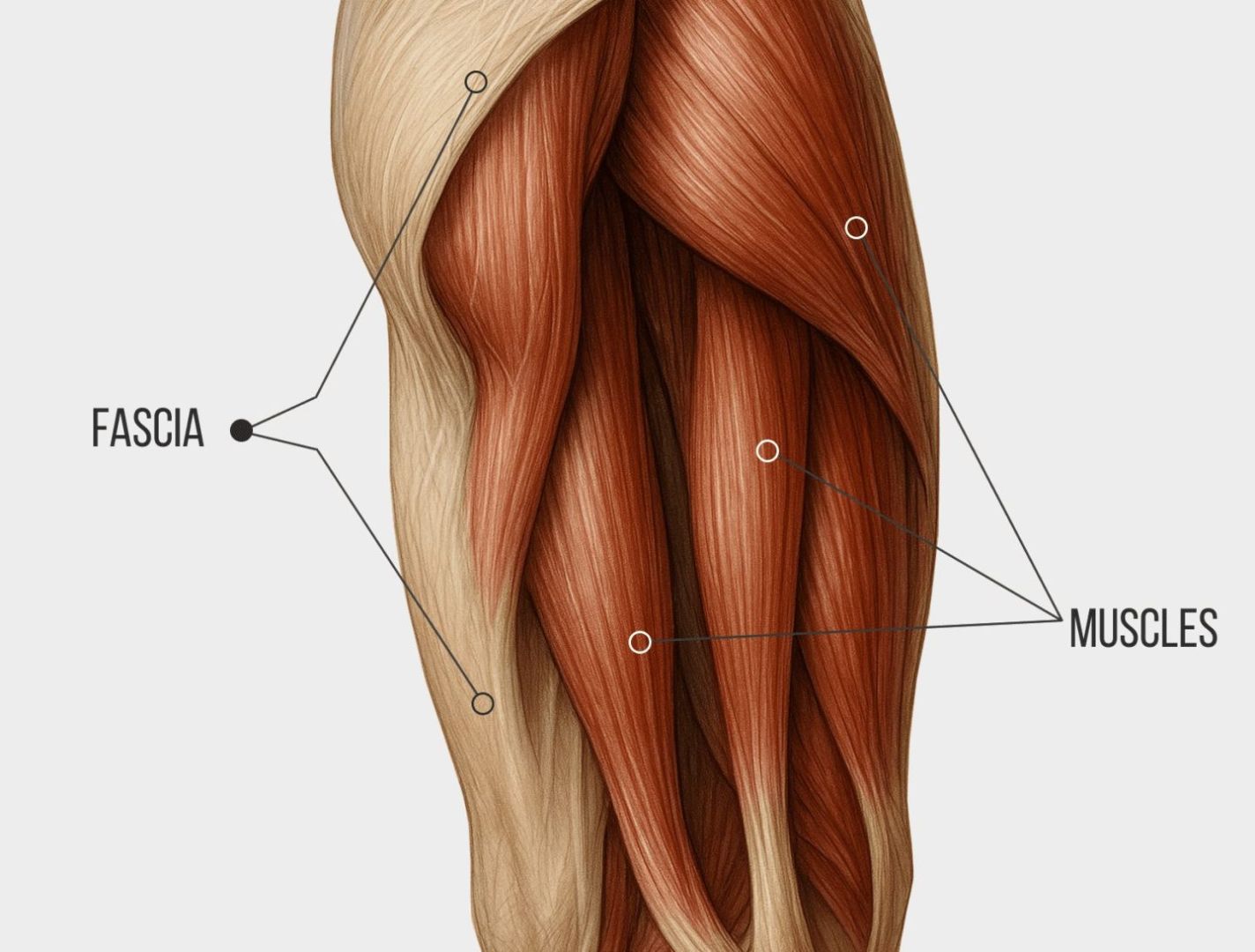
Fascia is a body-wide network of smooth, elastic connective tissue that encases and connects soft tissues, and anchors vital organs in place. Fascia works together with muscles to guide and control movement, transmit forces, and provide tensile support (biotensegrity). Healthy fascia is smooth, slippery, and elastic, enabling nerves and blood vessels to glide among other structures without friction.

When hamstring muscles and tendons are injured, fascia is also affected. Damaged fascia can become dense and sticky, adhering to other structures, inhibiting muscle action, and entrapping nerves and blood vessels. Fascia is densely embedded with proprioceptors that provide feedback to the brain about the body’s position, changes in muscle tension, temperature, pressure, and other factors.

Fascia is also richly innervated with nociceptors – nerve endings that transmit pain signals to the brain – making it a primary pain generator 10X stronger than muscle tissue. When fascia is damaged, biotensegrity is disrupted, resulting in inefficient movement strategies that increase the risk of injury. Prior to starting physical therapy, fascia affecting the hip and knee must be addressed to restore its functional properties.

Stages of Hamstring Tendinopathy Rehab

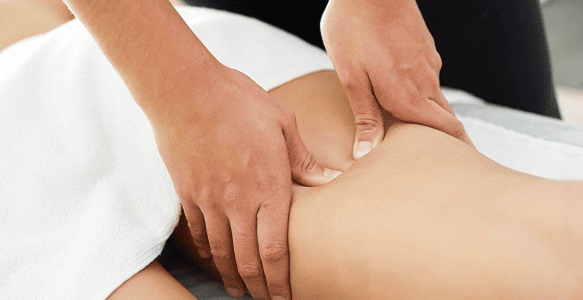
Physical therapy alone is not enough to rehabilitate the hamstring tendons. Prior to beginning physical therapy, we pretreat the tissues to address microtears and ruptures, eliminate myofascial trigger points, and restore biotensegrity throughout the lower kinetic chain. Regenerative technologies, orthobiologics, and manual therapies may all be used to prepare tissues prior to loading.

Phases of Achilles tendon rehab at NYDNRehab include:

  1. Acute phase, aimed at reducing pain and inflammation, and protecting the tendon from further damage or degeneration. Shockwave therapy may be introduced at this phase to reduce pain and inflammation, and jump-start the healing process.
  2. Pre-treatment phase, where we promote tissue regeneration and begin to restore functional mobility. Orthobiologics, regenerative technologies and manual therapies come into play, with the goal of repairing and healing tissues before subjecting them to force loads.
  3. Functional physical therapy phase, to restore joint range of motion and progressively introduce strengthening exercises. This phase includes eccentric loading, stretching, proprioception and balance, and progressive resistance training. At NYDNRehab, hamstring tendon physical therapy is personalized, based on the patient’s unique profile.
  4. Return-to-activity phase, to prepare the patient to return to their regular physical activities. This phase may include sport-specific skills training, plyometrics, functional drills, and enhancement of muscle coordination patterns.

We monitor every phase of your healing journey with high-resolution ultrasound. In addition to confirming tissue healing, ultrasound imaging helps to ensure proper load progression, and lets us closely monitor tissue responses to loading.

Advanced Therapies Accelerate Hamstring Tendinopathy Recovery

Our evidence-based regenerative technologies and cutting-edge therapies are designed to accelerate healing and restore functional mobility, without drugs or surgery. Our holistic approach means your therapy will include attention paid to other tissues and structures that affect – or are affected by – your injury.

Your hamstring tendon treatment protocol may include:

Multimodal Shockwave Therapy

Many clinics advertise shockwave therapy, but few offer radial, linear, focused and defocused shockwaves to address different tissue types. At NYDNRehab, we use ultrasound guided extracorporeal shockwave therapy (ESWT), to promote the healing of tendon and fascia tissue and restore biotensegrity. ESWT helps to realign collagen fibers, promotes hydration of the fascia tissues, and restores tissue gliding.

Ultrasound-Guided Dry Needling

The dry needling procedure inserts filament-thin needles through the skin to reach myofascial trigger points – fibrous knots of tightly contracted tissues that cause pain and disrupt myofascial function. When precisely inserted, the needles cause a twitch response that immediately releases the trigger point.

PENS

Percutaneous neuromodulation (PENS) is a therapeutic approach that uses electrical stimuli to calm and desensitize hyperactivated nerves. It involves the insertion of several filament-thin needles under ultrasound guidance into muscle tissue adjacent to the targeted nerve. PENS stimulates the nerve with varying waves of low frequency electrical current to help restore optimal neural function.

SM Neuromuscular Electrical Stimulation (SMNMES)

SM neuromuscular electrical stimulation (NMES) dynamically interacts with the patient during therapeutic exercises, providing real-time sensory, auditory and visual biofeedback to the patient. This breakthrough technology helps patients to recalibrate muscle actions, to optimize joint function. SMNMES has helped numerous patients to avoid unnecessary shoulder, knee and ankle surgeries, even in complex scenarios.

Stecco Myofascial Release

The Stecco method of fascial manipulation involves deep friction that heats up tissues and stimulates mechanical action. When performed by a trained professional, Stecco fascia manipulation is a fantastic and evidence-based methodology for breaking up scar tissue and releasing adhesions, to restore the integrity of fascial tissue. Patients often report immediate pain relief after a single Stecco session.

Blood Flow Restriction Training (BFRT)

Rebuilding muscle strength while joints and connective tissues are still healing is a challenge for athletes who need to return to sport in the shortest time possible. BFRT enables you to increase muscle size and strength at much lower training volumes, to reduce stress on still-healing structures while rapidly restoring muscle performance. In the case of Achilles tendinopathy, BFRT can help restore strength in adjacent muscles that have atrophied due to Achilles dysfunction.

Kineo Intelligent Load System

We use the Kineo intelligent loading system to create customized training and rehabilitation programs for our patients. With Kineo, we can customize variable load protocols for functional training, core training, agility drills and more. The Kineo variable resistance system lets us design a personalized variable load curve based on the needs of the individual patient.
icon

Orthobiologics Promote Rapid Tendon Healing

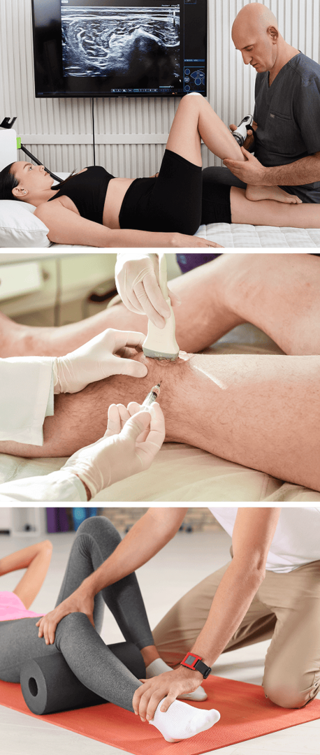
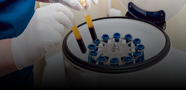
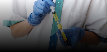
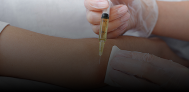
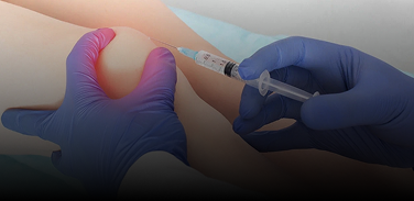
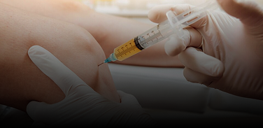
Orthobiologic injection therapies use natural/neutral solutions, injected with precision thanks to ultrasound guidance. The injected solutions stimulate cellular repair by either nourishing or irritating the targeted cells. However, it is important to note that orthobiologics are not a stand-alone solution – they only treat tissue biology, but cannot restore functional biomechanics.

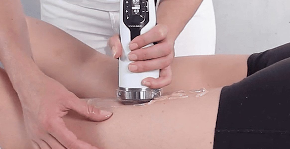
Full rehabilitation of the hamstring tendons requires restoration of biotensegrity and personalized physical therapy to complete the healing process. Treatment results are dramatically enhanced when tissues are pre-treated with focused extracorporeal shockwave therapy (fESWT), and myofascial release techniques.

Orthobiologic procedures available at NYDNRehab include:

Platelet Rich Plasma (PRP)

PRP therapy uses a sample of the patient’s own whole blood, spun in a centrifuge to extract a high concentration of platelets. When injected into damaged tissues, PRP initiates tissue repair by releasing biologically active agents such as growth factors, cytokines, lysosomes and adhesion proteins. To be effective, it is critical to use the right concentration and quality of platelets, and to and follow proper isolation techniques. When administered correctly, PRP can help to jump-start tissue healing in chronic injuries and accelerate repair in acute injuries.

Matrix PRP

For tendon ruptures, Matrix takes PRP therapy to the next level by creating a collagenous bridge between the walls of the tear and the rest of the tendon. Matrix is a highly concentrated PRP, diluted and mixed with fibrinogen. At the injection site, the solution becomes a gel-like collagenous substance that adheres to the walls of the tear and fills the space between them, creating a fibrin matrix that helps to stabilize growth factors and attract stem cell migration to the treatment site.

Platelet Releasate Therapy

Platelet releasate therapy involves injecting platelet releasate – a mixture of growth factors and biomolecules – into injured muscles and tendons to promote healing. Platelet releasate works by activating leukocytes and endothelial cells, and stimulating blood vessel growth, to increase the flow of oxygen, nutrients and growth factors to the damaged tissues.

Alpha-2-Macroglobulin (A2M)

Alpha 2 macroglobulin (A2M) is a naturally occurring blood plasma protein that acts as a carrier for numerous proteins and growth factors. As a protease inhibitor, A2M reduces inflammation in arthritic joints and helps to deactivate a variety of proteinases that typically degrade cartilage.

Prolotherapy and Prolozone

Prolotherapy uses a biologically neutral solution to irritate stubborn tissues, triggering the body’s innate healing mechanisms to grow new normal tendon, ligament and muscle fibers. Prolotherapy is often used for slow-to-heal tendon and ligament ruptures, where low vascularity inhibits tissue healing. Prolozone takes Prolotherapy to the next level by adding a combination of procaine, anti-inflammatory medications, vitamins, and minerals, followed by a mixture of ozone/oxygen gas, injected into targeted joints or tissues. When performed under ultrasound guidance, Prolozone therapy quickly reduces pain and inflammation while jump-starting the healing process.

Hyaluronic Acid Injections

Hyaluronic acid is a natural component of joint synovial fluid and fascial tissue. Its slippery gel-like properties provide lubrication that reduces friction, enabling joints, muscles and fascia to glide freely without pain. In most cases of Achilles tendinopathy, there is a loss of gliding among the muscles of the triceps surae. Hyaluronic acid injections can help to restore hydration to the fascia that encases the muscles, to revitalize its functional properties.

Interfascial Plane and Nerve Hydrodissection

The hydrodissection procedure injects a saline solution into densified fascial layers under ultrasound guidance, separating the layers and releasing entrapped nerves and blood vessels. Hydrodissection is often used in conjunction with manual fascial manipulation, to fully restore fascial integrity.

Tips for Preventing Hamstring Tendinopathy

There are several things you can do to reduce your risk of hamstring tendon injuries:
  • Optimize your nutrition for protein and collagen synthesis. Bone broth and collagen supplements provide the fundamental building blocks for post-exercise tendon recovery. High-quality protein from grass-fed, pasture-raised and wild-caught beef, poultry and fish are the best sources of amino acids essential to muscle and tendon repair.
  • Drink plenty of water. Dry tendons are less elastic and more likely to rupture.
  • Choose supportive footwear that provides shock absorption, and frequently replace worn athletic shoes.
  • Get a biomechanical analysis to trouble-shoot and retrain faulty biomechanics.
  • Allow for ample recovery time after sports and exercise, especially if you have delayed-onset soreness. Most people need at least 48-72 hours to recover from intense physical activity.
  • Stretch regularly to maintain the elastic properties of myofascial tissue.

Get the Most Advanced Hamstring Tendon Rehab in NYC

The recent emergence of advanced, evidence-based methodologies has dramatically changed the way tendinopathies are diagnosed, treated and rehabilitated. At NYDNRehab, we steer clear of invasive surgeries and pharmacological solutions that target symptoms without fixing the problem. Our holistic, non-invasive therapies work with the body’s own self-healing mechanisms to optimize treatment outcomes, restore pain-free movement, and return athletes to play at pre-injury performance levels.

Our personalized approach to patient care ensures you get the best one-on-one Physical Therapy, custom-designed based on your unique profile. Our expertise in tendon rehab combined with our advanced technologies and methodologies make NYDNRehab your clinic of choice for hamstring tendinopathy treatment in NYC.

Our Awards

    Request a Consultation

    We contact you during the day


    Clinical Case Studies
    NYDNRehab


    Case Study: Resolving Chronic Sit Bone Pain in a Female Runner

    Our Patient Our patient is a 45 year-old female runner complaining of left sit bone pain when running and sitting. She has a history of proximal hamstring tendinopathy, and had been treated with plasma injections about 5 years prior, along with physical therapy. Her pain was initially resolved by the injections, and she continued to […]

    learn more

    Case Study: Hamstring and SI Joint Dysfunction in Female Runner

    Our Patient Our patient is a 52 year-old female runner who came to us after unsuccessful treatment elsewhere, complaining of mild low back pain shooting down her right leg. She explained that she had never had low back pain before, and that her locus of pain was slightly lower than her lumbar spine and closer […]

    learn more

    Latest Research & Evidence

    Article

    2023

    Comparison of conservative interventions for proximal hamstring tendinopathy: A systematic review and recommendations for rehabilitation.

    • Dizon
    • Pilar
    Learn More

    Tendinopathy FAQs

    How can I tell if my tendon injury is serious enough to seek treatment?
    If you have chronic pain near the musculotendinous junction that never goes away or has gotten worse, it’s a good idea to see a tendon specialist. Failure to treat an injured tendon can result in degeneration of the tendon tissue, leading to reduced performance and undesirable compensation patterns.
    Can an old hamstring tendon injury be rehabilitated?
    Tendon injuries that were never properly rehabilitated can cause additional problems anywhere along the kinetic chain. Regenerative therapies have the potential to stimulate tissue healing and restore function, even when the injury is several years old.
    Can I return to sports after a serious hamstring tendon rupture?
    Prior to getting back on the playing field, you should make sure your tendon is completely rehabilitated. Not only do you need to fully heal damaged tissues, but you should also restore neurodynamics that were disrupted by your injury, to reestablish coordinated muscle firing patterns and reduce your risk of re-injury.
    My hamstring tendon stops hurting once I warm up. Is it OK to work out?
    Absence of pain does not always indicate that the tendon is functioning properly. If the pain returns once you cool down, you may have serious tendon damage. Loading a ruptured tendon can make it worse. Best to see a specialist.
    What is the treatment for a detached tendon?
    When a tendon tears away from bone, it must be surgically re-attached, followed by intensive physical therapy to restore its mechanical properties and strength. The clinic at NYDNRehab features some of the most advanced technologies available for rehabilitating tendon injuries.
    bg

    In this instance, an athlete was originally diagnosed with minor quadriceps muscle strain and was treated for four weeks, with unsatisfactory results. When he came to our clinic, the muscle was not healing, and the patients’ muscle tissue had already begun to atrophy.

    Upon examination using MSUS, we discovered that he had a full muscle thickness tear that had been overlooked by his previous provider. To mitigate damage and promote healing, surgery should have been performed immediately after the injury occurred. Because of misdiagnosis and inappropriate treatment, the patient now has permanent damage that cannot be corrected.

    The most important advantage of Ultrasound over MRI imaging is its ability to zero in on the symptomatic region and obtain imaging, with active participation and feedback from the patient. Using dynamic MSUS, we can see what happens when patients contract their muscles, something that cannot be done with MRI. From a diagnostic perspective, this interaction is invaluable.

    Dynamic ultrasonography examination demonstrating
    the full thickness tear and already occurring muscle atrophy
    due to misdiagnosis and not referring the patient
    to proper diagnostic workup

    Demonstration of how very small muscle defect is made and revealed
    to be a complete tear with muscle contraction
    under diagnostic sonography (not possible with MRI)

    image

    Complete tear of rectus femoris
    with large hematoma (blood)

    image

    Separation of muscle ends due to tear elicited
    on dynamic sonography examination

    Request Telehealth Request Telehealth Request in office visit Book now