Running Gait and Sports Injury Clinic

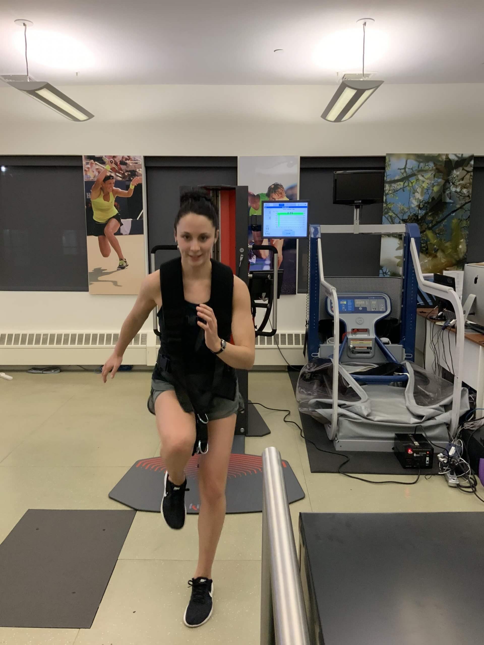
NYDNR’s running and sports injury clinic was developed tory, with a specialized treadmill, a 3D video analysis system with special software, a surface electromyography system, and ground reaction force plates.

What Is the Procedure?

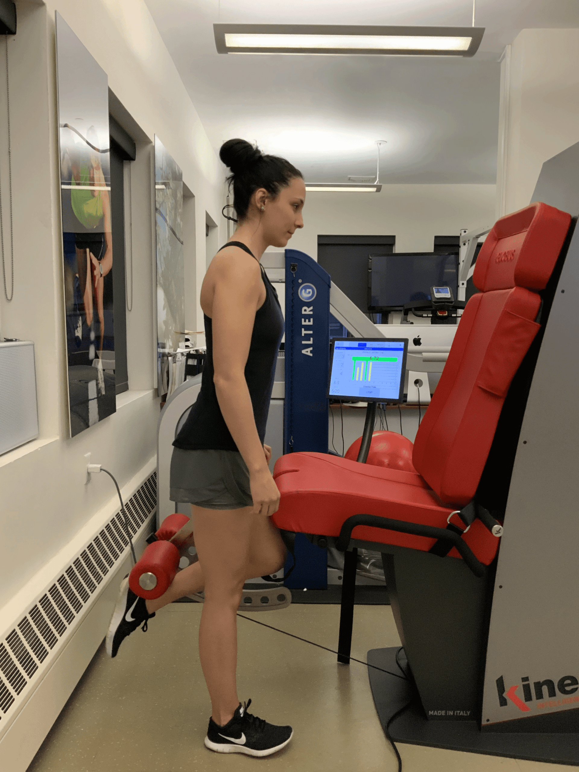
An initial evaluation is performed tory, assessing shoes and abnormal wear patterns, and assessing bodily structure and alignment, with a focus on the trunk and lower extremities.

During gait analysis, movement patterns are monito a computer, where joint angles and speed of motion are calculated from this information.

As part of the analysis, a force plate located at the center of the runway measures the runner’s ground reaction forces, providing information about balance and weight distribution, and revealing where excessive loading is occurring. Videotaping is done from the front, rear and side to provide a visual assessment of the runner. Muscular activity is recorded with a surface electromyography system.

Interpretation of Gait Analysis

Gait analysis gives us quantitative and qualitative data that can be integrated with results from the structural assessment, biomechanical analysis, and injury histo determine the best course of treatment for the individual athlete.

From this analysis, recommendations are typically made in the following areas:

  • Orthotics or braces to control and facilitate normal movement
  • Footwear recommendations and shoe modifications to optimize gait patterns
  • Therapeutic intervention to abnormal gait patterns
  • Correction of movement mechanics that may be contributing to abnormal gait

Be sure to see how these technologies can help you.

Should You Invest In A Gait Analysis?

Testimonials

img
Ibra morales thirty time nyc marathon participant

Dr. Kalika, I just want to my injuries! I’m ready!!

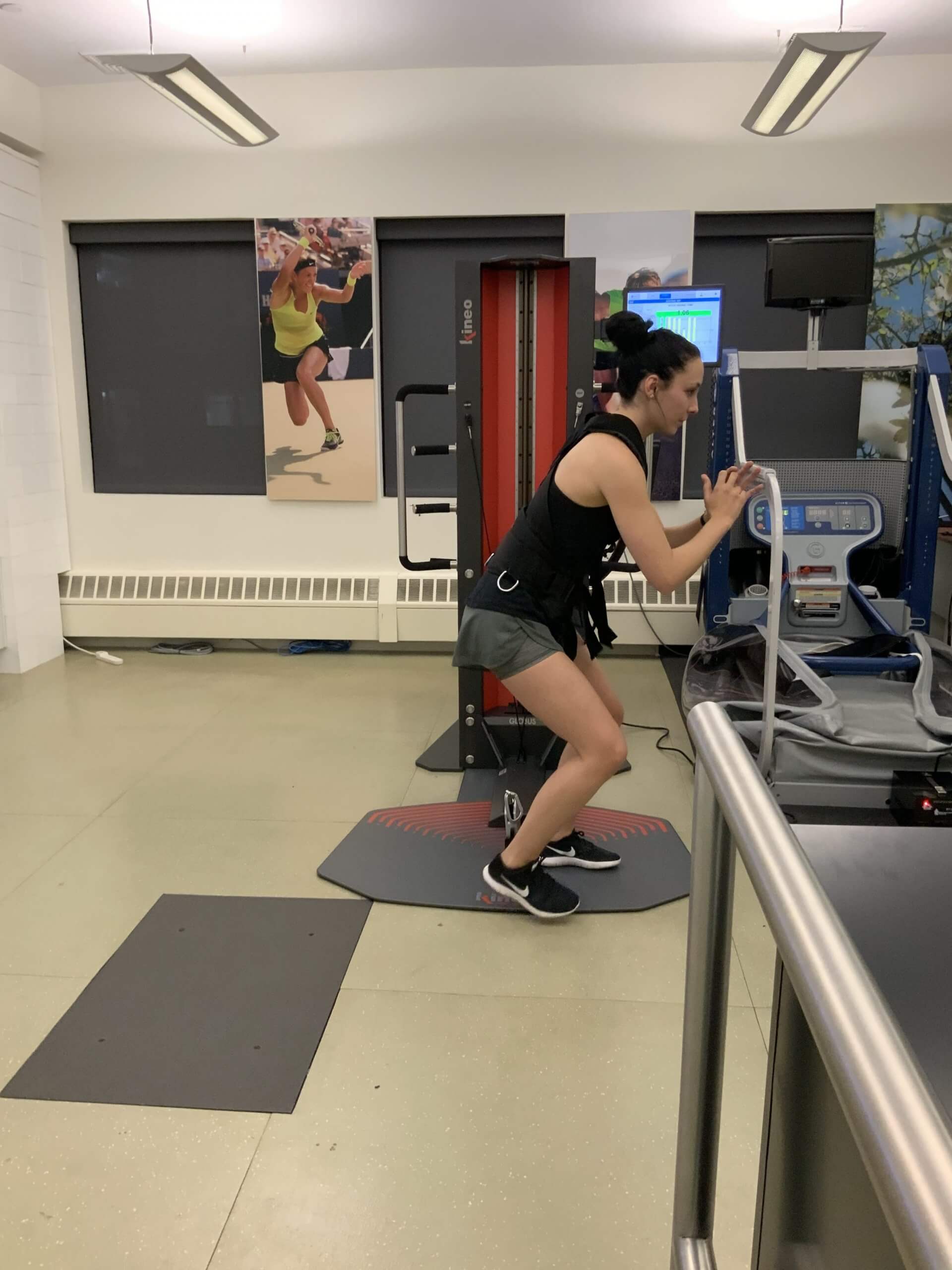
Reactive Neuromuscular Training on Kineo

pic

Kineo – the most versatile muscle testing using artificial intelegence

pic

Kineo – the most versatile muscle testing using artificial intelegence

pic

Kineo – the most versatile muscle testing using artificial intelegence

bg

In this instance, an athlete was originally diagnosed with minor quadriceps muscle strain and was treated for four weeks, with unsatisfactory results. When he came to our clinic, the muscle was not healing, and the patients’ muscle tissue had already begun to atrophy.

Upon examination using MSUS, we discovered that he had a full muscle thickness tear that had been overlooked by his previous provider. To mitigate damage and promote healing, surgery should have been performed immediately after the injury occurred. Because of misdiagnosis and inappropriate treatment, the patient now has permanent damage that cannot be corrected.

The most important advantage of Ultrasound over MRI imaging is its ability to zero in on the symptomatic region and obtain imaging, with active participation and feedback from the patient. Using dynamic MSUS, we can see what happens when patients contract their muscles, something that cannot be done with MRI. From a diagnostic perspective, this interaction is invaluable.

Dynamic ultrasonography examination demonstrating
the full thickness tear and already occurring muscle atrophy
due to misdiagnosis and not referring the patient
to proper diagnostic workup

Demonstration of how very small muscle defect is made and revealed
to be a complete tear with muscle contraction
under diagnostic sonography (not possible with MRI)

image

Complete tear of rectus femoris
with large hematoma (blood)

image

Separation of muscle ends due to tear elicited
on dynamic sonography examination

bg
Buy now 3D Gait
Payment Success
Request Telehealth Request Telehealth Request in office visit Book now