
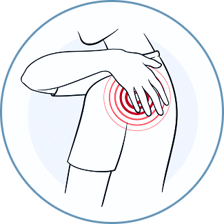
Shoulder Pain and Dysfunction
Personalized boutique services supported by cutting-edge integrative diagnostics and advanced holistic therapies.
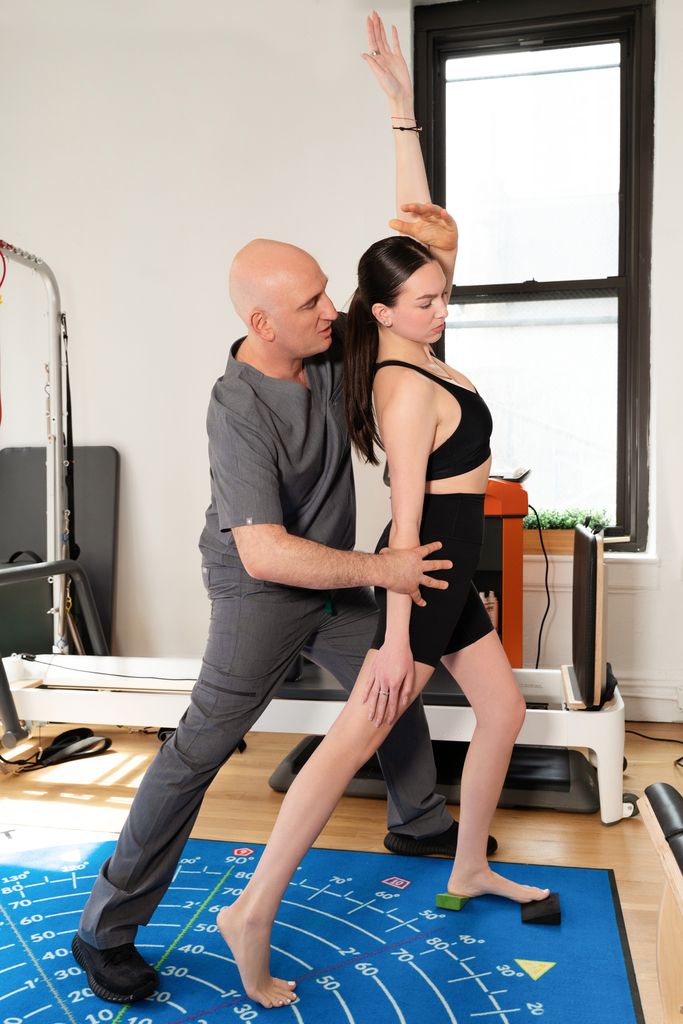
Our Unique Approach to Shoulder Pain and Dysfunction
At NYDNRehab, our holistic approach to shoulder pain and dysfunction addresses more than just your symptoms – we use advanced technologies and cutting-edge therapies to identify the underlying source of your shoulder pain and eliminate it for good.
![]()
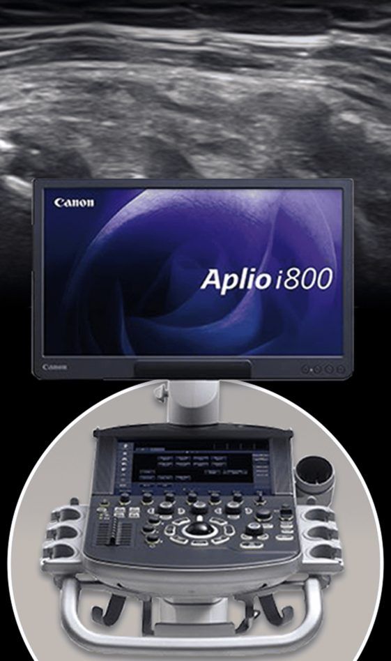
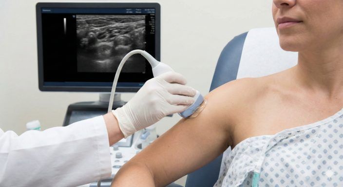
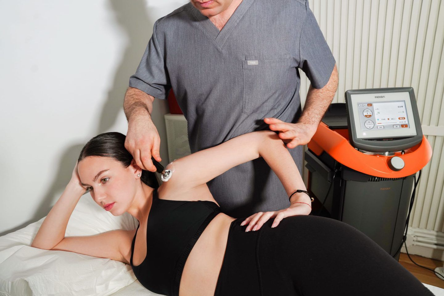
Step 1: Precision DiagnosticsOn your first visit, we conduct a thorough analysis of your condition using cutting-edge tools. Your clinical exam may include standardized tests for strength and range of motion, evaluation with high-tech equipment like ShowMotion and ForceFrame, and an on-site dynamic exam using high resolution diagnostic ultrasound. Dr. Kalika has over 20 years of experience in musculoskeletal ultrasonography, ensuring accurate
diagnostic results.
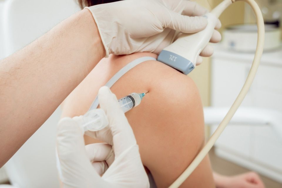
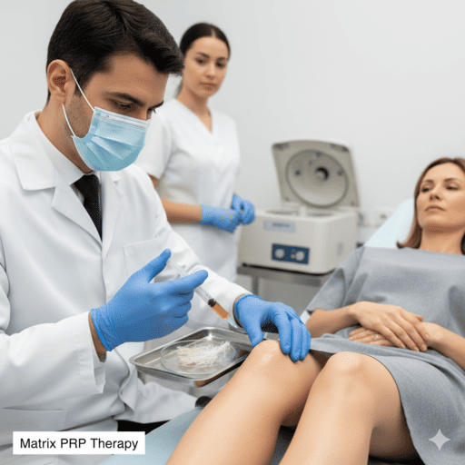
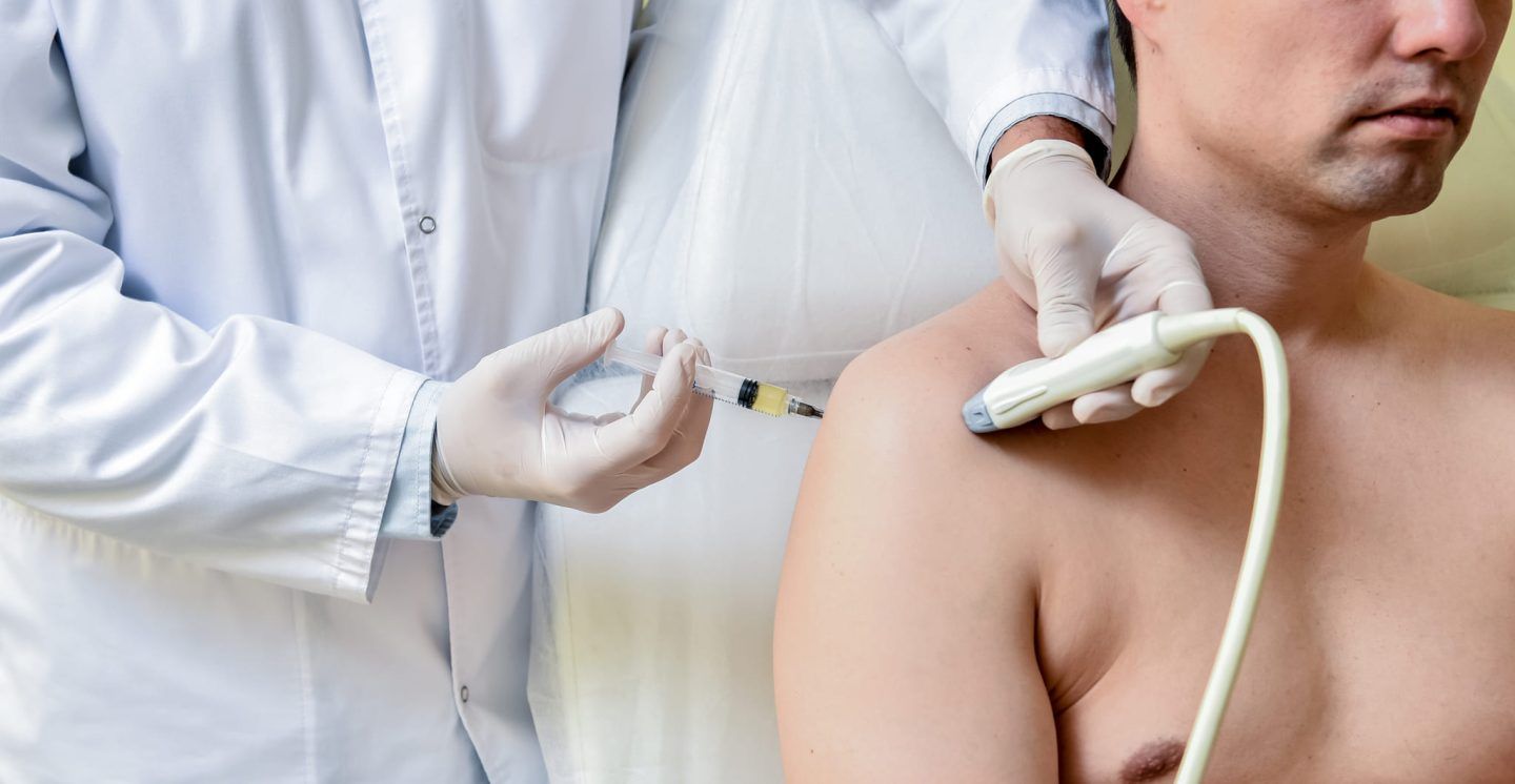
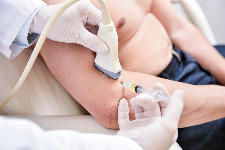
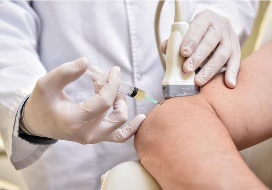
Step 2: Strategic ApproachWe use high resolution ultrasound to skillfully and precisely guide our various injection procedures. Unlike many sports and orthopedic doctors and physical therapists, we only use injections when we are confident they are needed, and strategically place them at the right time, to ensure the patient reaps maximum benefits from physical therapy.
In some cases, we precede injection therapy with physical therapy and soft tissue work to prepare the tissues in advance, to optimize the effects of injected solutions. However, when there is structural damage – e.g. tendon and fascial tears, nerve entrapments, degenerative changes or loss of fascial gliding – we use injections to repair damaged tissues and restore their functional properties before introducing load-bearing exercise.
Without a strategic approach and ultrasound guidance, injections can be a total waste of time and money.
Step 3: Advanced Personalized TreatmentUnlike conventional medical treatments that focus on pain management, our integrative approach addresses issues beyond the locus of pain that affect mobility and contribute to mechanical dysfunction. Prior to beginning physical therapy, we use the most advanced technologies and therapies currently available to regenerate and repair damaged tissues.
Step 4: Customized Functional RehabilitationConventional physical rehab clinics typically use standardized treatment protocols in a group setting – a one-size-fits-all approach that often falls short of restoring pain-free function. At NYDNRehab, we create a customized treatment plan based on your unique profile. Our one-on-one sessions ensure that you get targeted physical therapy that accelerates recovery, with the goal of fully restoring pain-free functional mobility.
Our Philosophy on Musculoskeletal Pathologies
- Dr. Lev Kalika
The human body is more than the sum of its individual parts – It is a dynamic machine made up of interdependent systems that work together to produce movement. Conventional medicine takes a reductionist approach to musculoskeletal pain, treating symptoms without connecting the dots that cause them. That approach often leads to misdiagnosis or underdiagnosis, creating life-long patients with unresolved problems.
For meaningful rehabilitation to take place, we first need to address structural, mechanical, neural, and metabolic issues that cause – or have been caused by – the patient’s complaint. Muscles, fascia, and connective tissues need to be balanced and strengthened, and damaged tissues must be healed before taking on load-bearing activities.
At NYDNRehab, we treat the whole patient, not just your symptoms. Dr. Kalika’s in-depth understanding of human anatomy and biomechanics, coupled with years of hands-on experience and training, form the foundation of our holistic approach to patient care. His approach to patient care includes subjective analysis derived from quantitative data, providing a baseline for measuring patient progress.
Dr. Kalika works closely with Dr. Yuri Brosgol, providing ultrasound-guided orthobiologic and injection procedures that heal damaged muscle, fascial, and connective tissues and restore tissue integrity. Their combined skills provide patients with cutting-edge therapies that are revolutionizing rehabilitative medicine.

Dr. Lev Kalika
Dr. Lev Kalika, DC, RMSK, clinical director of NYDNRehab, is an internationally recognized expert on the role of the scapula in shoulder pathology and its critical influence on pain, dysfunction, and performance. To perfect his skills, Dr. Kalika studied directly under Dr. Ben Kibler, world-renowned orthopedic surgeon and pioneer of scapular dyskinesis. He is one of the first certified US practitioners to be certified in Dynamic Neuromuscular Stabilization (DNS), a developmental approach to movement quality enhancement.
In addition to operating his Manhattan clinical practice, Dr. Kalika regularly publishes and presents research on the use of musculoskeletal ultrasonography, shoulder motion kinematic analysis. Dr. Kalika is one of few experts in the United States specializing in scapular diagnostic ultrasonography.
Dr. Kalika’s unique skill set has attracted multiple patients with complex shoulder cases — often after several attempts at treatment have failed. Such patients ultimately find solutions at NYDNRehab, thanks to Dr. Kalika’s focused, integrative approach.

Dr. Yuri Brosgol
Dr. Yuri Brosgol, MD, is a neurologist with 25+ years of experience in treating myofascial and chronic pain conditions. As a pioneer in orthobiologics and fascial release techniques, Dr. Brosgol learned fascial hydro release methodology directly from Dr. Carla Stecco, the world’s leading specialist in fascial science. Thanks to guidance by high resolution ultrasound, Dr. Brosgol’s approach to orthobiologic procedures ensures precision injections for optimal results.
Dr. Brosgol’s expertise provides a holistic and nuanced approach to common sports injuries and orthopedic issues. His expertise in neurology and functional medicine is especially geared to treating chronic pain syndromes. High-resolution ultrasound empowers Dr. Brosgol to precisely evaluate and treat fascia, peripheral nerves, tendons, ligaments, and joint-supporting structures – factors commonly overlooked in standard orthopedic care.
Together, Dr. Kalika and Dr. Brosgol are revolutionizing the way shoulder pain is diagnosed and treated. The clinic at NYDNRehab features some of the most advanced technologies and therapies available for musculoskeletal rehabilitation.
Symptoms and Causes of Shoulder Pain
Shoulder Rehabilitation for Athletes
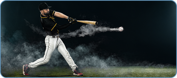
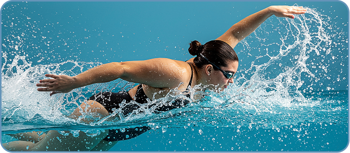
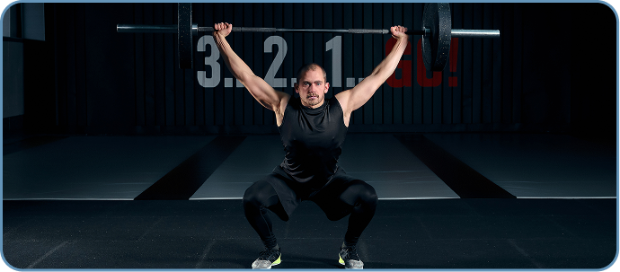
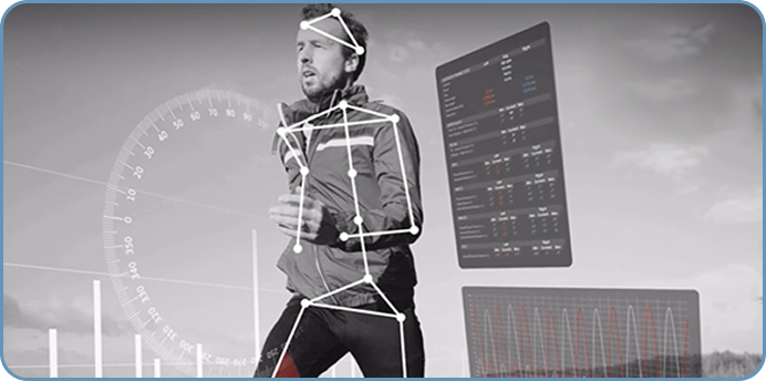
The shoulder’s ball-and-socket architecture allows for a broad range of movements in all 3 planes of motion, enabling a variety of sport-specific skills. But the price for mobility is reduced stability, making the shoulder highly vulnerable to injury and overuse during sports and exercise. Athletes in overhead sports like tennis and swimming, throwing sports like baseball, and athletes who lift weights are all at higher risk.
At NYDNRehab, we specialize in scapular and shoulder injuries. Our advanced diagnostic tools include dynamic high-resolution ultrasound, tech-driven biomechanical and kinematic analysis, and sport-specific shoulder evaluation, ensuring a thorough and accurate diagnosis. By creating an objective baseline, we are able to monitor your progress and fine-tune your rehab protocol.
Your customized treatment protocol is designed to heal and repair damaged tissues, re-establish coordinated muscle recruitment patterns, enhance strength and stability, and restore pre-injury performance levels. We offer a broad range of orthobiologics, regenerative energy technologies, and cutting-edge therapies – everything you need to return to sport with confidence.
Dr. Kalika is an internationally recognized expert on the role of the scapula in sports-related shoulder disorders. Our proprietary approach to sports-related shoulder injuries is second to none in NYC.
Read our case study to learn more about our unique approach to shoulder injuries.
Shoulder Conditions We Treat
at NYDNRehab
NYDNRehab Shoulder Diagnostic Model


The first-step bridge that brings together your symptoms, movement, and clinical findings into a clear functional map that directs all imaging, motion analysis, and treatment decisions.
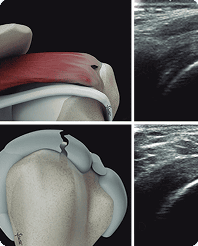
Dynamic shoulder ultrasound
Diagnosis of shoulder pathology typically relies on reported symptoms, findings from a clinical exam, and testing for strength and range of motion. While that approach can help to narrow down possible causes, it may overlook critical aspects of your injury, resulting in mistreatment, prolonged pain, and excess medical costs.
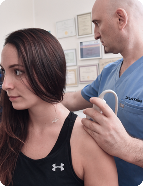
At NYDNRehab, we use high-resolution diagnostic ultrasound to view your shoulder in real time, enabling us to:
- Compare the injured and uninjured shoulders
- View nerves, muscles, fascia and bones along their entire path
- Identify fascial densifications and adhesions, and myofascial trigger points
- Detect the exact site of nerve compression or entrapment
- View multiple tissues and structures in a single session
- Elicit patient feedback during the imaging session
In addition to high-resolution imaging, our ultrasound gives us capabilities for sonoelastography to gauge muscle and tendon stiffness, and superior microvascular imaging to detect early signs of healing and monitor patient progress. Your ultrasound exam takes place on your very first visit, in the comfort of our clinic. Quick and accurate diagnosis means you can begin therapy right away, with no wait time for lab results.
Stecco fascial assessment
The Stecco Fascial Examination is a specialized diagnostic process designed to identify densification within the connective tissue, which is often a root cause of chronic shoulder stiffness. By carefully evaluating tissue glide and mobility, we can detect specific areas where the fascia has lost its essential functional properties. This examination allows our experts to target and restore the normal sliding mechanics of damaged fascia, facilitating pain-free movement and long-term joint health. It forms a critical component of our comprehensive shoulder diagnostic model, ensuring that no underlying fascial restriction goes unaddressed.
Identifying these restrictions is the first step toward recovery. This examination allows our experts to:
- Compare the injured and uninjured shoulders
- View nerves, muscles, fascia and bones along their entire path
- Identify fascial densifications and adhesions, and myofascial trigger points
- Detect the exact site of nerve compression or entrapment
- View multiple tissues and structures in a single session
- Elicit patient feedback during the imaging session

ForceFrame Max
When dealing with shoulder pain, traditional manual strength testing often relies on a clinician’s subjective feel, which can overlook subtle but critical deficits. The ForceFrame Max transforms this process into a high-precision digital assessment, providing objective data on muscle output and symmetry. It is a cornerstone of our diagnostic model, answering the vital question: “Are you strong and balanced?”.
Quantifying Muscle Output and Imbalances
Shoulder injuries often lead to compensatory movement patterns, where some muscles overwork while others weaken. The ForceFrame Max accurately measures your muscle output across various planes of motion, allowing us to compare the injured and uninjured shoulders with “side-to-side” precision. This data-driven approach identifies movement dysfunctions that are invisible to the naked
At NYDNRehab, we use high-resolution diagnostic ultrasound to view your shoulder in real time, enabling us to:
- Compare the injured and uninjured shoulders
- View nerves, muscles, fascia and bones along their entire path
- Identify fascial densifications and adhesions, and myofascial trigger points
- Detect the exact site of nerve compression or entrapment
- View multiple tissues and structures in a single session
- Elicit patient feedback during the imaging session
In addition to high-resolution imaging, our ultrasound gives us capabilities for sonoelastography to gauge muscle and tendon stiffness, and superior microvascular imaging to detect early signs of healing and monitor patient progress. Your ultrasound exam takes place on your very first visit, in the comfort of our clinic. Quick and accurate diagnosis means you can begin therapy right away, with no wait time for lab results.
Scapular Ultrasound
High-resolution ultrasound is an important tool for observing scapular movement relative to the humeral head of the shoulder joint. Combining scapular ultrasound with ShowMotion technology provides substantial information for comprehensive shoulder rehabilitation.
The scapula compensates for shoulder instability by acting as a stable base for the glenoid fossa, to optimize positioning of the humeral head. Dynamic scapular stabilization requires well-coordinated muscle activation patterns, especially of the rotator, deltoid, trapezius and rhomboid muscles. Scapular dysfunction can increase strain on the rotator cuff and labrum, contributing to pain and instability.
Dynamic scapular imaging lets us observe the complex interactions of the shoulder girdle kinetic chain, to pinpoint issues such as deficiencies in muscle activation, glenohumeral joint subluxation, and capsular laxity that contribute to shoulder instability. For JHS/EDS patients, scapular ultrasound can be a game-changer, ensuring they receive proper rehabilitation that enhances stability after a shoulder dislocation.
According to Dr. Kalika, shoulder pain is too often mismanaged, focusing on the rotator cuff while ignoring the scapula. Physical therapy is most effective when the structural integrity of the scapular muscles and nerves is restored. Unless the shoulder girdle/neck complex is addressed holistically, therapies targeting the rotator cuff deliver only mediocre results. However, few physical therapists know how to approach chronic shoulder blade pain and scapular dyskinesis.
As a life-long learner, Dr. Kalika keeps up-to-date with the latest therapeutic approaches and technologies. He frequently participates in international workshops and conferences, where he rubs elbows with some of the world’s most renowned orthopedic specialists in shoulder pain and scapular dyskinesis.
Identifying these restrictions is the first step toward recovery. This examination allows our experts to:
- Compare the injured and uninjured shoulders
- View nerves, muscles, fascia and bones along their entire path
- Identify fascial densifications and adhesions, and myofascial trigger points
- Detect the exact site of nerve compression or entrapment
- View multiple tissues and structures in a single session
- Elicit patient feedback during the imaging session
In addition to high-resolution imaging, our ultrasound gives us capabilities for sonoelastography to gauge muscle and tendon stiffness, and superior microvascular imaging to detect early signs of healing and monitor patient progress. Your ultrasound exam takes place on your very first visit, in the comfort of our clinic. Quick and accurate diagnosis means you can begin therapy right away, with no wait time for lab results.
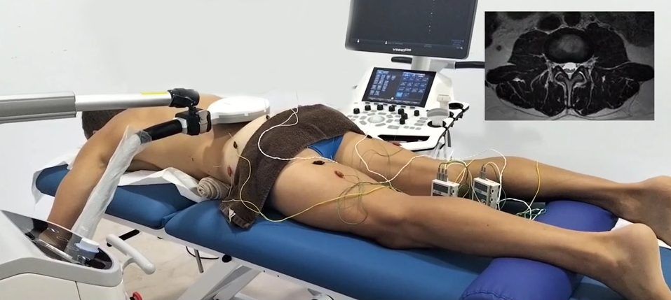
ShowMotion
ShowMotion is an objective tool for joint movement analysis that uses motion tracking sensors, placed on the patient’s skin to collect data about movement quality. The patient performs a series of joint-specific movements, and the data is analyzed by ShowMotion’s proprietary software. The collected information provides valuable insights about inefficient muscle recruitment patterns, compensation patterns, and improvements in response to therapy, enabling practitioners to personalize shoulder rehabilitation.
Quantifying Muscle Output and Imbalances
Shoulder injuries often lead to compensatory movement patterns, where some muscles overwork while others weaken. The ForceFrame Max accurately measures your muscle output across various planes of motion, allowing us to compare the injured and uninjured shoulders with “side-to-side” precision. This data-driven approach identifies movement dysfunctions that are invisible to the naked
At NYDNRehab, we use high-resolution diagnostic ultrasound to view your shoulder in real time, enabling us to:
- Compare the injured and uninjured shoulders
- View nerves, muscles, fascia and bones along their entire path
- Identify fascial densifications and adhesions, and myofascial trigger points
- Detect the exact site of nerve compression or entrapment
- View multiple tissues and structures in a single session
- Elicit patient feedback during the imaging session
In addition to high-resolution imaging, our ultrasound gives us capabilities for sonoelastography to gauge muscle and tendon stiffness, and superior microvascular imaging to detect early signs of healing and monitor patient progress. Your ultrasound exam takes place on your very first visit, in the comfort of our clinic. Quick and accurate diagnosis means you can begin therapy right away, with no wait time for lab results.
Shoulder nerve ultrasound
Reveals where nerves traveling from the neck through the shoulder and down the arm are being compressed, irritated, or stretched – a hidden cause of shoulder pain, weakness, and numbness.
The scapula compensates for shoulder instability by acting as a stable base for the glenoid fossa, to optimize positioning of the humeral head. Dynamic scapular stabilization requires well-coordinated muscle activation patterns, especially of the rotator, deltoid, trapezius and rhomboid muscles. Scapular dysfunction can increase strain on the rotator cuff and labrum, contributing to pain and instability.
Dynamic scapular imaging lets us observe the complex interactions of the shoulder girdle kinetic chain, to pinpoint issues such as deficiencies in muscle activation, glenohumeral joint subluxation, and capsular laxity that contribute to shoulder instability. For JHS/EDS patients, scapular ultrasound can be a game-changer, ensuring they receive proper rehabilitation that enhances stability after a shoulder dislocation.
According to Dr. Kalika, shoulder pain is too often mismanaged, focusing on the rotator cuff while ignoring the scapula. Physical therapy is most effective when the structural integrity of the scapular muscles and nerves is restored. Unless the shoulder girdle/neck complex is addressed holistically, therapies targeting the rotator cuff deliver only mediocre results. However, few physical therapists know how to approach chronic shoulder blade pain and scapular dyskinesis.
As a life-long learner, Dr. Kalika keeps up-to-date with the latest therapeutic approaches and technologies. He frequently participates in international workshops and conferences, where he rubs elbows with some of the world’s most renowned orthopedic specialists in shoulder pain and scapular dyskinesis.
Identifying these restrictions is the first step toward recovery. This examination allows our experts to:
- Compare the injured and uninjured shoulders
- View nerves, muscles, fascia and bones along their entire path
- Identify fascial densifications and adhesions, and myofascial trigger points
- Detect the exact site of nerve compression or entrapment
- View multiple tissues and structures in a single session
- Elicit patient feedback during the imaging session
In addition to high-resolution imaging, our ultrasound gives us capabilities for sonoelastography to gauge muscle and tendon stiffness, and superior microvascular imaging to detect early signs of healing and monitor patient progress. Your ultrasound exam takes place on your very first visit, in the comfort of our clinic. Quick and accurate diagnosis means you can begin therapy right away, with no wait time for lab results.
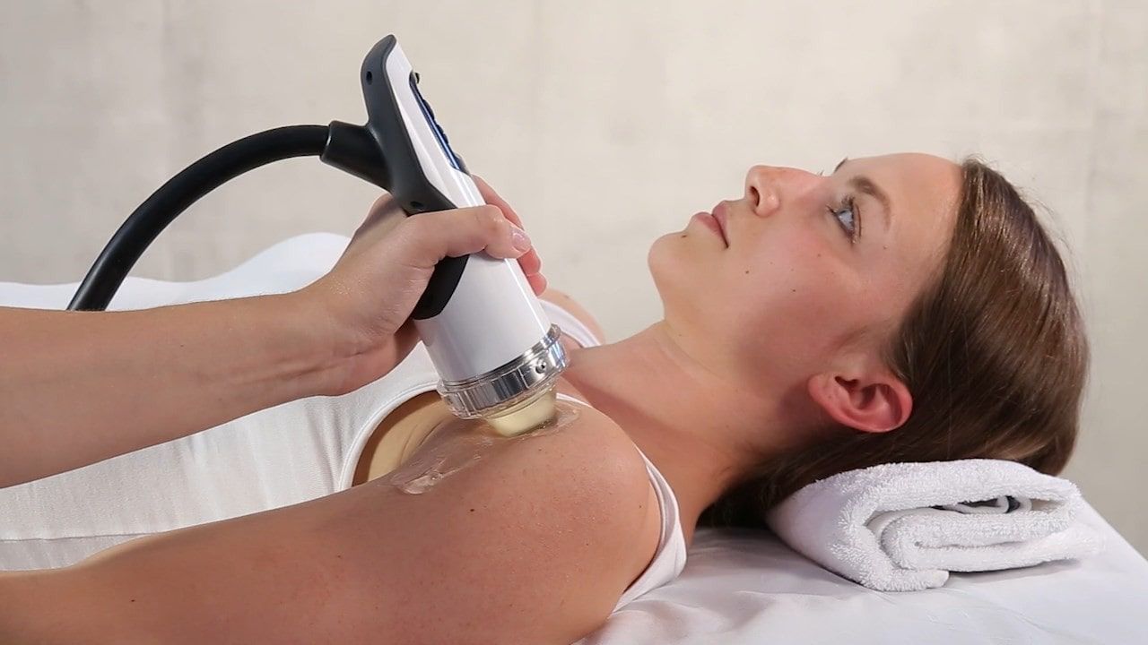
We Pre-Treat Your Tissues to Optimize
Physical Therapy Results
Physical therapy is a valuable and effective tool for restoring pain-free mobility after an injury, but it does not provide a stand-alone solution. We first need to address damaged muscle and connective tissues, myofascial dysfunction, and proprioceptive deficits that can lead to reinjury. At NYDNRehab, we pretreat your damaged tissues to accelerate healing and restore biotensegrity before introducing weight-bearing activities.
To reduce pain and inflammation and stimulate tissue neogenesis
To accelerate healing at the cellular level
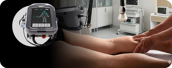
To restore the ionic charge of damaged cells, for faster injury healing
To eliminate myofascial trigger points
manipulation
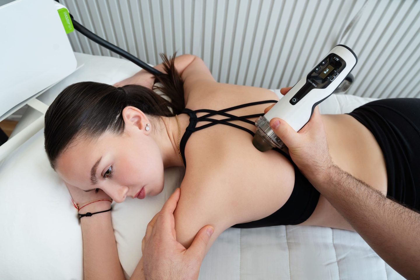
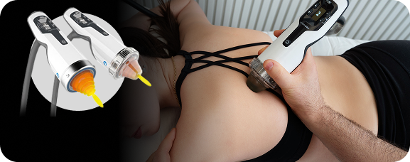
To restore the functional properties of damaged fascia
transduction therapy (EMTT)
To trigger a regenerative response in damaged tissues
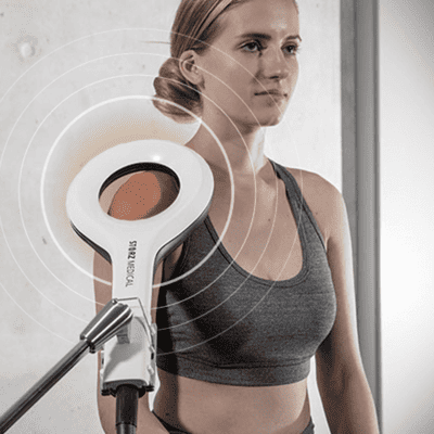
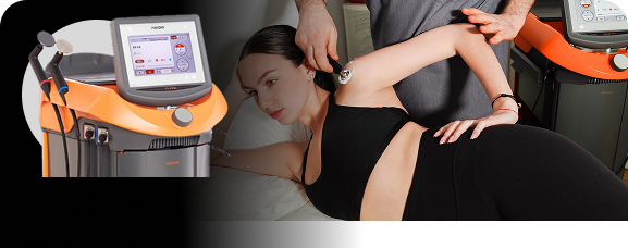
Therapy (HILT)
To reduces pain and inflammation, boosts circulation, and prepares tissues for movement and loading during therapy.

Orthobiologics and Regenerative Energy Therapies Optimize Rehabilitation
Our goal is to not only relieve symptoms, but to restore pre-injury performance levels and promote long-term functional mobility.
– Dr. Lev Kalika
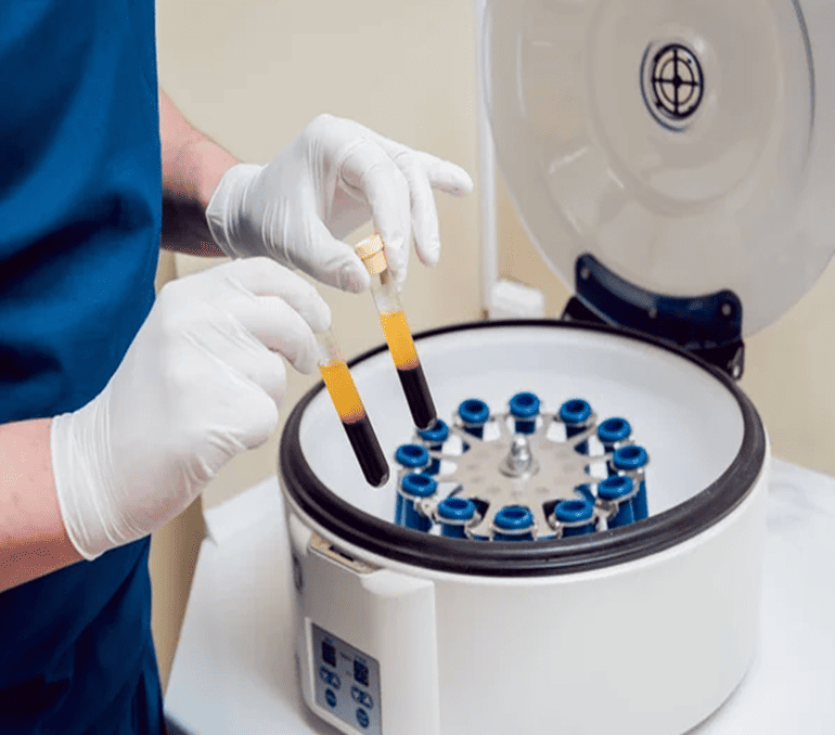
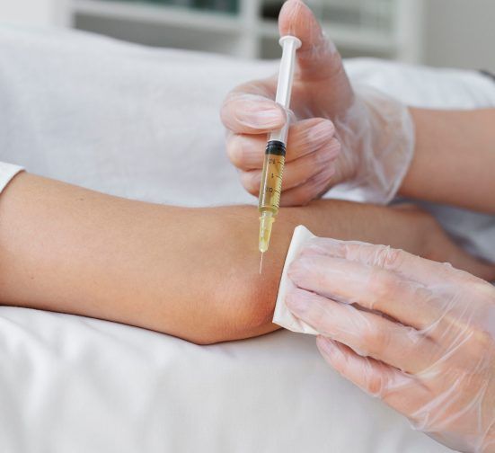
Injection therapies like PRP and Prolotherapy are often marketed as miracle cures for tissue healing, but orthobiologics is not a silver bullet for restoring functional mobility. Human biomechanics relies on multiple systems working in harmony to produce movement, and that requires a multifaceted, integrative approach.
At NYDNRehab, our customized treatment protocols incorporate injection therapies only when needed, as part of your overall rehabilitation plan. When appropriate, we combine energy therapies like shockwaves, electromagnetic pulses, and radiofrequency therapy with selected orthobiologic solutions, strategically timed to deliver the greatest synergistic benefits.
When delivered at the most advantageous time, orthobiologics and regenerative therapies support physical rehab, aimed at optimizing strength, mobility, and stability, and re-establish coordinated muscle recruitment patterns. Our goal is to not only relieve symptoms, but to restore pre-injury performance levels and promote long-term functional mobility.
Orthobiologics + Regenerative
Technologies = Optimal Tissue Healing
Regenerative therapies and orthobiologic procedures are often administered as stand-alone solutions to treat damaged tissues and pain syndromes. But while that approach may provide temporary relief, it rarely resolves the problem over the long term. By carefully selecting and combining the most appropriate therapies, we are able to harness the synergistic effects of both regenerative therapies and orthobiologics, for superior and enduring results.
- Stimulates resident tenocytes/chondrocytes/MSCs; complements PRP/BMAC’s cytokine effect.
- Promotes microvascular regeneration, opening biologic access to hypoxic tendons and their enthesis (point of attachment to bone)
- Upregulates angiogenic growth factors
- Induces nitric oxide and substance P modulation, to provide pain relief without blocking inflammation
- Supports bone–tendon junction healing
- Creates microtrauma and mechanotransduction, releasing growth factors that synergize with injected biologics.
- Induces neovascularization, improving nutrient and oxygen delivery to injected tissues.
- Enhances the homing and activation of stem cells through SDF-1 and VEGF pathways.
- Improves extracellular matrix remodeling, preparing a scaffold for PRP/stem cell integration.
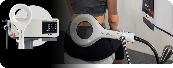
Therapy (EMTT)
- Synchronize with the body’s own magnetic fields
- Helps to extend PRP’s anti-inflammatory effects, prolonging matrix remodeling and angiogenesis
- Promotes remodeling of the extracellular matrix and collagen fiber reorganization
- Reduces fibrotic adhesions and minimizes scarring, especially in tendons, fascia and joint capsules
- Acts as a biological booster to prolong PRP regenerative effects
- Promotes long-term structural repair of tendons, ligaments and joints
- Enhances cell membrane permeability, improving migration, adhesion, and integration of injected PRP/stem cells.
- Stimulates ion channel activity (Ca²⁺, Na⁺, K⁺), supporting regenerative signaling.
- Promotes angiogenesis and microcirculation, improving survival of transplanted cells.
- Reduces oxidative stress and inflammation, creating a favorable niche for stem cell activity.
- Restores the ionic charge of damaged cells, for faster injury healing
- Boosts blood flow and enhances local circulation to improve delivery of nutrients and immune cells to the treatment site
- May improve PRP dispersal
- Promotes fibroblast activity and collagen remodeling at PRP treatment site
- Promotes lymphatic drainage of inflammatory byproducts
- Assists in eliminating fascial densifications and adhesions, and restoring tissue gliding
- Increases deep tissue perfusion and oxygenation, boosting injected cell survival.
- Enhances metabolic activity and waste clearance, optimizing the healing environment.
- Enhances cell membrane permeability, improving migration, adhesion, and integration of injected PRP/stem cells
- Stimulates ion channel activity (Ca²⁺, Na⁺, K⁺), supporting regenerative signaling.
- Promotes angiogenesis and microcirculation, improving survival of transplanted cells.
- Reduces oxidative stress and inflammation, creating a favorable niche for stem cell activity.
- Provides analgesia and reduction of muscle spasm, facilitating early functional rehab after injections.
- Improves viscoelasticity of tissues, allowing better integration of regenerative cells.
- Works via photobiomodulation (PBM), delivering light energy to specific tissues that is absorbed by mitochondria (the energy-producing organelles within cells)
- Triggers a cascade of biochemical events, including increased cellular metabolism, reduced inflammation, improved blood flow, and enhanced tissue regeneration
- Stimulates mitochondrial ATP production
- Upregulates growth factors (TGF-β, VEGF), enhancing the effects of PRP
- Enhances local blood perfusion and oxygenation to improve platelet distribution
- Reduces post-injection inflammation without suppressing the regenerative cascade
- Accelerates activation of fibroblast, chondrocyte, and tenocyte cells
- Promotes collagen repair
- Stimulates mitochondrial ATP production, energizing injected cells.
- Enhances cell proliferation and differentiation of PRP and stem cell populations.
- Reduces inflammation and pain, protecting the fragile early healing phase post-injection.
- Promotes collagen synthesis and angiogenesis, complementing regenerative injection effects.
When delivered at the most effective levels under ultrasound guidance, ESWT + EMTT prime the microenvironment via mechanical and electromagnetic stimulation. TECAR + Laser therapy optimizes tissue metabolism, circulation, and cellular energy. Together, they extend and amplify the biological effects of PRP and stem cells, accelerating tissue integration, repair, and functional recovery.
In addition, when used post-procedure, regenerative therapies dramatically reduce pain and discomfort, reinforce soft tissue healing, and promote collagen fiber realignment.
Despite the effectiveness of these cutting-edge therapies, without a multifaceted approach that addresses not only symptoms and tissue healing, but also causative and biomechanical factors, the patient is unlikely to restore and maintain efficient pain-free mobility over the long term. At NYDNRehab, our goal is to not only address symptoms and heal tissues, but to optimize mobility and prevent future injuries.
Our Shoulder Physical Therapy is Customized for Every Patient
At NYDNRehab, we treat the whole patient, not just your symptoms. We never use one-size-fits-all rehab protocols or standardized recovery timelines. We believe that every injury is unique, and treatment should be based on the patient’s unique profile.
The average physical therapist is unfamiliar with regenerative therapies and their proper use. When applied incorrectly, results can range from disappointing to disastrous. Many patients pay for regenerative procedures, only to have their effects undone by physical therapy.
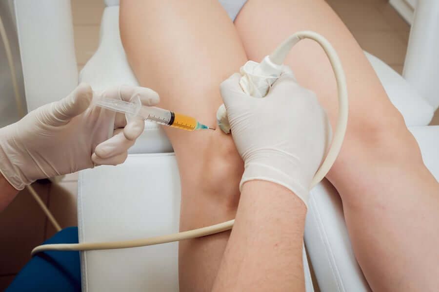
At NYDNRehab, we set a high bar for regenerative therapies and orthobiologic procedures. Guidance by high-resolution ultrasound ensures that the therapies and solutions are delivered precisely, maximizing their effectiveness. We only begin physical therapy once we are certain that your tissues are healed and ready to bear loads.
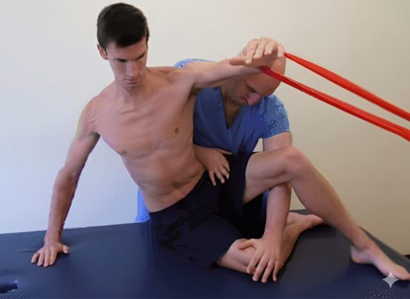
Once we have successfully pre-treated your damaged tissues, we can begin one-on-one physical therapy to restore strength and stability, optimize mobility, and re-establish optimal neuromuscular pathways and muscle coordination patterns.
manipulation
(AIM)

Our Advanced Therapies Quicky Restore
Physical Performance
Conventional treatment of shoulder pathologies often involves rest, anti-inflammatory drugs, NSAIDs, physical therapy exercises, steroid injections and surgery. However, conventional approaches often fall short of restoring full functionality, especially for athletes and physically active patients.
Advancements in technology are changing the game in rehabilitative medicine, enabling us to accelerate healing and restore performance at an unprecedented pace. The clinic at NYDNRehab features some of the most advanced therapeutic equipment currently available, and rarely found in private clinics.
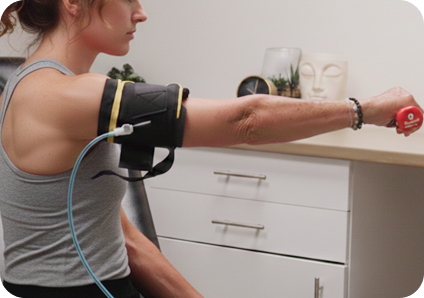
Blood Flow Restriction Training (BFRT)
Rebuilding muscle strength while joints and connective tissues are still healing is a challenge for athletes who need to return to sport in the shortest time possible. BFRT enables you to increase muscle size and strength at much lower training volumes, to reduce stress on still-healing structures while rapidly restoring muscle performance.
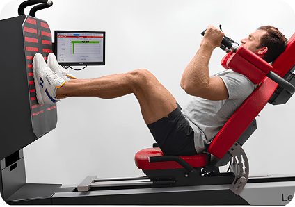
Kineo Intelligent Load System
We use the Kineo intelligent loading system to create customized training and rehabilitation programs for our patients. With Kineo, we can customize variable load protocols for functional training, core training, agility drills and more. The Kineo variable resistance system lets us design a personalized variable load curve based on the needs of the individual patient.
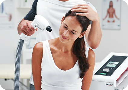
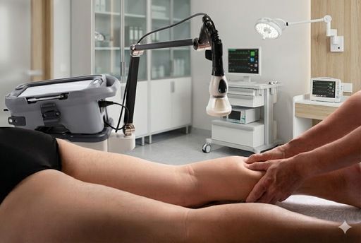
Cryotherapy
Cold therapy has long been used for injury treatment and recovery from sports and exercise. Modern cryotherapy has replaced ice baths and ice packs with a dramatically faster and more convenient technology using nitrogen gas, directed via a specialized device to target injured tissues.
Our team of sports medicine professionals will develop a personalized treatment protocol for you, based on the specifics of your injury. In addition to restoring shoulder strength and range of motion, we use specialized technologies to retrain the brain-body connection, to restore optimal muscle recruitment patterns that are often disrupted after an injury. Neuromuscular feedback training helps our athletes to return to sport safely and confidently, with reduced risk of re-injury.
Preventing Shoulder Pain and Injuries
Any physical activity comes with built-in risks, especially if you engage in sports or occupations that place repetitive demands on the shoulder complex. But sedentary populations are also at risk for rotator cuff and shoulder injuries due to muscle deconditioning, poor joint alignment and metabolic disorders that affect shoulder function.
There are multiple steps you can take to reduce your risk shoulder pain and dysfunction:
- Adopt a healthy lifestyle with a whole foods diet and regular exercise.
- Eliminate sugars and processed foods from your diet, and achieve a healthy weight to prevent diabetes and metabolic disorders.
- Be mindful of your posture and avoid rounding your shoulders, especially when using electronic devices.
- Allow for ample recovery time between sports events and exercise bouts, especially if you are a swimmer or play overhead sports like tennis, lacrosse or baseball.
- Do yoga or other stretching exercises to optimize mobility in the shoulder complex.
- Take measures to manage stress and reduce chronic systemic inflammation.
- Consider a biomechanical analysis to optimize shoulder movement and improve joint alignment.
- Begin a balanced resistance training program to strengthen the structures that support and stabilize the shoulder complex.

Complex Shoulder Architecture Calls for Advanced Expertise
The shoulder complex encompasses more than just the glenohumeral joint – it involves multiple joints, muscles, fascia, ligaments, and other tissues that work together to produce, guide, and control movement. Its complex architecture makes the shoulder the most mobile joint in the human body, but its mobility comes at the cost of stability, making it prone to injury.
Joints of the shoulder complex:
- The glenohumeral joint forms the ball-and-socket architecture that gives the shoulder its broad range of motion.
- The acromioclavicular joint connects the acromion of the scapula to the clavicle, allowing for limited gliding.
- The sternoclavicular joint attaches the clavicle to the sternum, allowing for elevation, depression, protraction, retraction, and some rotation of the shoulder girdle.
- The scapulothoracic articulation, although not a true joint, allows the scapula to glide over the posterior thoracic wall, enabling it to tilt, rotate, and slide.
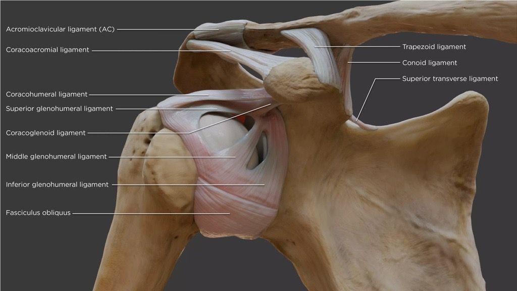
The glenohumeral joint’s shallow socket makes it vulnerable to dislocations and subluxations, especially when stabilizing structures are weak, damaged or imbalanced. Stability is provided by the rotator cuff muscles and tendons, the labrum of the glenoid, and ligaments. Fascia also plays a critical role in protecting the shoulder during physical activity, helping to guide and distribute forces via a network of elastic tension.
The rotator cuff is made up of four muscles and their tendons that compress the humeral head into the glenoid fossa, keeping it in place during movement. The infraspinatus, teres minor, subscapularis help to initiate and control shoulder rotation, while the supraspinatus aids in shoulder abduction. The rotator cuff works in concert with the deltoid, trapezius, and pectoralis major to produce smooth, coordinated motion.
Scapular stability is also important for optimal shoulder function. The scapular stabilizer muscles like the serratus anterior and trapezius hold the scapulae in position during movement, to prevent issues like winging or impingement. Multiple bursae provide shock absorption and reduce friction between tissues and bony structures. They also facilitate smooth gliding of the rotator cuff tendons beneath the acromion during arm elevation.
A complex network of neurovascular structures surround and embed the shoulder region, including the brachial plexus that innervates the arm and shoulder, and major blood vessels like the axillary artery and vein. On their path from the neck and chest to the arm, a bundle of major nerves and blood vessels passes through a narrow space beneath the clavicle called the thoracic outlet. In some cases, due to injury or repetitive stress, neurovascular bodies within the thoracic outlet can be compressed, causing pain, numbness and weakness in the arm and shoulder.
The shoulder’s complex architecture and relative instability makes it vulnerable to a broad range of injuries, such as:
- Overuse injuries like tendinitis
- Dislocations and subluxations of the glenohumeral joint
- Impingement syndromes, where nerves and blood vessels are compressed by other structures
- Compression of the rotator tendons or the subacromial bursa
- Rotator cuff tears
- Tears in the glenoid labrum
- Injury to the acromioclavicular joint, where the clavicle meets the acromion process
Without advanced knowledge of functional anatomy and the ability to visualize the shoulder in motion, it is nearly impossible to arrive at an accurate and comprehensive diagnosis based on symptoms alone. Dr. Kalika’s 20+ years of experience in diagnostic ultrasonography takes the guesswork out of shoulder diagnosis, meaning patients get the most appropriate treatment necessary for fast and effective recovery.

Get the Best Shoulder Treatment in NYC
Conventional medicine attempts to treat and manage shoulder pain symptoms before sending the patient to physical therapy. Corticosteroid injections are often used to relieve shoulder pain, but repeated treatment with steroids can increase your risk of rotator cuff injuries. Patients often suffer through multiple rounds of steroid injections, narcotic pain medications and even surgeries before they finally come to NYDNRehab for help.
The causes and symptoms of shoulder pain can vary greatly from one patient to the next, and each case requires a specialized treatment approach. We provide one-on-one Physical Therapy, custom-designed for the individual patient. We use high-tech diagnostic tools and integrative treatment approaches to get to the root cause of your shoulder pathology and eradicate it for good.
Don’t waste your time and money on treatments that don’t optimize shoulder mobility and stability – contact NYDNRehab today, and get the best shoulder pain treatment in NYC!
Our Awards


Clinical Case Studies
NYDNRehab
Shoulder Pain FAQs
Post-exercise soreness will go away after a few days. If your pain persists, you should see a shoulder specialist. There is no way to fully understand what is going on without dynamic imaging, interpreted by a seasoned professional.
Your rate of healing depends on the type and severity of your injury, and other factors unique to your condition. We personalize our treatment protocols based on your individual patient profile. Our regenerative technologies and orthobiologic procedures dramatically speed up the healing process, and ultrasound imaging confirms when your injury is fully healed.
While your damaged tissues will eventually heal on their own, you may develop scar tissue and muscle imbalances that reduce your shoulder mobility and stability. In many cases, collagenous connective tissues self-heal in erratic patterns that reduce their functionality. It’s best to fully rehabilitate your shoulder injury with the help of a professional.
Most people become less active over time, and many acquire metabolic disorders as they age. Lifestyle factors, including nutrition and exercise, play a role. Research shows that certain medications likestatin drugs, often prescribed to older adults, are associated with a greater risk of tendinopathy. Staying active and adopting a healthy lifestyle can decrease your injury risk and improve your overall quality of life.
Metabolic diseases like diabetes and hypertension are closely linked to obesity and other lifestyle factors. According to research, obesity increases the risk of shoulder tendinopathy due to excess mechanical stress. Metabolic disorders include chronic systemic inflammation that promotes shoulder osteoarthritis and impairs healing.
Certificates and Continuing Education


 Lev Kalika
  Rostyslav Bubnov
  Lev Kalika


