Ultrasound Guided Injection Therapies for Joints and Tendons

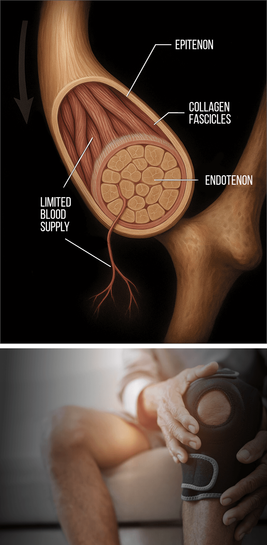
Musculoskeletal injuries typically involve multiple tissue types, including muscles, tendons, ligaments, fascia, nerves and bony structures. Each type of tissue has its own unique makeup, and each responds differently to therapy. The tissues that make up joints and tendons tend to be avascular – meaning they have a very limited blood supply – making them slow to heal due to a meager influx of oxygen and nutrients.

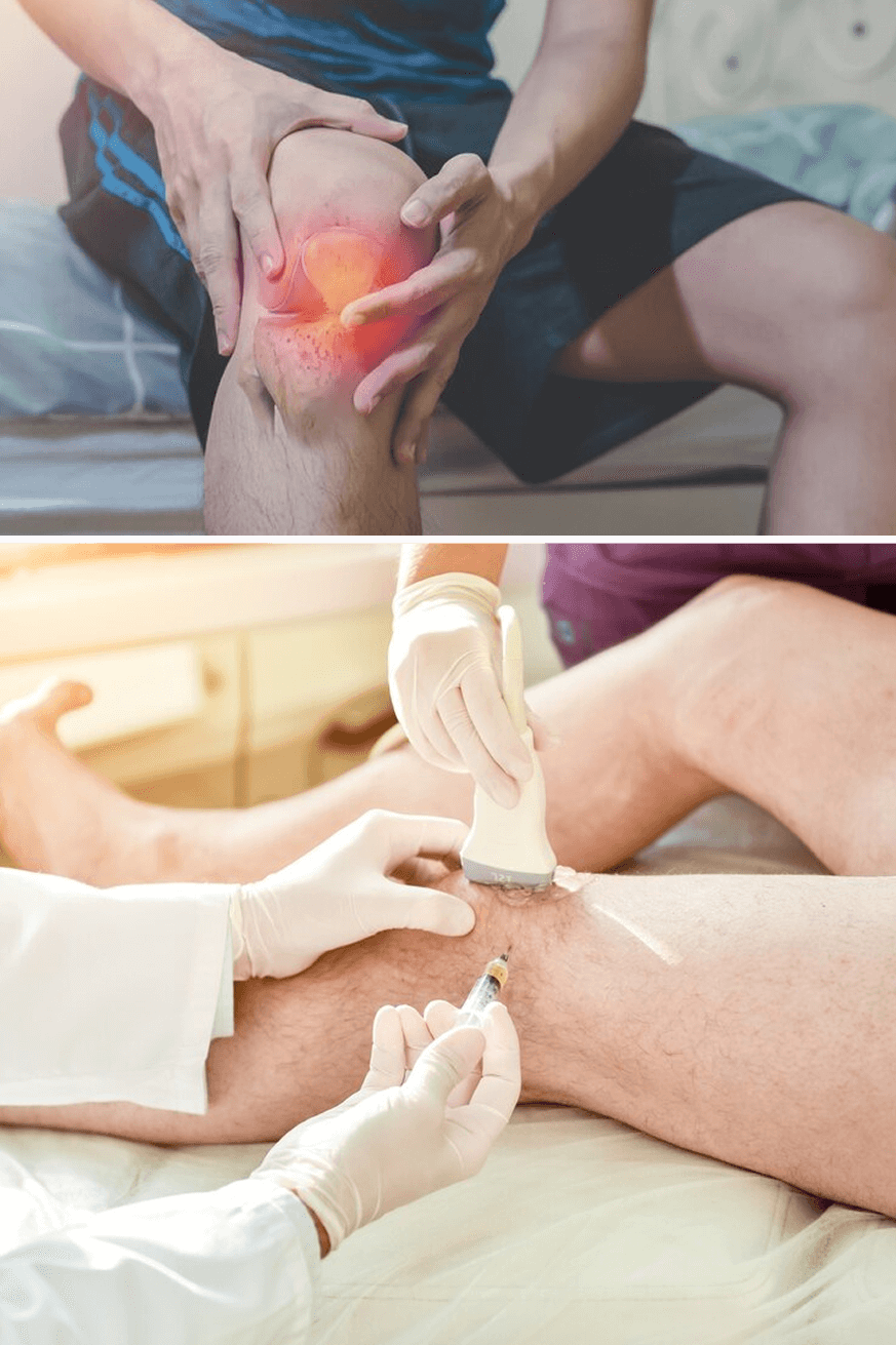
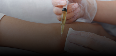
Injection therapies use orthobiologic and/or neutral solutions to stimulate and accelerate healing in stubborn joints and tendons. At NYDNRehab, our innovative breakthrough injection therapies for joints and tendons are changing the game for injury rehab and return-to-sports.

Contact us »

We’re located on 25th street in Manhattan NYC.
Open Monday-Friday, 10am-8pm.

Dr. Kalika and Dr. Brosgol are Leading the Charge in Advanced Injury Rehab

Dr. Lev Kalika
Dr. Lev Kalika

Clinical director & DC RMSK

Dr. Lev Kalika, DC clinical director of NYDNRehab, is an internationally recognized expert in diagnostic and musculoskeletal ultrasound imaging, with multiple research papers to his credit. Dr. Kalika has studied with some of the world’s most prestigious experts in diagnostic, fascia, and nerve ultrasonography, and has presented his research at multiple international professional conferences.

Dr. Kalika is an active member of the American Institute of Ultrasound in Medicine (AIUM), and has developed his own unique approach to Dynamic Functional and Fascial Ultrasonography.

Dr. Yuri Brosgol

Orthobiologic specialist

Dr. Yuri Brosgol, MD is a neurologist with 20+ years of experience in treating pediatric and adult myofascial pain. When emerging research on the critical role of fascia in human mobility captured Dr. Brosgol’s interest, he pursued training in orthobiologics and fascial release techniques. Dr. Brosgol learned fascial hydro release techniques directly from Dr. Carla Stecco, the world’s leading specialist in fascial science.

Dr. Brosgol has become a pioneer in the use of orthobiologic solutions, paving the way for transformative advancements in the treatment of myofascial disorders.

Together, Dr. Kalika and Dr. Brosgol are combining their skills to revolutionize the way musculoskeletal pain and injuries are treated. Dr. Kalika’s successful track record of rehabilitating musculoskeletal injuries combined with Dr. Brosgol’s expertise in treating myofascial pain makes NYDNRehab the clinic of choice for injury rehab in NYC.

Why Joints and Tendons are Slow to Heal

A combination of factors cause joints and tendons to heal more slowly than other body tissues. In today’s fast-paced culture, prolonged healing times can have a dramatic impact on health and productivity. For athletes, the more time spent in injury recovery, the greater the career risks.

Multiple factors can slow down joint recovery:

  • Limited blood supply to collagen-rich tissues like tendons, ligaments and joint cartilage inhibits the delivery of oxygen, nutrients and growth factors that promote tissue healing.
  • The collagenous architecture of tendons and ligaments makes them tough and elastic, but reduced metabolic activity in connective tissues prolongs collagen regeneration and remodeling.
  • Lower cell density compared to other tissues means chondrocytes and tenocytes are more spread out, making healing less efficient.
  • Ongoing mechanical stress can slow the healing process, as joints are subjected to force loads throughout the day, meaning inadequate rest and new microtears in damaged tissues. For many people, total immobilization is not an option, and too much rest can cause stiffness and tissue atrophy.
  • Chronic tendinopathies and cartilage tears are difficult to heal due to reduced regenerative resources.
  • Age-related factors like reduced collagen production, increased systemic inflammation and limited stem cell activity can dramatically delay healing.

At NYDNRehab, we use advanced regenerative therapies and orthobiologic procedures to override factors that slow the healing process. These holistic approaches tap into the body’s innate healing mechanisms to stimulate and accelerate cell regeneration in stubborn tissues. Addressing damaged joint tissues is the first step toward restoring functional mobility.

Biotensegrity is Fundamental to
Injury Healing

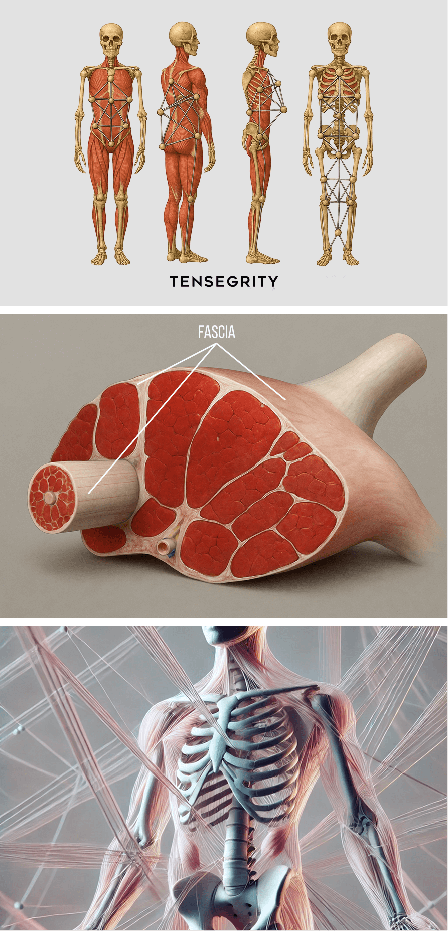
Mobility and stability are key principles of human movement, requiring a balance of tissue strength, elasticity, and optimal joint range of motion. Biotensegrity describes an optimal state of internal tension, where muscles and fascia work together to guide and control movement, and to mediate outside forces.

Until very recently, human movement science focused on muscles, joints and connective tissues, and the physiological factors that drive energy metabolism and force production. Little recognition was given to the important role of fascia in uniting the body’s systems. But over the past decade, mountains of research are confirming that fascia is the key to biotensegrity.

Fascia is a complex web of tough and elastic connective tissue that surrounds and connects muscles, engulfs the visceral organs, and holds the body’s structures in place during dynamic movement. Fascia is made up of collagen fibers, lubricated by hyaluronic acid – a slippery gel-like substance able to attract 100X its mass in water.

Fascia promotes biotensegrity via the following mechanisms:

  • Forms a protective and nourishing covering for muscles, bones, nerves and blood vessels.
  • Covers the abdominal and urogenital viscera and anchors them in place.
  • Provides elastic tension that guides and controls movement.
  • Has slippery and elastic properties that enable nerves, blood vessels and other structures to glide in harmony, without friction.
  • Works together with muscles, tendons and ligaments to stabilize joints and hold them in alignment.

Healthy fascia is smooth, slippery and elastic, but when overworked or depleted due to trauma, mechanical stress, or lack of fluids and nutrients, it can become dehydrated and fibrous. Trigger points – fibrous knots of tightly contracted muscle or collagen fibers – can form in myofascial tissue, causing pain and interfering with muscle function.

Damaged or dehydrated fascia can become dense and sticky, creating friction and impeding the ability of nerves and blood vessels to glide among other structures. Densified fascia undermines biotensegrity, disrupts muscle coordination patterns, reduces physical performance, and increases risk of injury.

Without biotensegrity, the joints lose their ability to absorb, mitigate and transfer force loads. During injury rehabilitation, it is not enough to heal muscle and connective tissues – fascial densifications and adhesions must be addressed in order to restore biotensegrity.

At NYDNRehab, we use advanced interventions to restore fascia’s slippery and elastic properties:

1
Ultrasound guided interfascial plane and nerve hydrodissection, an injection procedure that "unsticks" fascial layers and liberates nerves and blood vessels.
2
Stecco myofascial release, an evidence-based manual approach to restoring fascial integrity.
3
Ultrasound guided extracorporeal shockwave therapy, to realign collagen fibers, promote fascial hydration, and restore tissue gliding.
4
Orthobiologic injection therapies to promote collagen production and eliminate fascial densifications and adhesions.

Our fascia-first approach to injury rehab lays the groundwork for restoring biotensegrity, optimizing mobility and enhancing physical performance.

Our Advanced Orthobiologic and Regenerative Therapies Promote Fast and Effective Healing

Advanced treatment approaches are changing the game in injury rehab, and NYDNRehab is on the cutting edge. Our clinic features the latest evidence-based technologies and innovative methodologies that are only now emerging in the injury rehab space. Dr. Kalika and Dr. Brosgol are pioneers in holistic rehabilitative medicine, prompting the body’s own healing mechanisms to renew and repair tissues without drugs or surgery.

At NYDNRehab, our goal is to fully restore functional pain-free mobility that meets or exceeds the patient’s pre-injury condition. Patient diagnosis, treatment and release are based on our extensive experience and expertise. Our decision-making is backed by scientific evidence, objective data, and expertise in a broad range of regenerative and advanced methodologies.

Our personalized one-on-one approach to patient care ensures that you receive the most effective treatment available, based on your unique anatomy and diagnostic profile. We continually monitor your progress using advanced technologies and ultrasound imaging, to keep you on the path to fast and complete recovery.

Accurate Diagnosis Means Faster Recovery

As holistic practitioners, we know that it’s not enough to diagnose your injury based on symptoms alone. Most injuries involve multiple tissue types, and every injury has distinct characteristics.

Our diagnostic process includes:

  • A comprehensive review of your health history, including details of your injury onset.
  • A clinical exam to identify the exact location of your injury, and how it affects your mobility.
  • Gold-standard assessments for joint mobility and range of motion.
  • On-site exam using high resolution diagnostic ultrasound, to explore the site of injury and its effects on peripheral tissues and structures.

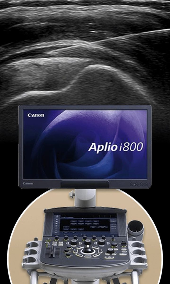
High resolution diagnostic ultrasound gives us dynamic images in real time, presenting a clear picture of the extent and severity of damage. We are able to differentiate between tissue types, examine long bodies like muscles, bones, and nerves, and compare injured structures to their uninjured counterparts on the opposite side of the body.

Accurate data-based diagnosis ensures that you get the most appropriate treatment for your injury, so you can begin your healing journey right away.

icon

Our Ultrasound Guided Orthobiologic Procedures

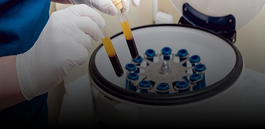
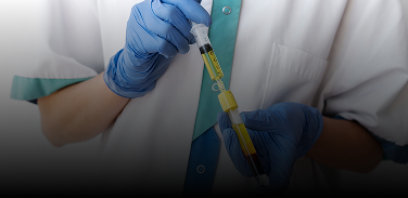
Orthobiologic injection therapies use natural/neutral solutions, injected with precision thanks to ultrasound guidance. The injected solutions stimulate cellular repair by either nourishing or irritating the targeted cells. Needling procedures like dry needling and PENS use filament-thin non-medicated needles to target myofascial trigger points and normalize neural activity.

For ultrasound-guided needling procedures, Dr. Kalika partners with orthobiologic specialist Dr. Brosgol to ensure that the needles hit their mark. Treatment results are dramatically enhanced when combined with focused extracorporeal shockwave therapy (fESWT), another area of expertise for Dr. Kalika.

Orthobiologic procedures available at NYDNRehab include:

Platelet Rich Plasma (PRP)

PRP therapy uses a sample of the patient’s own whole blood, spun in a centrifuge to extract a high concentration of platelets. When injected into damaged tissues, PRP initiates tissue repair by releasing biologically active agents such as growth factors, cytokines, lysosomes and adhesion proteins. To be effective, it is critical to use the right concentration and quality of platelets, and to and follow proper isolation techniques. When administered correctly, PRP can help to jump-start tissue healing in chronic injuries and accelerate repair in acute injuries.

Matrix PRP

For tendon ruptures, Matrix takes PRP therapy to the next level by creating a collagenous bridge between the walls of the tear and the rest of the tendon. Matrix is a highly concentrated PRP, diluted and mixed with fibrinogen. At the injection site, the solution becomes a gel-like collagenous substance that adheres to the walls of the tear and fills the space between them, creating a fibrin matrix that helps to stabilize growth factors and attract stem cell migration to the treatment site.

Platelet Releasate Therapy

Platelet releasate therapy involves injecting platelet releasate – a mixture of growth factors and biomolecules – into injured muscles and tendons to promote healing. Platelet releasate works by activating leukocytes and endothelial cells, and stimulating blood vessel growth, to increase the flow of oxygen, nutrients and growth factors to the damaged tissues.

Alpha-2-Macroglobulin (A2M)

Alpha 2 macroglobulin (A2M) is a naturally occurring blood plasma protein that acts as a carrier for numerous proteins and growth factors. As a protease inhibitor, A2M reduces inflammation in arthritic joints and helps to deactivate a variety of proteinases that typically degrade cartilage.

Prolotherapy and Prolozone

Prolotherapy uses a biologically neutral solution to irritate stubborn tissues, triggering the body’s innate healing mechanisms to grow new normal tendon, ligament and muscle fibers. Prolotherapy is often used for slow-to-heal tendon and ligament ruptures, where low vascularity inhibits tissue healing. Prolozone takes Prolotherapy to the next level by adding a combination of procaine, anti-inflammatory medications, vitamins, and minerals, followed by a mixture of ozone/oxygen gas, injected into targeted joints or tissues. When performed under ultrasound guidance, Prolozone therapy quickly reduces pain and inflammation while jump-starting the healing process.

Hyaluronic Acid Injections

Hyaluronic acid is a natural component of joint synovial fluid. Its slippery gel-like properties provide lubrication that reduces friction, enabling joints, muscles and fascia to move freely without pain. In arthritic patients, synovial hyaluronic acid tends to break down, causing bony structures to rub against one another. Hyaluronic acid replenishes synovial fluid to reduce friction and relieve pain. Hyaluronic acid is also a primary component of fascial tissue, providing its slippery gliding properties. Hyaluronic acid injections can help to restore hydration to densified fascia, to revitalize its functional properties.

Interfascial Plane and Nerve Hydrodissection

Injuries often involve damaged fascial tissue that has thickened and become sticky, often adhering to other structures. In the process, nerves and blood vessels can become entrapped, causing pain and restricting mobility. The hydrodissection procedure injects a saline solution into densified fascial layers under ultrasound guidance, separating the layers and releasing entrapped nerves and blood vessels. Hydrodissection is often used in conjunction with manual fascial manipulation, to fully restore fascial integrity.

Ultrasound Guidance Makes Our Injection Therapies More Accurate and Effective

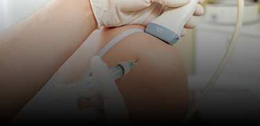
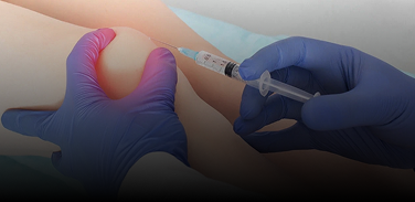
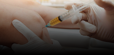
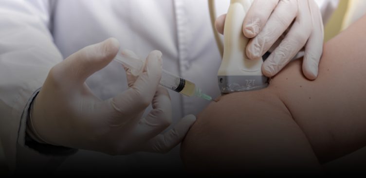
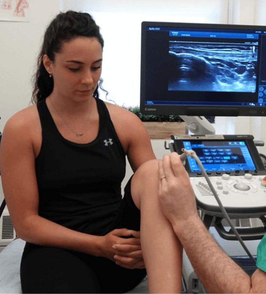
Without ultrasound imaging, therapeutic procedures are hit-or-miss, often failing to achieve their goals. Guidance by high resolution ultrasound ensures that injected solutions reach their intended tissues, without bleeding over into other structures. This means faster pain relief and accelerated healing, often with fewer treatment sessions.

During needling procedures, ultrasound guidance protects nerves and blood vessels from accidental needle penetration while ensuring that injected substances hit their target. It is important to note that only advanced high resolution ultrasound shows us minute details that cannot be seen with regular ultrasound imaging. High resolution imaging is critical for fascial and nerve injections, and for treating tendon tears.

The safety and efficacy of ultrasound guidance for needling procedures is backed by the latest research:

1
A 2018 study of injections into the pes anserine bursa – a fluid-filled sac located at the inner knee – affirmed that performing the procedure under ultrasound guidance resulted in superior patient outcomes in terms of pain relief and enhanced mobility.
2
A 2025 study compared needle placement based on palpation (aka “blind” needling) versus placement under ultrasound guidance. The ultrasound-guided procedure was associated with significantly greater accuracy and safety, achieving 100% accuracy in reaching the targeted tissues, compared to 40% with the palpation-guided procedure. Moreover, sensitive structures were pierced in 38% of blind procedures, compared to only 4% with ultrasound guidance.
3
Another 2025 study compared the efficacy and safety of ultrasound guided needling to non-guided procedures. The authors concluded that ultrasound guidance enhances treatment accuracy, reduces adverse events, and improves clinical outcomes.
4
A new 2025 study by Dr. Kalika and his colleague, Dr. Bubnov, highlights the distinctive role of ultrasound imaging in distinguishing intricate nerve patterns and accurately diagnosing specific changes within the brachial plexus, noting that ultrasound is a highly informative and indispensable tool in injury assessment and management.

Physical Therapy Puts the Icing on the Cake

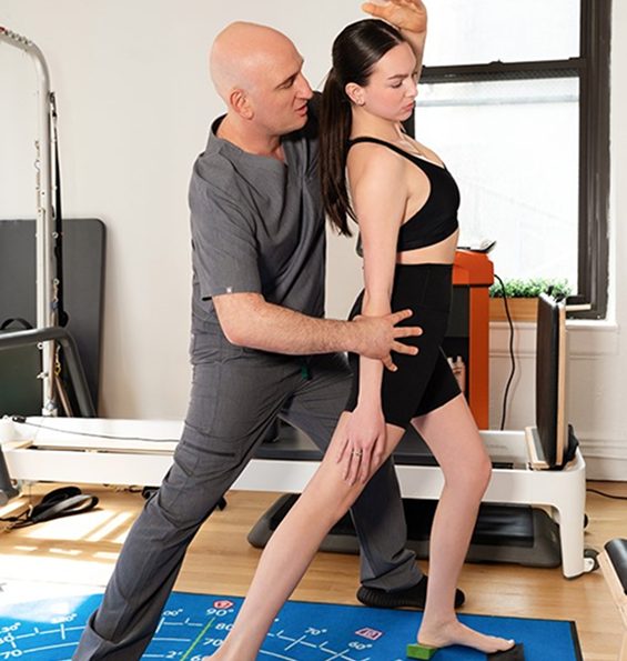
Physical therapy is an important final step on your healing journey, but it should not be started prematurely. For physical therapy to provide effective and lasting results, we must first address structural issues and restore biotensegrity to the body’s systems. Advanced orthobiologic injection therapies play a critical role in injury rehab that cannot be filled by physical therapy alone.

Once your pretreatment is complete, we can begin a customized physical therapy program with your specific goals in mind. Whether you want to return to sport as quickly as possible and perform at your peak, or you simply want to get back to your daily routine without pain or limitations, our personalized one-on-one approach ensures that your physical therapy prepares you for whatever lies ahead

icon

Get the Most Advanced Injury Rehab Available in NYC

There are dozens of clinics in Manhattan that offer physical rehabilitative services, but many of them rely on antiquated recovery timelines and one-size-fits-all treatment protocols that fail to restore biotensegrity and mobility. At NYDNRehab, we keep up to speed with the most advanced technologies and methodologies for diagnosis and treatment. Our goal is to fully restore pain-free functional mobility, so you can get back to doing the things you love.

Our Awards

    Get Advanced Holistic Therapy

    Provide your contact details and we will contact you during our operating hours


    Joint and Tendon FAQs

    What’s the difference between a joint injury and a tendon injury?
    Joint injuries affect the structures within or around a joint, like ligaments, cartilage and bursae. They are often caused by twisting, impact, or overuse. Tendon injuries involve the connective tissue linking muscle to bone. They typically arise from overuse, repetitive strain, or acute trauma. If left untreated, a damaged tendon can degenerate over time, losing its functional properties and impairing mobility.
    How long will it take to rehabilitate my injury?
    Injury healing varies greatly from one patient to the next. Beware of doctors who are quick to quote a specific healing timeline based on the type or location of injury. Premature release from rehab can set you up for re-injury. Look for a doctor who offers personalized patient care. Factors that can affect your rate of healing include:
    • Location, extent and severity of injured tissues
    • Types of tissues affected by the injury
    • Health, fitness status, and age of the injured patient
    • Patient compliance and consistency with treatment protocols
    • Lifestyle factors like nutrition, hydration and sleep
    • Practitioner experience and expertise
    • Access to advanced treatment methodologies
    Can I work out during rehab?
    During the first few weeks when pain and inflammation are present, it’s a good idea to stick to your rehab protocol, with no extra physical activity. You may be able to exercise the unaffected body parts – for example, if you have a knee injury, you may be able to work your upper body, but avoid standing exercises that require knee stability. Before you get back to your regular workout routine, consult with your doctor or therapist, to ensure you don’t do further damage to healing tissues.
    When should I seek intervention for an injury?
    Some injuries self-heal given adequate rest and time, but if pain and inflammation persist, or if your injury gets worse, you should seek treatment. Early intervention can help to prevent chronic injuries You should seek professional help if:
    • Pain persists beyond a week, despite rest
    • You hear a “pop” at the time of injury, indicating a possible rupture
    • You have severe swelling, bruising, or instability
    • Normal weight-bearing and movement are painful
    How can I tell if I need surgery?
    For most injuries, advanced conservative care is enough to promote injury healing, and surgery should only be considered as a last resort. However, severe injuries like an ACL tear may require surgery, especially for athletes who want to return to sports. Tendinopathies rarely need surgery, although a severe tendon rupture may need surgical intervention. Imaging by MRI or high-resolution ultrasound can help your doctor determine if surgery is the best path for you.

    Research at NYDNRehab

    Bubnov, Rostyslav, and Lev Kalika. "P199 CHEMOTHERAPY-INDUCED PLEXOPATHY: ROLE OF ULTRASOUND IN MULTIDISCIPLINARY MANAGEMENT STRATEGY." Neuromodulation: Technology at the Neural Interface 28.1 (2025): S286.Kim, Kwangho, et al. "Ultrasound-Guided vs Non-Guided Pharmacopuncture for Cervical Myofascial Pain Syndrome: A Multi-Center Prospective Comparative Study Protocol." Journal of Pain Research (2025): 1417-1431.https://www.sciencedirect.com/science/article/abs/pii/S1094715924011498
    Kim, Kwangho, et al. "Ultrasound-Guided vs Non-Guided Pharmacopuncture for Cervical Myofascial Pain Syndrome: A Multi-Center Prospective Comparative Study Protocol." Journal of Pain Research (2025): 1417-1431.Ultrasound-Guided vs Non-Guided Pharmacopuncture for Cervical Myofascial Pain Syndrome: A Multi-Center Prospective Comparative Study Protocol
    Lee, Jong H., Jae U. Lee, and Seung W. Yoo. "Accuracy and efficacy of ultrasound‐guided pes anserinus bursa injection." Journal of Clinical Ultrasound 47.2 (2019): 77-82.Accuracy and efficacy of ultrasound‐guided pes anserinus bursa injection
    Rodríguez-Sanz, Jacobo, et al. "Anatomical study of ultrasound vs landmark guidance for needle placement in the obliquus capitis inferior." Scientific Reports 15.1 (2025): 11667.Anatomical study of ultrasound vs landmark guidance for needle placement in the obliquus capitis inferior
    bg

    In this instance, an athlete was originally diagnosed with minor quadriceps muscle strain and was treated for four weeks, with unsatisfactory results. When he came to our clinic, the muscle was not healing, and the patients’ muscle tissue had already begun to atrophy.

    Upon examination using MSUS, we discovered that he had a full muscle thickness tear that had been overlooked by his previous provider. To mitigate damage and promote healing, surgery should have been performed immediately after the injury occurred. Because of misdiagnosis and inappropriate treatment, the patient now has permanent damage that cannot be corrected.

    The most important advantage of Ultrasound over MRI imaging is its ability to zero in on the symptomatic region and obtain imaging, with active participation and feedback from the patient. Using dynamic MSUS, we can see what happens when patients contract their muscles, something that cannot be done with MRI. From a diagnostic perspective, this interaction is invaluable.

    Dynamic ultrasonography examination demonstrating
    the full thickness tear and already occurring muscle atrophy
    due to misdiagnosis and not referring the patient
    to proper diagnostic workup

    Demonstration of how very small muscle defect is made and revealed
    to be a complete tear with muscle contraction
    under diagnostic sonography (not possible with MRI)

    image

    Complete tear of rectus femoris
    with large hematoma (blood)

    image

    Separation of muscle ends due to tear elicited
    on dynamic sonography examination

    Request Telehealth Request Telehealth Request in office visit Book now