The arches of your feet provide support and stability for your body, and they control the transfer of forces up and down your legs during walking and running. The arches, along with other structures in your feet, provide important feedback to your brain about your body’s position relative to the ground.
Foot arches work like springs during physical activity, storing and releasing elastic energy to help propel you upward and forward with each foot strike. Considering the work they do, it is not surprising that the arches experience pain from time to time.
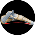
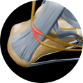
We commonly speak of the foot arch as a single structure, but there are in fact three distinct arches in your foot, the medial and lateral longitudinal arches and the anterior transverse arch. The arches help to absorb shock, and they conform to your foot shape as you walk. Together, the arches form a triangle when viewed from the bottom of your foot.
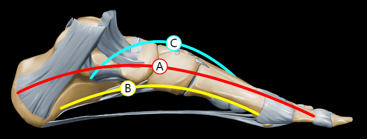
Medial longitudinal arch: The medial arch is the highest arch, spanning from your heel to the metatarsal bones of the first three toes. Elasticity is a primary characteristic of the medial arch, owing to its height and to the number of small joints between its parts.
Lateral longitudinal arch: The lateral arch is flatter and more stable than the medial arch, and spans your foot from the heel to the bases of the fourth and fifth metatarsals.
Transverse arch: The transverse arch runs across the width of your foot at the bases of the metatarsals.
The movement and stability of your foot arches are controlled by your intrinsic and extrinsic foot muscles, with support from a network of ligaments, bones and muscles in your lower leg. When you take a step, you create a stretch along the bottom of your foot that elicits an elastic response from the medial arch.
Due to the foot’s complex structure, there are a number of potential issues that can manifest as arch pain. Some of the most common causes of foot arch pain include:
Plantar fasciitis: Possibly the most common cause of foot arch pain, plantar fasciitis usually begins as pain under the heel that can spread along the bottom of the foot. Pain is often worse first thing in the morning, and lessens as the foot warms up.
Tarsal tunnel syndrome: This painful condition is caused by pressure on the posterior tibial nerve as it passes through the tarsal tunnel, located just below the the inside of the ankle. It is most often felt as a burning pain that radiates to the arch of the foot.
Heel spur: A bony growth that protrudes from the heel bone, a hell spur sometimes occurs along with plantar fasciitis, and while symptoms are similar, the two conditions are not the same.
Navicular stress fracture: One of the most common athletic stress fractures, it most often affects athletes in explosive sports involving sprinting and jumping.
Tibialis posterior tendinopathy: The tibialis posterior tendon runs from the back of the leg to the inside of the ankle and under the foot. Pain often originates where the tendon inserts into the foot bones and may radiate along the length of the tendon on the inside of the foot.
Plantar fascia strain: The plantar fascia is the band of ligament that runs from under your heel to your toes. Strains or ruptures may occur from a single traumatic incident, or may be caused by overuse over a period of time.
Abductor hallucis strain: The abductor hallucis muscle runs from the heel bone along the inside of the foot to the side of the big toe.
Medial calcaneal nerve entrapment: Symptoms of this condition are similar to that of tarsal tunnel syndrome, with pain radiating from the inside of the heel towards the center of the heel.
Careful diagnosis is critical to helping us identify the exact cause of foot arch pain so we can prescribe appropriate treatment.
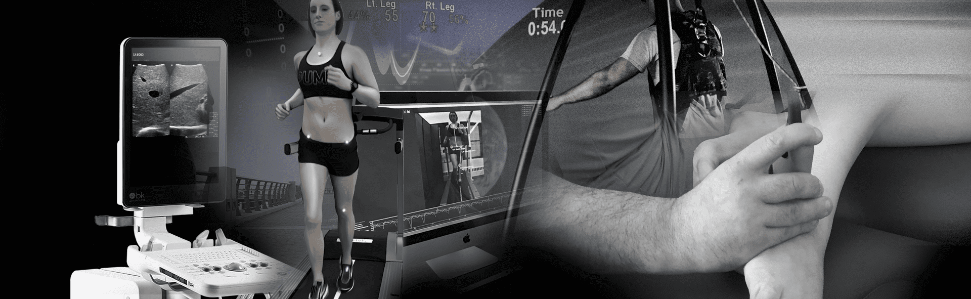
Some diagnostic approaches at NYDNRehab include:
Dynamic real-time ultrasonography: Dr. Kalika uses his own carefully researched methodology, allowing for quantification of intrinsic foot function through the use of dynamic ultrasonography, elastography and M-Mode.
Video gait analysis: By analyzing your gait, we can identify mechanical deficiencies and faulty movement patterns that cause you to place excessive loads on the structures within your feet, leading to injury and pain.
Pressure analysis of the foot: Using force plate technology, we are able to detect inefficient loading of your foot during standing and walking that may lead to foot arch pain.
Postural, structural and biomechanical assessment: The way you distribute your body weight as you stand and move can place excessive force loads on your feet, making your feet work overtime to maintain stability.
Jumping video analysis: Using a force plate, we are able to analyze movement at the knee, hip, and ankle/foot complex during a bilateral jump.
Traditional treatments for foot arch pain often center on immobilization, rest and pain management. While those strategies may temporarily relieve pain, they do not get to the root cause of your foot pain and correct it. Foot pain is usually related to muscle weakness or imbalance, faulty gait or movement mechanics, or postural issues that place excessive loads on the foot’s structures.
Some treatment approaches for resolving foot arch pain at NYDNRehab include:
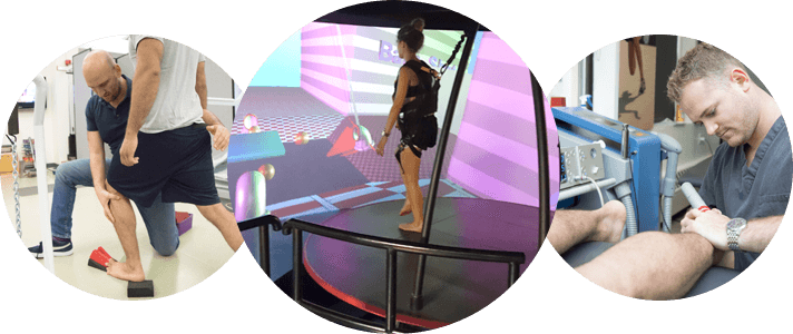
The foot pain specialists at NYDNRehab are dedicated to getting to the source of your foot pain and resolving it. We analyze and retrain your walking and running gait patterns, and restore the function of your foot to adapt to the forces imposed by walking, running, sprinting and jumping. We base our treatment on sophisticated diagnostic results, and target it to the individual needs of each patient.

Verified Expert Profiles
Dr. Lev Kalika is a world-recognized expert in musculoskeletal medicine. with 20+ years of clinical experience in diagnostic musculoskeletal ultrasonography, rehabilitative sports medicine and conservative orthopedics. In addition to operating his clinical practice in Manhattan, he regularly publishes peer-reviewed research on ultrasound-guided therapies and procedures. He serves as a peer reviewer for Springer Nature.
Dr. Kalika is an esteemed member of multiple professional organizations, including:
Below is a prime example of how ultrasound can take the guesswork out of diagnosis.

A bad physical therapy experience is one of the primary causes of unnecessary surgery
In this instance, an athlete was originally diagnosed with minor quadriceps muscle strain and was treated for four weeks, with unsatisfactory results. When he came to our clinic, the muscle was not healing, and the patients’ muscle tissue had already begun to atrophy.
Upon examination using MSUS, we discovered that he had a full muscle thickness tear that had been overlooked by his previous provider. To mitigate damage and promote healing, surgery should have been performed immediately after the injury occurred. Because of misdiagnosis and inappropriate treatment, the patient now has permanent damage that cannot be corrected.
The most important advantage of Ultrasound over MRI imaging is its ability to zero in on the symptomatic region and obtain imaging, with active participation and feedback from the patient. Using dynamic MSUS, we can see what happens when patients contract their muscles, something that cannot be done with MRI. From a diagnostic perspective, this interaction is invaluable.
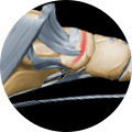
Dynamic ultrasonography examination demonstrating
the full thickness tear and already occurring muscle atrophy
due to misdiagnosis and not referring the patient
to proper diagnostic workup
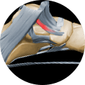
Demonstration of how very small muscle defect is made and revealed
to be a complete tear with muscle contraction
under diagnostic sonography (not possible with MRI)

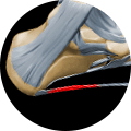
Complete tear of rectus femoris
with large hematoma (blood)

Separation of muscle ends due to tear elicited
on dynamic sonography examination


