About Knee Meniscus Injuries
Each of your knees has two menisci – horseshoe-shaped spongy disks made of fibrocartilage that work like shock absorbers between the femur and tibia bones. The menisci can become torn or ruptured from trauma, or from everyday wear and tear over time, and even a small meniscus tear can be painful, making it difficult to walk. Traumatic meniscus tears are common in high intensity sports, especially contact sports like rugby and football. Degenerative tears are common among middle aged and older adults, especially those with sedentary lifestyles.
Up to 98% of meniscus tears can be successfully treated with conservative care and do not require surgical intervention. In fact, meniscus surgery has been shown to increase the risk of knee osteoarthritis later in life. Moreover, research confirms that patients who undergo surgical repair fare no better than patients treated with physical therapy alone.
Surgery may be necessary in cases of unstable tears that produce knee locking or catching, but a partial meniscectomy (removal of meniscus tissue) should be avoided and only considered as a last resort, as it can reduce knee stability and produce early-onset osteoarthritis.
Rehabilitative physical therapy at NYDNRehab is custom-designed to accelerate meniscus healing and help to prevent recurring meniscus injuries.
Meniscus tissue is categorized by its relative vascularity. The meniscus “red zone” encompasses the outer edge that has a rich blood supply, making it faster to heal. The inner portion of the meniscus is the”white zone,” with low vascularity and minimal blood supply, making it slow to heal. Regenerative therapies are particularly effective for stimulating and accelerating meniscus healing in the white zone.
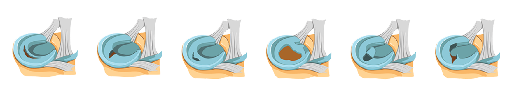
There are 6 main types of meniscus tears:
Clinical director & DC RMSK
Verified Expert Profiles
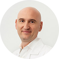
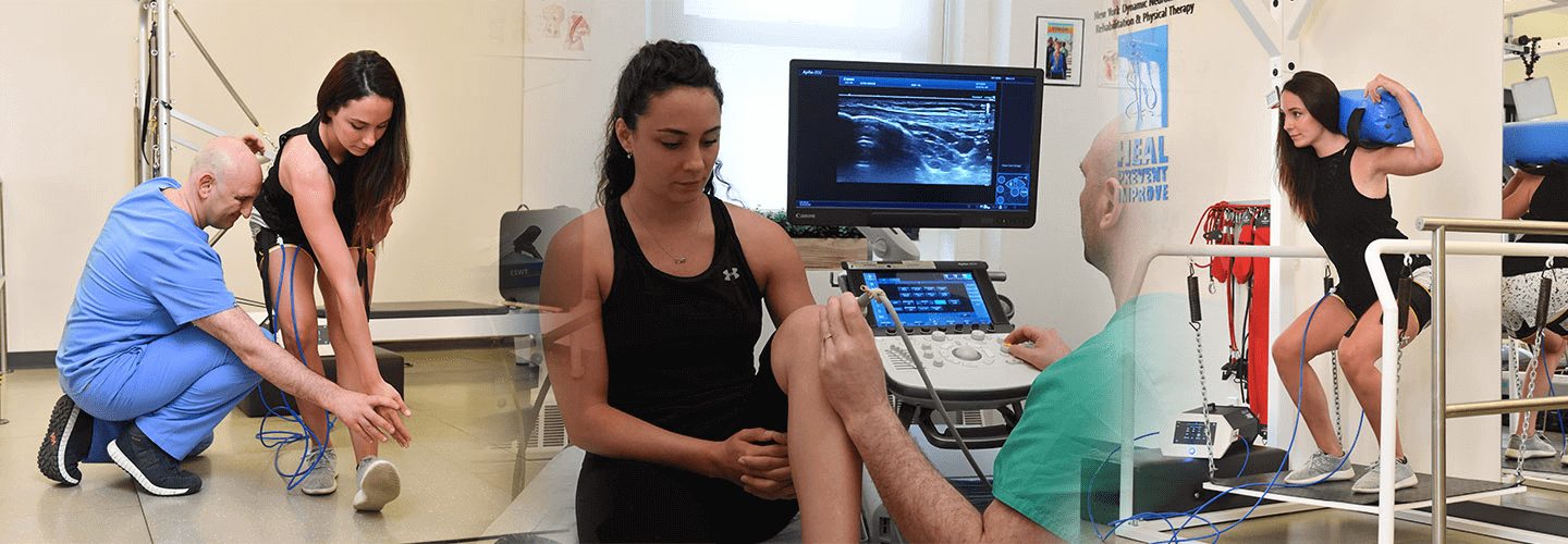
Dr. Kalika has revolutionized knee pain treatment by introducing high resolution diagnostic ultrasonography for structural diagnosis, combined with gait and motion analysis technology to visualize and objectify the functional movement of the knees and lower extremities. We conduct an on-site diagnostic ultrasound exam on your first visit.
Dr. Kalika is a recognized expert in diagnostic ultrasonography, with over two decades of experience and multiple research publications to his credit. He has developed his own proprietary methodology for repairing and rehabilitating meniscus tears without surgery. His treatment approach leverages regenerative technologies and targeted physical therapy based on the patient’s gait analysis results.
For orthobiologic procedures like Prolotherapy and PRP, Dr. Kalika teams up with Dr. Uri Brozgol, an experienced neurologist and orthobiologic injection specialist. Their combined expertise ensures that patients get the most accurate and effective treatment possible.
The motion and gait analysis lab at NYDNRehab features research-grade technologies that are rarely found in private physical therapy clinics.

Orthobiologic specialist
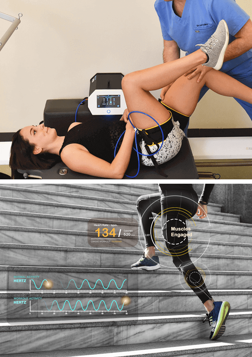
SM neuromuscular electrical stimulation (NMES) dynamically interacts with the patient during therapeutic exercises, providing real-time sensory, auditory and visual biofeedback to the patient. This breakthrough technology helps patients to recalibrate muscle actions, to optimize joint function. SMNMES has helped numerous patients to avoid unnecessary shoulder, knee and ankle surgeries, even in complex scenarios.
During PENS treatment, filament-thin needles are inserted through the skin into muscle tissue adjacent to the targeted nerve. A low frequency electrical current is then delivered via the inserted needles to stimulate the dysfunctional nerve. PENS normalizes nerve activity, improves brain plasticity and optimizes muscle recruitment patterns. This therapy is so effective that patients typically need only 4-6 treatment sessions.
Traumatic knee injuries often involve multiple tissue types, and meniscus tears can be easily overlooked when other structures are damaged. At NYDNRehab, we use high-resolution diagnostic ultrasound to dynamically visualize the structures of the knee in real time, ensuring that nothing is missed.
Early detection of a meniscus tear with high resolution ultrasound can dramatically reduce the need for surgery. Research shows that diagnostic ultrasonography conducted by an experienced professional is as accurate as MRI in identifying dynamic meniscus extrusion, where the meniscus moves out of its normal position during knee flexion, altering knee mechanics.
For the patient, on-site ultrasound imaging offers multiple advantages over MRI:
We use sophisticated 3D gait and motion analysis technology to identify sub-optimal gait mechanics and inefficient movement patterns that contribute to knee pain. Our high-tech motion and gait analysis lab features some of the most advanced technologies available for quantifying knee mechanics. Systems like ForceFrame and Kineo Intelligent Load equip us to objectively and accurately measure knee strength and range of motion.
Our rigorous diagnostic process involves:
After visualizing the knee in motion and gathering quantitative data about knee biomechanics, we are able to establish a baseline to measure your progress. Once we pinpoint the exact cause of your knee pain, we customize the most effective treatment plan, based on your unique profile.
Most people take everyday mobility for granted until an injury occurs or pain sets in. Sometimes pain and reduced mobility seem to arise out of nowhere, with no apparent cause. When it comes to knee stability, tensegrity plays a key role.
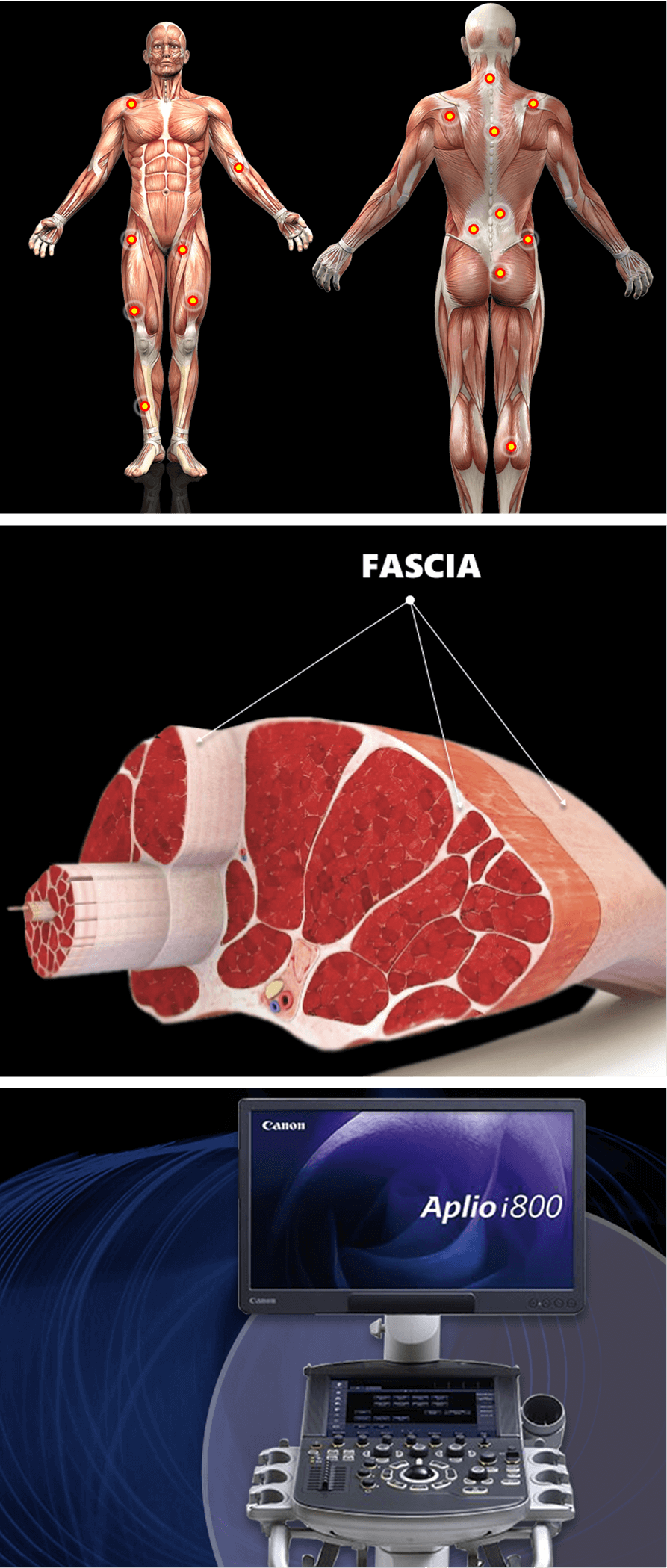
Tensegrity refers to tensile integrity – a state where individual structures are held in place by elastic tension generated by the myofascial system. The myofascia is a network of muscles and fascia that work together to produce, control, and guide forces, and to hold the body’s various organs and structures in place during movement.
When myofascial tissues are injured or damaged, tensegrity can be disrupted, altering muscle coordination patterns and inhibiting the gliding capabilities of nerves and blood vessels.
Factors that disrupt myofascial tensegrity include:
Most medical doctors do not understand the crucial role of the myofascial system in pain syndromes and movement disorders. They are trained to treat musculoskeletal pain symptoms with medications, without considering their root cause.
At NYDNRehab, we understand that the body’s systems work together as an integrated whole, and that treating pain is not enough to eliminate its source. We use dynamic high-resolution ultrasound to explore the myofascial system in real time. Ultrasound imaging lets us visualize muscles, fascia, nerves and other structures in motion, to identify places where tensegrity has been disrupted. Once we identify the problem, we use the most advanced therapeutic approaches to restore tensegrity and promote tissue healing.
Identifying and treating underlying issues prior to beginning physical therapy is key to getting fast and effective results. Failure to pre-treat your tissues can completely undermine your treatment protocol, and in some cases, your condition may even worsen.
Obstacles to physical therapy success include:
At NYDNRehab, we use a broad range of regenerative technologies and integrative therapeutic approaches to resolve issues that can stand in the way of successful physical therapy. Our staff is certified in a diverse array of holistic methodologies, and our one-on-one therapy sessions are personalized, based on your unique diagnostic profile.
Once we pre-treat your damaged tissues and eliminate compensation patterns, your body will be ready to begin physical therapy.
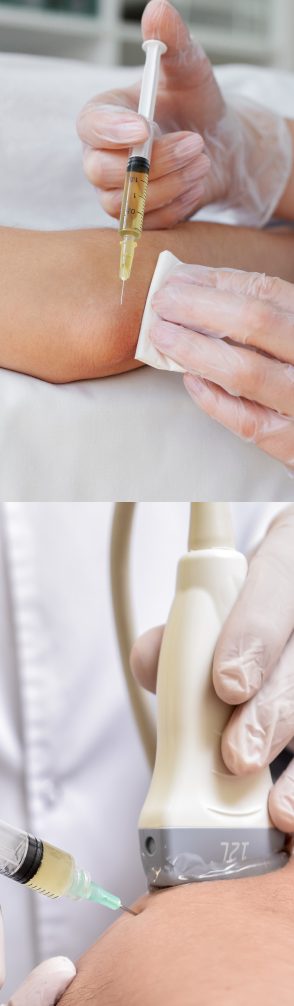
Injection therapies use orthobiologic or neutral solutions that stimulate cellular repair by either nourishing or irritating the targeted cells. Dr. Kalika teams up with an orthobiologic injection specialist, guiding needling procedures by ultrasound to ensure that the injected substances hit their mark, for maximum effectiveness.
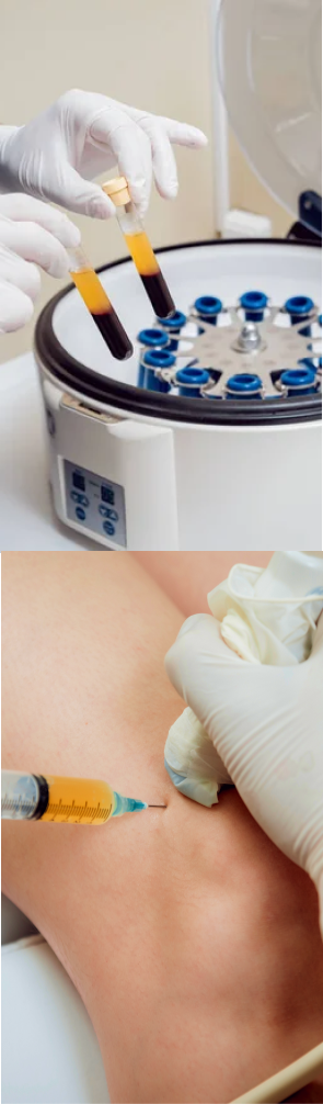
PRP therapy uses a sample of the patient’s own whole blood, spun in a centrifuge to extract a high concentration of platelets. When injected into damaged tissues, PRP initiates tissue repair by releasing biologically active factors such as growth factors, cytokines, lysosomes and adhesion proteins. The injected solution stimulates the synthesis of new connective tissues and blood vessels. PRP can help to jump-start meniscus healing.
Alpha 2 macroglobulin (A2M) is a naturally occurring blood plasma protein that acts as a carrier for numerous proteins and growth factors. As a protease inhibitor, A2M reduces inflammation in arthritic joints and helps to deactivate a variety of proteinases that contribute to cartilage degradation.
Prolotherapy uses a biologically neutral solution to irritate stubborn tissues, triggering the body’s innate healing mechanisms to grow new normal tendon, ligament and muscle fibers. Prolotherapy has been shown to repair and replenish eroded knee cartilage.
Knee injuries often involve fascial tissue that has become densified and/or formed adhesions, entrapping nerves and blood vessels, causing pain and restricting movement. During the hydrodissection procedure, a saline solution is injected into densified fascia under ultrasound guidance. The solution works by separating fascial layers and freeing up entrapped nerves and blood vessels. We often use hydrodissection in conjunction with manual fascial manipulation.
The human body has its own innate healing mechanisms, but stubborn tissues like the knee meniscus sometimes need a nudge to accelerate the healing process. Regenerative technologies help to jump-start healing by stimulating tissue repair at the cellular level. Our outpatient regenerative therapies expedite recovery with minimal discomfort for the patient.
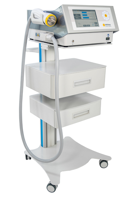
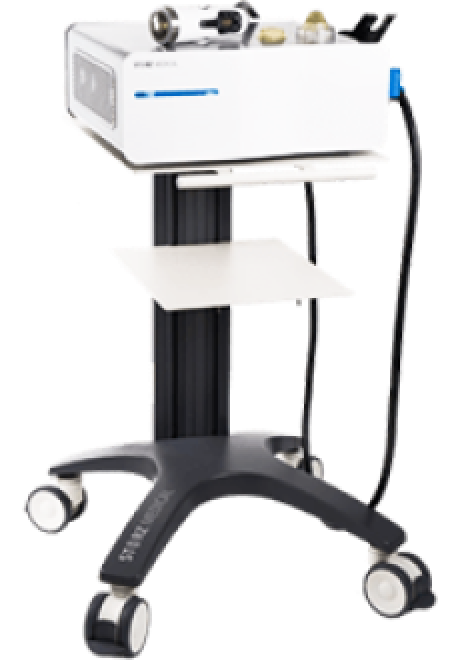
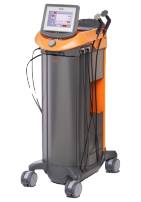
SoftWave is a regenerative mechanotransduction technology that delivers high-speed soundwaves to damaged tissues. SoftWave’s defocused and linear focused shockwaves recruit maximum stem cells to the treatment site to promote healing. According to recent research, SoftWave’s defocused waves combined with focused and radial shockwaves have maximum regenerative potential.
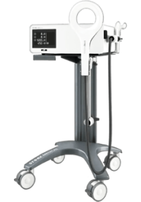
MyACT is a new type of focused shockwave technology that allows for deeper compression of the focused waves. Its higher frequency allows for precise neuro modulation under ultrasound guidance, with a special linear head for treating myofascial pain. MyACT transforms the mechanical energy of shockwaves into biochemical signals that precisely target damaged tissues. Most injuries involve more than one tissue type. When used together, our advanced shockwave technologies enable us to specifically target multiple tissue types in the same session.
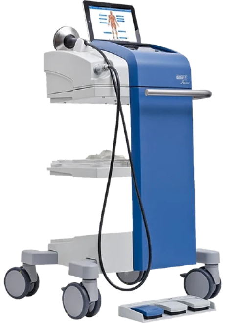
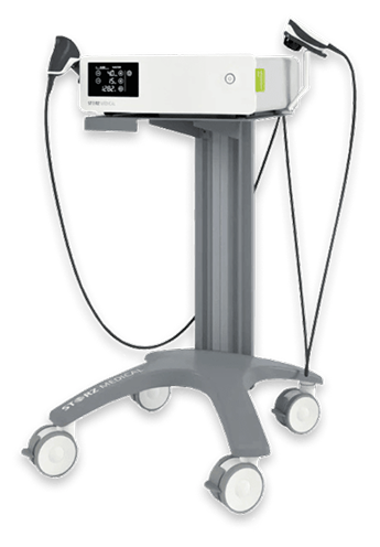
Focused ESWT is used as a regenerative treatment for damaged tendon, muscle and bone tissue. This technology produces high frequency sound waves to stimulate the body’s own reparative mechanisms. It is especially effective for chronic degenerative tendon disorders and myofascial pain syndrome.
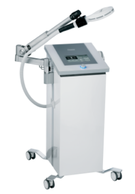
EMTT transmits high energy magnetic pulses to targeted tissues that synchronize with the body’s own magnetic fields, triggering a regenerative response. EMTT waves can penetrate deep tissues to target difficult-to-reach tendons, muscles, bones and nerves.
EPAT, sometimes called defocused shock wave therapy, is not a true shockwave. It uses mechanical pressure waves to enhance blood circulation, improving oxygen and nutrient delivery to muscle and fascia tissues. EPAT has minimal regenerative properties, but it can be especially effective when used in combination with focused shockwaves during fascial manipulation.
HEIT delivers high-intensity magnetic pulses to peripheral nerve tissues, to stimulate neuroplasticity. We leverage this FDA-approved methodology to treat pain and regenerate nerve fibers, for enhanced motor control.
INDIBA is a form of TECAR therapy that helps to restore the ionic charge of damaged cells, for faster injury healing and rehabilitation. We use INDIBA in conjunction with other regenerative technologies.
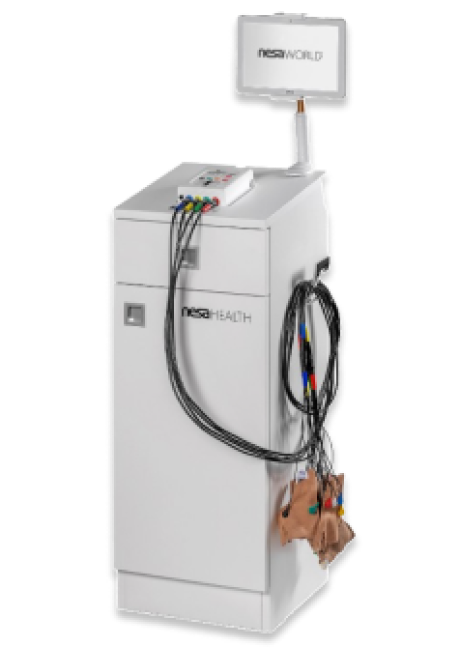
NESA generates a low-frequency electrical current that soothes hypersensitized nerves and restores optimal signaling between the autonomic nervous system and the brain. We leverage this FDA-approved methodology to treat pain and regenerate nerve fibers, to enhance motor control.
At NYDNRehab, we treat the whole patient, not just your symptoms. We never use one-size-fits-all rehab protocols or antiquated recovery timelines. We believe that every injury is unique, and treatment should be based on a holistic approach that factors in the patient’s unique profile.
Once we have successfully pre-treated damaged tissues, we can begin one-on-one physical therapy to restore strength and stability, optimize mobility, and re-establish optimal neuromuscular pathways and muscle coordination patterns.
Athletes can avoid meniscus tears by training for both strength and flexibility, staying hydrated, and regularly analyzing and correcting mechanical skills execution. Older adults can avoid knee problems in general by staying hydrated, eating a whole foods diet and remaining physically active. Weight management reduces load on the knees and lowers wear-and-tear of the knees’ structures.
Certain medications like statins, estrogen drugs, antidepressants, osteoporosis medicines and blood pressure drugs can affect the quality and performance of muscle, fascia and connective tissues. Making healthy lifestyle choices on a daily basis can steer you away from harmful prescription medications that can damage your joints and increase your risk of injury.
A nutrient-dense, protein-rich diet that promotes collagen production and muscle growth can help to maintain the integrity of the knee joints. Steer clear of sugar, refined carbohydrates and seed oils, all of which contribute to joint inflammation that can set you up for knee injuries and osteoarthritis.

If you are experiencing knee pain and suspect you have a torn meniscus, you should not delay seeking help. Early treatment can prevent your meniscus tear from getting worse and reduce your risk of needing surgery.
The knee pain specialists at NYDNRehab have 20+ years of experience in meniscus tear diagnosis and treatment, with a stellar track record of success. We will design a personalized knee meniscus rehab plan that keeps you moving toward your goal. We measure your progress every step of the way, to ensure your therapy delivers effective results.
Don’t settle for mediocre knee pain treatment that fails to restore pain-free functional movement. Contact NYDNRehab today, and get the best knee meniscus rehabilitation in NYC.


Independent peer-reviewed research relevant to this treatment approach.

Below is a prime example of how ultrasound can take the guesswork out of diagnosis.

A bad physical therapy experience is one of the primary causes of unnecessary surgery
In this instance, an athlete was originally diagnosed with minor quadriceps muscle strain and was treated for four weeks, with unsatisfactory results. When he came to our clinic, the muscle was not healing, and the patients’ muscle tissue had already begun to atrophy.
Upon examination using MSUS, we discovered that he had a full muscle thickness tear that had been overlooked by his previous provider. To mitigate damage and promote healing, surgery should have been performed immediately after the injury occurred. Because of misdiagnosis and inappropriate treatment, the patient now has permanent damage that cannot be corrected.
The most important advantage of Ultrasound over MRI imaging is its ability to zero in on the symptomatic region and obtain imaging, with active participation and feedback from the patient. Using dynamic MSUS, we can see what happens when patients contract their muscles, something that cannot be done with MRI. From a diagnostic perspective, this interaction is invaluable.
Dynamic ultrasonography examination demonstrating
the full thickness tear and already occurring muscle atrophy
due to misdiagnosis and not referring the patient
to proper diagnostic workup
Demonstration of how very small muscle defect is made and revealed
to be a complete tear with muscle contraction
under diagnostic sonography (not possible with MRI)

Complete tear of rectus femoris
with large hematoma (blood)

Separation of muscle ends due to tear elicited
on dynamic sonography examination


