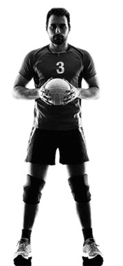
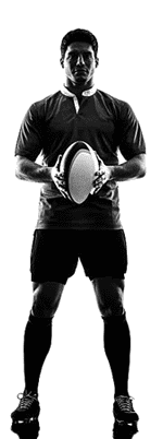
Myositis Ossificans (MO) is a condition that occurs following a direct traumatic injury that results in a contusion, or bruise. Such injuries are common in contact sports like rugby or football, where an athlete takes a direct hit from an opponent. However, anyone can sustain a serious contusion from a fall, or from running into an obstacle.
Not all contusions lead to MO, and the condition is more likely to arise from a contusion to the large muscles of the thigh or upper arm. In some instances, MO arises as a result of repetitive minor trauma. For example, it sometimes occurs on the inner thighs of equestrians. While it is more common in the tissue of large muscles, MO sometimes occurs in fat, tendons, ligaments, and fascia.
Initially, an injury leading to MO presents as a painful bruise, where a pool of blood forms a hematoma, or blood clot, over the injury. Unlike a normal contusion, pain and inflammation are not responsive to traditional soft tissue treatments like NSAIDs, icing, and immobilization of the muscle. MO is most commonly seen in teens and young adults, although older adults can develop MO after an injury or surgery. The condition is rare in children.
MO arises when the site of injury forms a benign lesion containing active fibroblasts and osteoblasts that begin to form bone within the soft tissue. Early damage is followed by the swelling of soft tissues that grows over the course of several weeks into a painful solid mass. MO is often not discovered until several weeks after the initial injury occurs, when pain, loss of function and reduced range of motion persist.
What causes myositis ossificans is poorly understood. Many believe it is due to a malfunction in repair mechanisms that cause abnormal fibroblasts to become osteogenic cells.
When your injury does not begin to heal after one to two weeks of traditional soft tissue treatment of NSAIDs, rest, ice, and elevation, you should consider the possibility of MO. During the clinical exam, your health care provider will ask for details about when and how you became injured, and what treatment measures were taken, and they will examine and palpate the injured area.
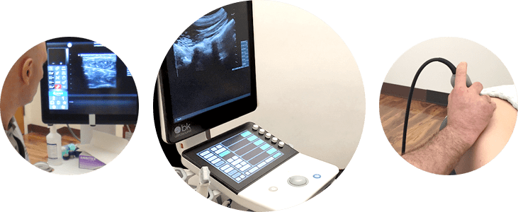
If your provider suspects MO, they will confirm it with imaging, such as X-ray, MRI, or diagnostic ultrasound. Real time diagnostic ultrasound such as that used at NYDNR can provide real time images in motion while you provide feedback, to help your clinician make an accurate diagnosis and assess the severity of your injury.
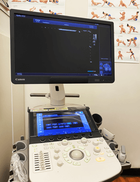
Aplio i800 ultrasound
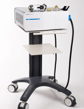
STORZ MEDICAL shockwave
In the early aftermath of injury, traditional treatment focuses on controlling pain and inflammation with NSAIDs, elevation, rest, and immobilization of the damaged area.
If pain persists and function and range of motion continue to deteriorate after four to six months, surgery is often considered to remove the mass. However, if surgery is performed prematurely, calcium deposits may remain and continue to grow. Moreover, surgical removal of the mass can result in permanent damage to the muscle, preventing it from regaining full strength and function.
Recently, a new treatment approach, ESWT (extracorporeal shockwave therapy), is showing overwhelming success in treating MO.
A study conducted by Buselli et al. (2010) of 24 young male athletes who incurred MO subsequent to traumatic contusion found that the condition was not responsive to traditional treatment methods that included icing, physical therapy and analgesics, and that as the condition persisted, the subjects experienced increasing pain, reduced function and reduced range of motion in the affected area.
The subjects were then treated with three sessions of ESWT combined with physical therapy. Although researchers observed only a partial reduction of ossification, functional improvements and reduced pain were seen immediately after ESWT therapy. Two months after therapy, range of motion and muscle strength were completely restored, and three months after treatment, 87.5% of subjects returned to their pre-injury level of play.
At NYDNR, we treat MO with ESWT and physical therapy, with the aim of eliminating pain and restoring strength, range of motion and optimal function of the affected tissues. To date, we have experienced 100% effectiveness in eliminating pain in patients suffering from MO.

Verified Expert Profiles
Dr. Lev Kalika is a world-recognized expert in musculoskeletal medicine. with 20+ years of clinical experience in diagnostic musculoskeletal ultrasonography, rehabilitative sports medicine and conservative orthopedics. In addition to operating his clinical practice in Manhattan, he regularly publishes peer-reviewed research on ultrasound-guided therapies and procedures. He serves as a peer reviewer for Springer Nature.
Dr. Kalika is an esteemed member of multiple professional organizations, including:
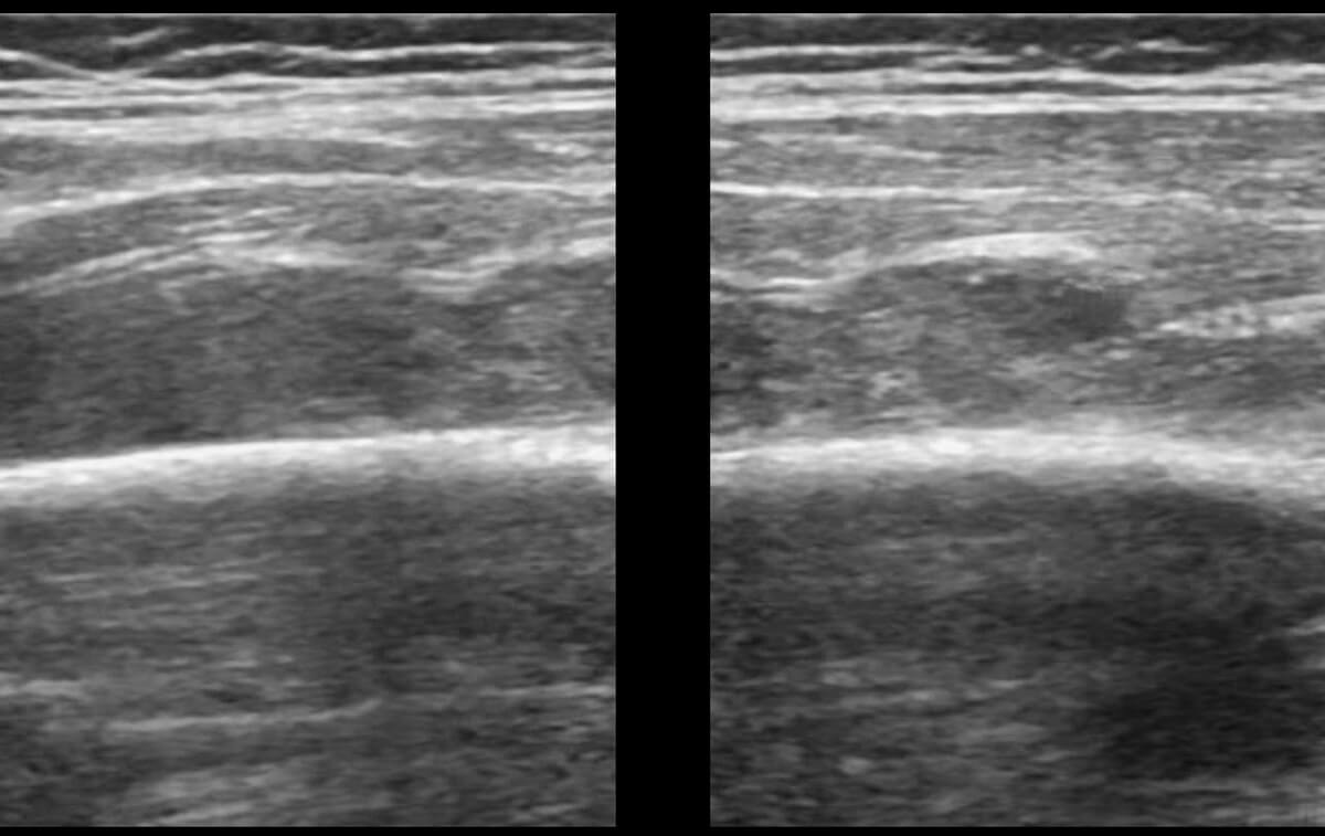
Below is a prime example of how ultrasound can take the guesswork out of diagnosis.

A bad physical therapy experience is one of the primary causes of unnecessary surgery
In this instance, an athlete was originally diagnosed with minor quadriceps muscle strain and was treated for four weeks, with unsatisfactory results. When he came to our clinic, the muscle was not healing, and the patients’ muscle tissue had already begun to atrophy.
Upon examination using MSUS, we discovered that he had a full muscle thickness tear that had been overlooked by his previous provider. To mitigate damage and promote healing, surgery should have been performed immediately after the injury occurred. Because of misdiagnosis and inappropriate treatment, the patient now has permanent damage that cannot be corrected.
The most important advantage of Ultrasound over MRI imaging is its ability to zero in on the symptomatic region and obtain imaging, with active participation and feedback from the patient. Using dynamic MSUS, we can see what happens when patients contract their muscles, something that cannot be done with MRI. From a diagnostic perspective, this interaction is invaluable.
Dynamic ultrasonography examination demonstrating
the full thickness tear and already occurring muscle atrophy
due to misdiagnosis and not referring the patient
to proper diagnostic workup
Demonstration of how very small muscle defect is made and revealed
to be a complete tear with muscle contraction
under diagnostic sonography (not possible with MRI)

Complete tear of rectus femoris
with large hematoma (blood)

Separation of muscle ends due to tear elicited
on dynamic sonography examination


