HomeBlogA Return to Athletic Power: Six Keys to ACL Rehabilitation
A Return to Athletic Power: Six Keys to ACL Rehabilitation
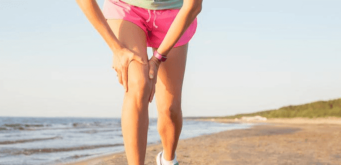
ACL rehabilitation, especially for men and women who are actively engaged in sports or other physical activities, must progress in a rigorous manner. When the early stages of rehab go well, individuals will be able to recover full mobility and strength rather quickly. Here are six of the most important keys to ACL rehab:
Eliminate Discomfort and Swelling
Because ACL surgery is considered major, there will be a fair amount of inflammation in the aftermath. Rehabilitation programs should not begin until the primary pain is gone. Discomfort can be eliminated in the following ways:
stimulation with electrical impulses
ice-packs
high-quality compression wraps
bed rest
elevation of the lower legs
Recover Knee Extension
Home exercise routines that focus intensely on the calf muscles and the hamstrings will be especially important in any rehab program. The exercises are designed to increase knee extension so that full mobility can be recovered. Once individuals have been cleared to perform these exercises by the medical professionals, the exercises should be performed several times throughout the day at low intensity. Once the muscles that control the motion of the knee have been strengthened readily for several weeks, a more intense physical regimen can be tried. The drive to restore knee extension will also help ward off arthritis and other chronic conditions.
Pay Attention to Knee Flexion
Knee flexion, which refers to the knee in a fully bent position, is just as important as knee extension. Controlled lunges and other exercises will encourage knee flexion and help people avoid the stiffness that is so common with rehabilitation. Flexion can usually be recovered in its entirety after about five weeks in most patients. All flexion exercises should be done with an attention toward very gentle motion.
Restore Patella Viability
The patellar joint itself will be crucial to the recovery of mobility. In fact, many patients find that the patellar area feels noticeably “heavy,” and failure to rehabilitate it can lead to scar tissue forming. If a tendon graft from the patella area has been used for the ACL surgery itself, then extra attention must be given to the area in the aftermath. Physical therapists can design targeted exercises that will strengthen every part of the knee.
Restore Quadriceps Control
Quadriceps control is usually best reestablished through the use of targeted electrical stimulations. Specialized machines send electrical impulses to certain nerve groups so that the related muscle groups can relearn how they are supposed to move. Electrical stimulation may be used in tandem with the following exercises to restore quad mobility and strength:
leg raises
narrow-stance lunges
specialized exercises that focus on knee flexion and extension
Return to Walking
While crutches are fine during the first week or two, patients should be placing weight on the reconstructed area as soon as possible. To focus the body on shifting weight between both legs, special cone-weaving exercises can be set up. The goal is to restore the body to normal walking posture before too much time has passed.
Ultimately, men and women will best be able to progress through their ACL rehabilitation program when all parts of the affected area are given attention at the same time. Athletes and non-athletes alike will be capable of returning to their normal regimen of physical activity, perhaps even more powerful than they were before.
In this instance, an athlete was originally diagnosed with minor quadriceps muscle strain and was treated for four weeks, with unsatisfactory results. When he came to our clinic, the muscle was not healing, and the patients’ muscle tissue had already begun to atrophy.
Upon examination using MSUS, we discovered that he had a full muscle thickness tear that had been overlooked by his previous provider. To mitigate damage and promote healing, surgery should have been performed immediately after the injury occurred. Because of misdiagnosis and inappropriate treatment, the patient now has permanent damage that cannot be corrected.
The most important advantage of Ultrasound over MRI imaging is its ability to zero in on the symptomatic region and obtain imaging, with active participation and feedback from the patient. Using dynamic MSUS, we can see what happens when patients contract their muscles, something that cannot be done with MRI. From a diagnostic perspective, this interaction is invaluable.
Dynamic ultrasonography examination demonstrating the full thickness tear and already occurring muscle atrophy due to misdiagnosis and not referring the patient to proper diagnostic workup
Demonstration of how very small muscle defect is made and revealed to be a complete tear with muscle contraction under diagnostic sonography (not possible with MRI)
Complete tear of rectus femoris with large hematoma (blood)
Separation of muscle ends due to tear elicited on dynamic sonography examination