Extracorporeal Shockwave Therapy (ESWT) is a popular non-invasive tool used in sports medicine to treat musculoskeletal pain and injuries in both athletes and non-athletes. It works by delivering shock waves to injured soft tissues, to reduce pain and promote healing.
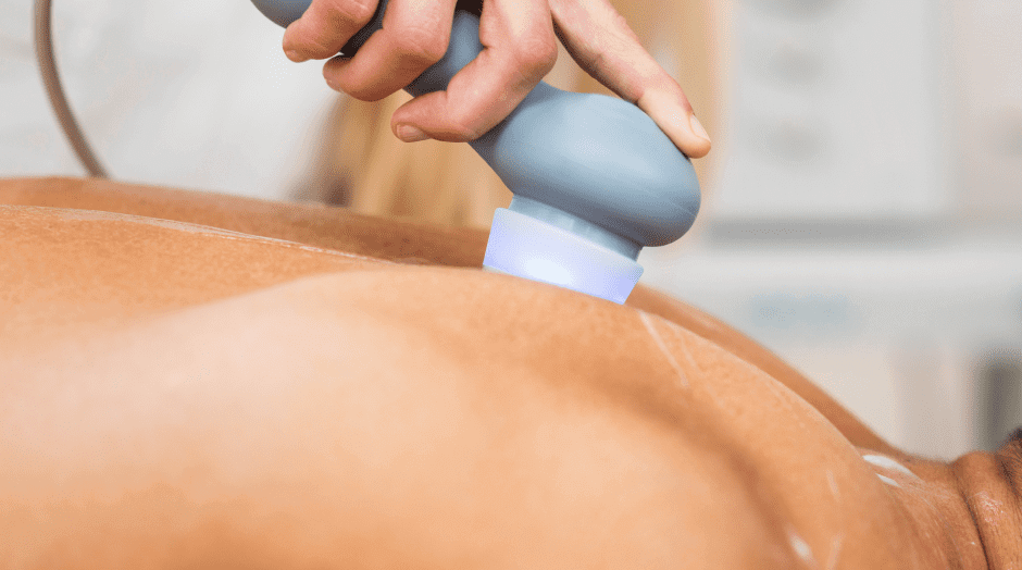
Shockwave therapy is often done “blind,” where the practitioner takes an educated guess to target damaged tissues. Ultrasound guidance takes the guesswork out of ESWT by providing real-time images of tissues buried deep beneath the skin’s surface.
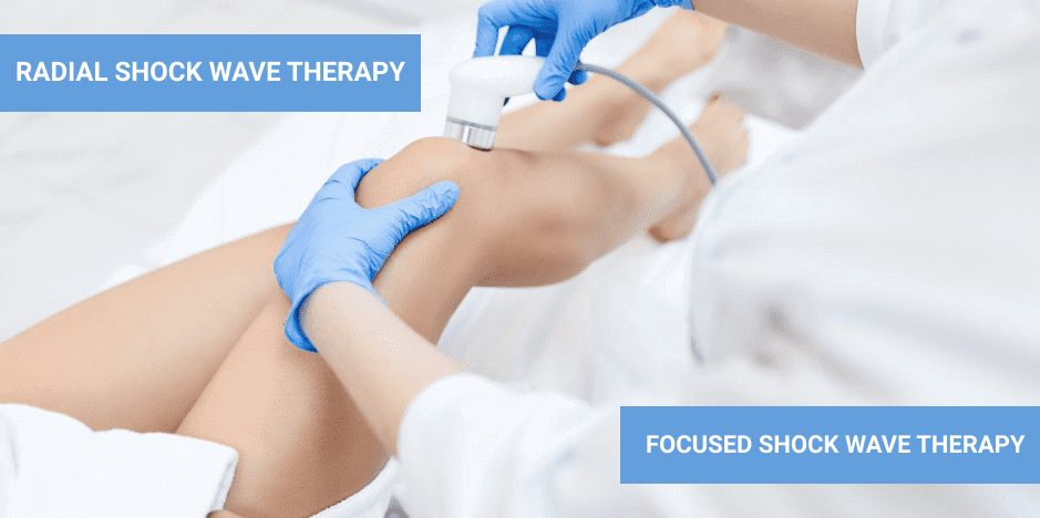
There are two basic types of ESWT: Radial (soft) and focused (hard).
Radial shock wave therapy is the most common form of ESWT, used for a larger treatment area at a more superficial level. Radial shock waves emit their greatest energy at the skin’s surface, and lose power as their energy penetrates deeper tissues. Radial shock waves are most often used for superficial tendinopathies and trigger points, myofascial pain, plantar fasciitis and hip pain at the greater trochanter.
Focused shock wave therapy uses a cylindrical coil to generate electromagnetic waves that penetrate deeper tissues. They cause tissue membranes to vibrate and create pressure waves in their surrounding fluids without losing energy. Focused shock waves target a smaller focal area. They are used to treat acute injuries, bone fractures, shin splints, groin pain, Achilles tendon injuries, tibial pain, ankle sprains and healing wounds.
Focused and radial shock waves are often used in combination to treat acute and repetitive overuse injuries in athletes.
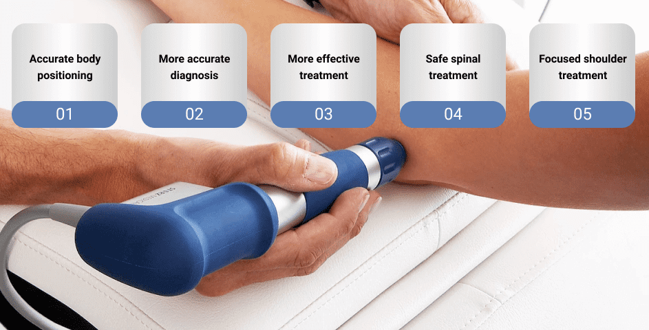
Using ultrasound technology to help guide ESWT has many advantages:
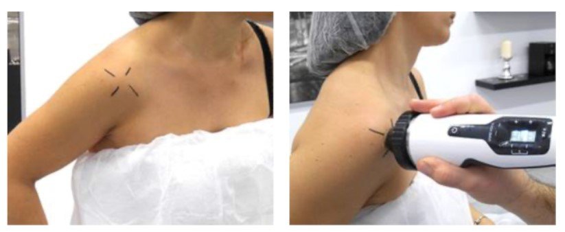
If you are athletic or physically active, you are no stranger to sore muscles and overuse injuries. Ultrasound guided ESWT can get to the source of pain and accurately target affected tissues, to speed up healing and recovery. ESWT offers a safe and effective treatment option for conditions like plantar fasciitis, carpal tunnel syndrome, tennis elbow, golfer’s elbow, and rotator cuff pain and dysfunction.
ESWT is not recommended for children, pregnant women, people with blood clots or taking blood thinners, people with pacemakers, anyone with tumors or infections at the treatment site, or if you have had a steroid injection in the past 6 weeks.
If you suffer from chronic pain, tendinopathies, athletic overuse injuries or other musculoskeletal issues, ESWT may be an effective solution for relieving pain and accelerating the healing process. At NYDNRehab, we have the most sophisticated technologies available in a private rehab clinical setting. We provide a number of innovative treatments geared to the unique needs of each individual patient. Contact us today, and discover what it feels like to move like a pro at NYDNRehab.

Verified Expert Profiles
Dr. Lev Kalika is a world-recognized expert in musculoskeletal medicine. with 20+ years of clinical experience in diagnostic musculoskeletal ultrasonography, rehabilitative sports medicine and conservative orthopedics. In addition to operating his clinical practice in Manhattan, he regularly publishes peer-reviewed research on ultrasound-guided therapies and procedures. He serves as a peer reviewer for Springer Nature.
Dr. Kalika is an esteemed member of multiple professional organizations, including:
Below is a prime example of how ultrasound can take the guesswork out of diagnosis.

A bad physical therapy experience is one of the primary causes of unnecessary surgery
In this instance, an athlete was originally diagnosed with minor quadriceps muscle strain and was treated for four weeks, with unsatisfactory results. When he came to our clinic, the muscle was not healing, and the patients’ muscle tissue had already begun to atrophy.
Upon examination using MSUS, we discovered that he had a full muscle thickness tear that had been overlooked by his previous provider. To mitigate damage and promote healing, surgery should have been performed immediately after the injury occurred. Because of misdiagnosis and inappropriate treatment, the patient now has permanent damage that cannot be corrected.
The most important advantage of Ultrasound over MRI imaging is its ability to zero in on the symptomatic region and obtain imaging, with active participation and feedback from the patient. Using dynamic MSUS, we can see what happens when patients contract their muscles, something that cannot be done with MRI. From a diagnostic perspective, this interaction is invaluable.
Dynamic ultrasonography examination demonstrating
the full thickness tear and already occurring muscle atrophy
due to misdiagnosis and not referring the patient
to proper diagnostic workup
Demonstration of how very small muscle defect is made and revealed
to be a complete tear with muscle contraction
under diagnostic sonography (not possible with MRI)

Complete tear of rectus femoris
with large hematoma (blood)

Separation of muscle ends due to tear elicited
on dynamic sonography examination


