HomeBlogCan Low Pressure Exercise Help Resolve Abdominal Diastasis?
Can Low Pressure Exercise Help Resolve Abdominal Diastasis?
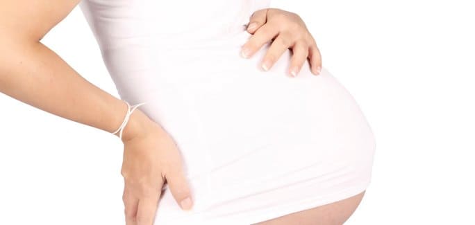
Diastasis rectus abdominis (DRA), the separation of the right and left sides of the long rectus abdominis (RA) muscle that forms the “six pack,” is most commonly seen in pregnancy, although it does occur tough vertical line of connective tissue that connects the two sides of the RA, and naturally expands with the growing fetus.
Because during DRA the two sides of the RA muscle are farther apart than normal, it has long been assumed that exercises like traditional crunches that draw the sections closer to resolving DRA than traditional abdominal exercises.
Effect of Crunches on DRA
In a recent study by Lee and Hodges (2016), 26 women with DRA and 17 control participants performed abdominal curl-ups using two distinct techniques. The first technique was a traditional curl-up, or crunch, that draws the rib cage toward the hips. In the second technique, activation of the transverse abdominal muscle preceded the curl-up.
Ultrasound images recorded the actions of RA and LA during the two crunch techniques, and at rest. The LA was rated on a distortion index during the three tasks.
The study’s authors concluded:
During a traditional crunch the right and left RA sections drew closer, but the LA was distorted.
During the transverse activation crunch, right and left sides of the RA were more separated, but the force transfer between the two sides was greater, and the LA was not distorted.
Reduced IRD narrowing by transverse contraction may positively impact abdominal mechanics.
It is thought that tension on the fascia during transverse contraction may stimulate tissue repair to the LA.
Low Pressure Exercises to Resolve DRA
Hypopressive exercise is increasingly being used to treat post-partum pelvic issues like incontinence, pelvic pain and uterine prolapse. To activate the tranverse abdominal muscles, perform the following steps:
Lie on your back, knees hip width at 90°, feet flexed in toes-up position, arms flat at sides at 45°.
Elongate your spine by tucking your chin and pulling your crown upward.
Inhale deeply through your nose and expand your ribcage (do not do diaphragmatic or “belly” breathing).
Exhale slowly through your mouth and allow your ribs to separate as you hollow out your midsection, creating a vacuum.
Hold for several seconds until you are forced to take a breath.
Perform two cycles of breathing-only, then create the vacuum on the third cycle.
Practice tranverse abdominal muscle recruitment until you feel confident, then add an abdominal crunch in the vacuum phase.
Long-term benefits include:
Flatter abdomen
Narrower waist
Less pelvic pressure
Improved posture
Diminished RA gap
Improved continence
DRA Treatment in NYC
If you are suffering from Diastasis Rectus Abdominis or other post-partem pelvic issues like incontinence, prolapse or pelvic pain, the sports medicine team at NYDNRehab can help. We use the latest technologies, therapies and treatments to your active life.
Source
Lee, D., & Hodges, P. W. (2016). Behavior of the linea alba during a curl-up task in diastasis rectus abdominis: an observational study. journal of orthopaedic & sports physical therapy, 46(7), 580-589.
In this instance, an athlete was originally diagnosed with minor quadriceps muscle strain and was treated for four weeks, with unsatisfactory results. When he came to our clinic, the muscle was not healing, and the patients’ muscle tissue had already begun to atrophy.
Upon examination using MSUS, we discovered that he had a full muscle thickness tear that had been overlooked by his previous provider. To mitigate damage and promote healing, surgery should have been performed immediately after the injury occurred. Because of misdiagnosis and inappropriate treatment, the patient now has permanent damage that cannot be corrected.
The most important advantage of Ultrasound over MRI imaging is its ability to zero in on the symptomatic region and obtain imaging, with active participation and feedback from the patient. Using dynamic MSUS, we can see what happens when patients contract their muscles, something that cannot be done with MRI. From a diagnostic perspective, this interaction is invaluable.
Dynamic ultrasonography examination demonstrating the full thickness tear and already occurring muscle atrophy due to misdiagnosis and not referring the patient to proper diagnostic workup
Demonstration of how very small muscle defect is made and revealed to be a complete tear with muscle contraction under diagnostic sonography (not possible with MRI)
Complete tear of rectus femoris with large hematoma (blood)
Separation of muscle ends due to tear elicited on dynamic sonography examination