Carpal tunnel syndrome (CTS) is a very frequent nerve compression condition among working adults. It leads to numbness, pain and weakness in the hands, wrists and arms, which can significantly affect the ability to perform daily activities, but is often overlooked, under-treated, or delayed until surgical intervention is considered.
This article will tell you what CTS actually is, who it affects, how it is properly diagnosed and what treatments such as shock wave therapy and ultrasound guided hydrodissection can do for you.

The carpal tunnel is a narrow space in the wrist, created by a small bone called a carpal and protected by the transverse carpal ligament. The median nerve is one of the primary nerves that supplies the hand, and runs through this tunnel with nine flexor tendons. As pressure increases in the tunnel, the nerve becomes inflamed, causing pain, numbness, tingling and weakness in the thumb, index, middle and ring fingers.
Symptoms are slow to appear and progressively get worse. The symptoms are more likely to occur at night when the wrist is flexed during sleep, which puts more pressure on the nerve.
The early warning sign of CTS is a numb or “asleep” hand in the morning that requires shaking to wake up. Don’t ignore it.

The pressure in the carpal tunnel may come from several sources other than the wrist. To decide on the appropriate therapy, it is crucial to grasp the entire situation.
Anatomical Factors

Functional and Fascial Factors
The median nerve is formed in the neck and passes through the shoulder, arm, elbow and forearm and into the hand. Any blockage on this pathway can cause the nerve to become compressed or stretched, resulting in symptoms that are similar to those of classic CTS. This is a blind spot in the standard orthopedic exam, as the diagnostic process is likely to end at the wrist when a person has carpal tunnel surgery. Studies of nerve mechanics always find that there are multiple levels of compression in many patients, and just treating the wrist doesn’t address these problems.
The main functional contributors are: scapular dysfunction, which is a poor control of the shoulder blade that causes an increase in mechanical load on the median nerve before it reaches the wrist; lacertus fibrosus entrapment, a fibrous band at the elbow that compresses the median nerve proximal to the wrist, and is often overlooked in the standard evaluation; and retinaculum stiffness, characterized by reduced movement of the transverse carpal ligament, which decreases the capacity of the tunnel to accommodate nerve movements. Another recent study by Carla Stecco and Carmelo Pirri also shed light on another important factor that can cause restriction of the median nerve in its course: thickening of the antebrachial fascia, a measurable increase in the density and stiffness of the deep forearm fascia, which is a diagnosable condition and can be specifically targeted for treatment with high-resolution diagnostic ultrasound. Last but not least, entrapment of the median nerve between the two heads of the pronator teres muscle in the forearm is a common and often overlooked cause of symptoms, either as an isolated process mimicking carpal tunnel syndrome or as an important upstream cause of double crush syndrome when wrist-level compression is also involved.
 Diabetes
 Hypothyroidism
 Rheumatoid arthritis
 Pregnancy
 Obesity
 Inflammatory conditions
Double crush syndrome is when the median nerve is compressed at two or more points at the same time. A patient might experience mild cervical compression at their neck, tightness in their lacertus fibrosus at their elbow, and a narrowed carpal tunnel at their wrist, each of which is minor, but collectively cause significant symptoms that are debilitating.
This pattern is not detected by standard work-ups, which only assess the wrist. The tunnel is opened during surgery, the nerve is released, and symptoms continue to occur because the nerve was also compressed upstream at a location that was not evaluated.
Persistent symptoms following carpal tunnel surgery sometimes aren’t a surgical failure, it’s a diagnostic one. The best way to avoid this is to have a full nerve pathway evaluation prior to treatment.


Women are diagnosed with CTS about three times more often than men, in part because hormones and the smaller size of the tunnel.
When symptoms are more advanced, many patients do not seek help. At this stage, nerve damage may have already started. The earlier the intervention, the better the results.
Early signs, take action on these now:
Severe symptoms, urgent assessment:


When symptoms are more advanced, many patients do not seek help. At this stage, nerve damage may have already started. The earlier the intervention, the better the results.
1. Clinical Examination
2. Nerve Conduction Studies (NCS/EMG)
NCS/EMG is the gold standard for measuring the efficiency of the median nerve and is important for grading the severity and determining surgical indications. When applied alone, however, it has its limits: it only assesses electrical function, and may return normal results in early CTS, and it does not detect double crush syndrome or proximal compression sites.
3. High-Resolution Diagnostic Ultrasound
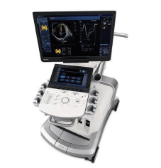
Ultrasound is a must-have addition to NCS/EMG. Nerve conduction studies provide information about the electrical activity of the nerve, but ultrasound provides information about the problem, by directly imaging the nerve and surrounding tissue in real time.
Consider them a power pair for diagnosis: NCS/EMG is the physiological power couple, while ultrasound is the structural power couple.
What ultrasound adds:


Treatment will vary based on the severity, length and structural findings of the symptoms. This staged framework is a representation of the progression of evidence based CTS care:
Stage 1: Initial Conservative Management
Indications: mild or early symptoms of the disease
Wrist splinting at night to keep the wrist in neutral position, along with ergonomic change of work station and anti-inflammatory measures. In truly early CTS, this phase may be the only one that yields significant improvement.
Stage 2: Advanced Non-Invasive Intervention
For: symptoms persisting despite splinting and ergonomic changes
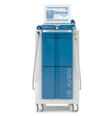
Extracorporeal Shockwave Therapy (ESWT) to stimulate tissue repair, reduce synovial inflammation and improve nerve circulation with radial shockwave applied specifically to the antebrachial fascia and myofascial densification points where fascial thickening has been identified as a contributing factor. Structured physical therapy, such as neurodynamic nerve mobilization, forearm strengthening and scapular rehabilitation, addresses the mechanical contributors that splinting cannot reach, and manual fascial manipulation using the Fascial Manipulation methodology is used to systematically release densified fascial points along the pathway of the median nerve.
Stage 3: Image-Guided Mechanical Release
If: structural nerve adhesions, fascial entrapment or multi-level compression
Hydrodissection is a procedure that uses ultrasound to separate the median nerve from the surrounding constricting connective tissue, allowing the nerve to glide freely again. FLUSH (Fascial Liquid Ultrasound-guided Hydrodissection) is a specialized technique that utilizes ultrasound-guided fluid injection to release the fascial layers along the nerve’s path, which goes beyond the reach of a typical surgical release.
Stage 4: Surgical Decompression
For: severe CTS with progressive muscle wasting, profound axonal loss, or failure of well-executed conservative care
Surgical release of the transverse carpal ligament is still indicated in patients with thenar atrophy, significant axonal loss on NCS/EMG, and failure of conservative treatment for a prolonged period. Surgery should not be the first step if compression is severe and progressive.


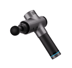
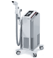
ESWT is a technique that sends concentrated pulses of acoustic energy into the targeted tissue, from the outside. It is non-invasive, does not involve anaesthetic and is done in an outpatient environment.
How it works:

The findings of the research:
Seok and Kim (American Journal of Physical Medicine and Rehabilitation, 2013) conducted a randomized controlled trial that showed that ESWT is as effective as corticosteroid injection in treating CTS, but without the invasiveness.
This is now more strongly supported by more recent evidence:
A systematic review in 2020 (Journal of Orthopaedic Surgery and Research) found that there were significant improvements in pain, hand function, and nerve conduction parameters, with continued improvement at follow-up.
In a randomized controlled trial conducted in 2021, it has been shown that ESWT is better than splinting alone for treating pain and functional improvement at 3 months.
Patients with mild to moderate CTS who are not improved with splinting benefit most.
There are limitations in that there was some variability in the protocol across the studies and that there was a need for longer-term follow-up data. ESWT works best when used along with a complete, personalized treatment program.
Before treatment, a complete nerve pathway ultrasound is necessary, which is dynamic.
Movement habits:
Targeted exercises:


At NYDNRehab, we will assess the entire trajectory of the median nerve from the neck to the fingertips, and determine all the factors involved before creating a treatment plan. The aim is not just to ease the symptoms of the wrist, but to get the nerve to work and glide along its entire course
1. Diagnostic Ultrasound:
High resolution imaging to measure nerve swelling, evaluate tissue stiffness, assess blood flow and map compression sites along the entire nerve pathway. Complementary to NCS/EMG when clinically indicated for a full picture of structure and function.
2. Shockwave Therapy (ESWT):
Shockwaves are applied to the carpal tunnel area to decrease inflammation, encourage healing and enhance blood flow. Usually 3-5 sessions, 1 session every week, and improvement is seen after the second or third session.
3. Hydrodissection:
Procedure in which sterile saline is injected around the nerve with ultrasound guidance, to physically separate the nerve from the adherent tissue and restore gliding mobility. It is unlike a steroid injection which is chemically acting, not structurally. It is effective at addressing the mechanical source of nerve gliding problems, rather than just reducing inflammation, making its effects more lasting, especially if the main clinical finding is adhesions.
4. FLUSH (Fascial Liquid Ultrasound-guided Hydrodissection):
A specialized hydrodissection technique that focuses on fascial restrictions throughout the entire nerve pathway, not just at the wrist. The fluid is injected under ultrasound guidance directly into the layers of the tethers to unblock them at several points at once. Very useful for patients who have had carpal tunnel surgery but have not completely resolved their symptoms, or who have been found to have multiple levels of nerve involvement on imaging.
5. Neurodynamic Nerve Mobilization:
A progressive movement technique to restore the ability of the nerve to glide through surrounding tissues. The median nerve is meant to move freely and adjust with each arm and hand movement, and if it becomes compressed or “stuck” because it cannot move, tension builds up along the nerve, even after the structure is treated. The active rehabilitation (neurodynamic mobilization) component of treatment is designed to consolidate and maintain structural changes resulting from other interventions.
6. Myofascial Release:
Manual therapy to the forearm, elbow and shoulder to relieve restrictions in the fascia that create an upstream tension on the median nerve at the wrist. The removal of these restrictions decreases the combined mechanical load on the nerve and helps make more specific treatments more effective.
7. Joint Mobilization:
Applied when limited mechanics of the wrist or forearm joints are a factor in loading the carpal tunnel. Combined with soft tissue and nerve-directed treatments in a coordinated, individualized treatment plan instead of used separately.
8. Acupuncture:
As a complimentary modality to modulate pain and regulate the nervous system, especially in those patients with increased pain sensitivity, marked night time symptoms, and/or a prominent central sensitization component to their presentation.


Yes, mild to moderate CTS can be relieved significantly or completely in a large percentage of patients with splinting, physical therapy, ESWT, and hydrodissection. The sooner treatment is started, before nerve damage occurs, the better the results. Surgery is best indicated when the compression is significant, worsening and not improved by good conservative treatment.
Patients do not feel any sharp pain while undergoing treatment, instead it is a mild discomfort. It usually feels something like a deep rhythmic pressure at the treatment area. No anaesthetic is needed and any post-treatment soreness is usually gone within 24 – 48 hours.
Patients undergo three to five sessions about 1 week apart. ESWT is a biologically mediated tissue remodeling, and the full benefits can continue for weeks after the last treatment. Your clinician will review your response during and make adjustments as needed.
Yes, there is a possibility of recurrence, especially when underlying factors are not treated along with structural treatment. This risk can be greatly diminished with a comprehensive plan, which involves medical management of contributing conditions, targeted strengthening and ergonomic modification. CTS is often multifactorial in etiology and recovery is a long-term process that must involve all factors.
The use of high-resolution musculoskeletal ultrasound is very useful for diagnosis of CTS and the identification of the structural etiology of CTS. It directly visualises nerve swelling, maps compression sites, identifies adhesions and evaluates nerve blood flow, which cannot be reproduced by nerve conduction studies. NCS/EMG is most effective when used in combination: NCS/EMG helps determine the electrical conduction of the nerve, while ultrasound helps determine the structural problem causing the nerve to have difficulty conducting electricity.
As the compression is progressive, the numbness worsens, the weakness becomes more pronounced, and eventually, the hand function is lost. If the thenar atrophy is present, there is wasting of the muscle at the base of the thumb, which is a sign of severe and possibly irreversible nerve damage. The longer the compression continues, the less able the median nerve is to heal itself, making it important to get an early evaluation.
Yes, for many patients with mild to moderate CTS. Many patients have avoided surgery altogether by undergoing physical therapy, ESWT and hydrodissection, if treatment is started before irreversible nerve damage occurs. Physical therapy for CTS restores nerve mobility, frees up fascial restrictions, corrects joint mechanics, rehabilitates proximal muscle imbalances and changes the movement patterns that are causing excessive load on the median nerve.
A corticosteroid injection is a chemical that is used to reduce the swelling in the tunnel. Hydrodissection involves the use of fluid to physically separate the nerve from tissue that it has become attached to, in a mechanical manner, restoring the gliding ability of the nerve. It is done under the precise and safe supervision of real-time ultrasound. Hydrodissection is effective not just at reducing inflammation, but also at addressing the structural cause of the restricted nerve mobility, making the results more long-lasting, especially if the dominant clinical finding is nerve adhesions.
Carpal tunnel syndrome is not simply a wrist problem. It is a problem with the mechanics of the nerves, the health of the fascia, the system and frequently multiple areas of compression along the median nerve from neck to fingertips. Taking one wrist procedure as the sole measure is only looking at a part of a much larger picture.
The good news is that most patients, especially those who come in for evaluation before they’ve developed significant nerve damage in the structure, have a real chance of a full recovery without surgery. The reasons it may or may not be realized are simple: a proper and complete diagnosis, which goes beyond the wrist, and a treatment plan that treats the structural and functional causes of the individual patient’s symptoms.
Early diagnosis means you have options for treatment. After the appearance of thenar atrophy or substantial axonal loss on NCS/EMG, the opportunity for complete recovery diminishes. The warning signs listed in this article are there for a reason: to give you time to take action before it closes.
Your nerve is protected in comprehensive treatment. A single ligament release at the wrist will relieve tunnel pressure at one point along a nerve that runs the entire length of the arm. A durable recovery will demand that the nerve’s ability to move, circulate and function freely along its entire course be restored, not just decompressed at the obvious site.
If you have any of the early signs mentioned in this article, including numbness, wrist pain, tingling fingers, the best thing you can do now is a comprehensive exam that is structurally complete, before symptoms worsen and options are limited.
NYDNRehab provides specialized non-surgical evaluation and treatment for carpal tunnel syndrome, nerve entrapment disorders and musculoskeletal conditions in NYC.
Request an appointment»

Our location: 11 West 25th Street 5th floor, New York, NY 10010

Dr. Lev Kalika is a world-recognized expert in musculoskeletal medicine. with 20+ years of clinical experience in diagnostic musculoskeletal ultrasonography, rehabilitative sports medicine and conservative orthopedics. In addition to operating his clinical practice in Manhattan, he regularly publishes peer-reviewed research on ultrasound-guided therapies and procedures. He serves as a peer reviewer for Springer Nature.
Dr. Kalika is an esteemed member of multiple professional organizations, including:

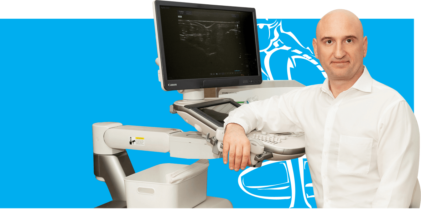
Below is a prime example of how ultrasound can take the guesswork out of diagnosis.

A bad physical therapy experience is one of the primary causes of unnecessary surgery

In this instance, an athlete was originally diagnosed with minor quadriceps muscle strain and was treated for four weeks, with unsatisfactory results. When he came to our clinic, the muscle was not healing, and the patients’ muscle tissue had already begun to atrophy.
Upon examination using MSUS, we discovered that he had a full muscle thickness tear that had been overlooked by his previous provider. To mitigate damage and promote healing, surgery should have been performed immediately after the injury occurred. Because of misdiagnosis and inappropriate treatment, the patient now has permanent damage that cannot be corrected.
The most important advantage of Ultrasound over MRI imaging is its ability to zero in on the symptomatic region and obtain imaging, with active participation and feedback from the patient. Using dynamic MSUS, we can see what happens when patients contract their muscles, something that cannot be done with MRI. From a diagnostic perspective, this interaction is invaluable.
Dynamic ultrasonography examination demonstrating
the full thickness tear and already occurring muscle atrophy
due to misdiagnosis and not referring the patient
to proper diagnostic workup
Demonstration of how very small muscle defect is made and revealed
to be a complete tear with muscle contraction
under diagnostic sonography (not possible with MRI)
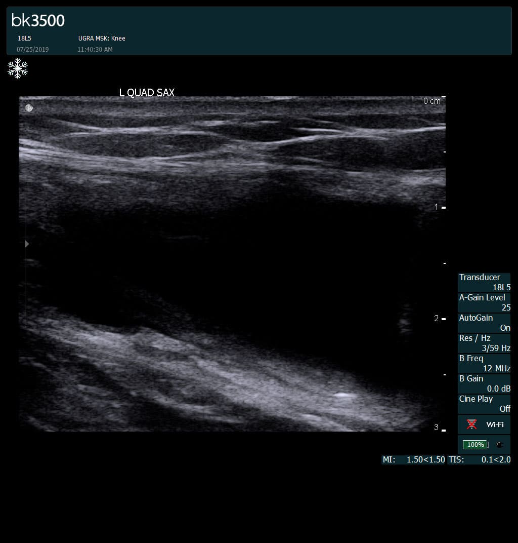
Complete tear of rectus femoris
with large hematoma (blood)
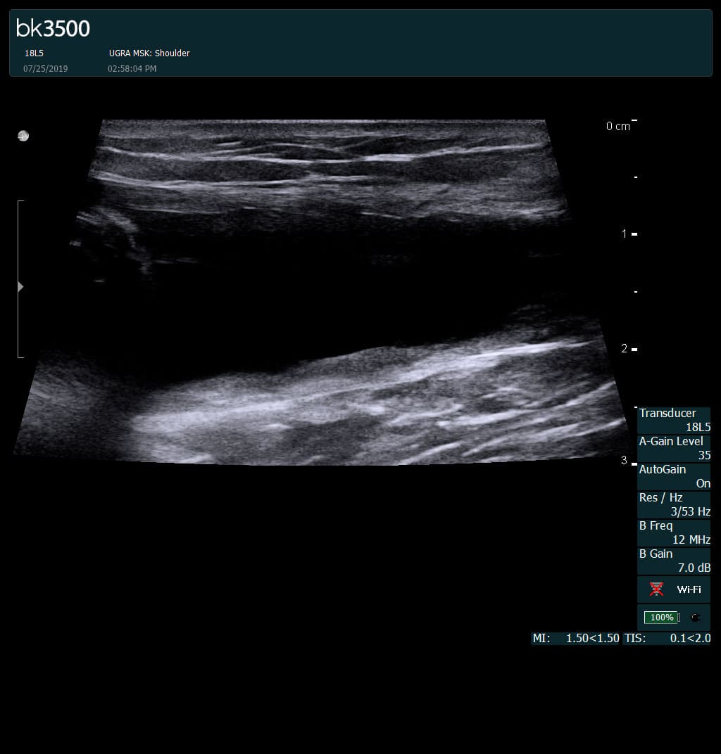
Separation of muscle ends due to tear elicited
on dynamic sonography examination


 Request Telehealth