Causes of Hip and Groin Pain

Causes of Hip and Groin Pain

Physiotherapists need to pay attention to the demographics of each patient when pain is present in the hips or groin. Age, gender, and activity level are key factors one should consider in diagnosis, as is the speed of onset. If the pain appears to be a result of an injury, the physiotherapist should ask the patient if he or she can supply any remembrance of how, precisely, the injury occurred. If the patient was playing sports, which sport was he playing? Did the injury happen outdoors? Did she remember turning suddenly at a sharp angle? Did he hear a popping sound, or feel something tearing? Does she feel something catching while she tries to walk? Palpate the entire area carefully and gently, and always respectfully, remembering that the patient and any direct observations are your primary resources for information.

If the patient reports a soccer or football injury, that is, kicking while running, suspect a flexor tear. The patient will feel pain upon lifting the leg upward, stretching the flexor muscle, or suspending in that position. Compare to the non-injured side as well if possible. These tears can also runners, particularly sprinters.

An adductor tear occurs when kicking hard, or sudden, unexpected, or forceful use of that muscle occurs. This can happen on an unlit staircase or a sidewalk edge. Palpate the area firmly but carefully, or try stretching it to see if the patient reports increased pain.

A labral tear is not a cartilage tear per se but damage to the lining of the cartilage in the hip socket. The patient will report a clicking sensation or perhaps a feeling that something is catching while he is walking. The pain is deep inside or within the groin, but could be referred as vague groin pain. Patients usually report increased pain while weight-bearing or walking.

Sometimes, hip or groin pain is referred from elsewhere, such as the lower back. Often this might present as a “pins and needles” sensation or numb feeling.

Rarely, physiotherapists may encounter pain that is referred from another area, but in fact, is felt in the groin. Often, the source is in the lower back, but always check the patient’s footwear, and the condition of the knees and lower legs as well.

With elderly patients, postmenopausal women especially, osteoarthritis can be a factor in hip pain. The onset is usually gradual. Elderly patients with poor bone density need to learn to strengthen their muscles to avoid any falls in the future.

Almost always, a fractured hip is so painful that the patient cannot bear weight nor walk on her own. Since dementia may also complicate matters, this type of injury is often rather stressful for families and patients alike.

Physiotherapists should be culturally aware and realize that not all patients express pain the same way. Pay careful attention to athletic concerns and the particularities of the sport and movements involved. Athletes may have concerns about how soon they can return to full functioning. Be sure to understand what this means, as for each patient, functionality is an individual matter that must be respected.

Diagnosis

app

A clinical exam and diagnostic ultrasound imaging can help your therapist pinpoint the exact location and cause of your hip and groin pain.

Ultrasound enables you and your therapist to view the hip and groin region in real time, while in motion. In addition to ultrasound, video gait analysis can help us identify faulty movement mechanics that contribute to hip and groin pain. Once the exact cause is determined, an effective treatment plan can be initiated.

img

Explore more advanced diagnostic tools available only at NYDNRehab:

img

Hip dysfunction and pain can be a complex issue due to interactions of the trunk, pelvis, low back, groin and hip joint. Physical therapy and rehabilitation that is based only on subjective clinical analysis often addresses the symptoms without resolving the underlying cause.

icons

At NYDNRehab, our groundbreaking motion analysis technology and high resolution diagnostic ultrasonography have enabled us to develop a battery of tests that perfectly reveal the dynamic functional pathology of the hip joint and pelvis. Our tests are evidence-based protocols that are considered to be the gold standard in the world of research.

Our testing protocol includes:

1

Combined lumbopelvic hip stability test using DLEST methodology with C.A.R.E.N., our computer assisted rehab environment

2

Hip joint stability test using DLEST methodology with C.A.R.E.N.

3

3D star excursion banner test (SEBT) for assessing the involvement of the hip joint and muscles in postural stability

4

3D gait or running analysis

5

3D kinematic joint angle analysis during a squat, lunge, drop jump and pelvis on hip rotation

6

Rehabilitative ultrasonography for viewing intrinsic hip stabilizing muscle activation patterns

bg

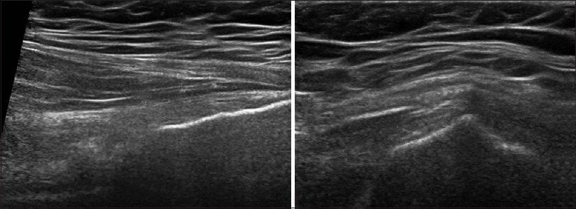
In this instance, an athlete was originally diagnosed with minor quadriceps muscle strain and was treated for four weeks, with unsatisfactory results. When he came to our clinic, the muscle was not healing, and the patients’ muscle tissue had already begun to atrophy.

Upon examination using MSUS, we discovered that he had a full muscle thickness tear that had been overlooked by his previous provider. To mitigate damage and promote healing, surgery should have been performed immediately after the injury occurred. Because of misdiagnosis and inappropriate treatment, the patient now has permanent damage that cannot be corrected.

The most important advantage of Ultrasound over MRI imaging is its ability to zero in on the symptomatic region and obtain imaging, with active participation and feedback from the patient. Using dynamic MSUS, we can see what happens when patients contract their muscles, something that cannot be done with MRI. From a diagnostic perspective, this interaction is invaluable.

Dynamic ultrasonography examination demonstrating
the full thickness tear and already occurring muscle atrophy
due to misdiagnosis and not referring the patient
to proper diagnostic workup

Demonstration of how very small muscle defect is made and revealed
to be a complete tear with muscle contraction
under diagnostic sonography (not possible with MRI)

image

Complete tear of rectus femoris
with large hematoma (blood)

image

Separation of muscle ends due to tear elicited
on dynamic sonography examination

bg
Buy now 3D Gait
Payment Success
Request Telehealth Request Telehealth Request in office visit Book now