HomeBlogCould Spinal Stenosis Be The Cause Of Your Pain?
Could Spinal Stenosis Be The Cause Of Your Pain?
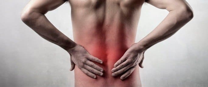
As the body ages, bones, nerves and muscles age as well, and it can be difficult to tell what the root cause of back and neck pain may be. Spinal stenosis is one possible cause for discomfort and pain in the back, neck, numbness and cramping in the legs. Understanding more about this condition can aid in choices for spinal stenosis treatment.
Spinal Cord Anatomy
Knowledge of spinal cord anatomy aids in understanding the causes of stenosis.
The spinal cord is a long, thin bundle of nerves running from the base of the skull to protect them and provide stability for the back. In between each vertebra are cushions, called disks, which are like shock absorbers. When the canal that houses these nerves narrow, the nerves can become compressed or pinched by the vertebrae and cause spinal stenosis. The disc material can also become diseased or damaged, resulting in pain when the nerves are compressed.
Compressed nerves can cause a range of sympto all parts of the body, a nerve pinch in the spine may not even cause back pain. The pain can show up in an extremity, hands or feet.
Top Three Spinal Stenosis Causes
Doctoms and pain depending on the underlying cause of the compression of the spinal canal.
The Aging Process: With age comes decreased activity and decreased range of motion and flexibility for joints, muscles and bones. Degenerative changes take places, as well, leading to conditions like arthritis and slipping of the vertebral bones.
Injury: A fall, break or strong blow to one of the vertebra can cause narrowing of the spinal canal and pinch the nerves inside causing pain.
Degenerative Spine Issues: Aging and injury can lead to the nerves, that the injured or degenerating material can put pressure on the nearby nerves.
Sprains, strains, infections or tumors are other, less likely spinal stenosis causes, but they do need to consideration.
Spinal Deterioration
Any area along the spine can deteriorate, but this problem is most prevalent in the neck (cervical) and back (lumbar) regions where the most weight and force are required for the body’s movement. These regions of the spine are at the highest risk for degenerative diseases. Common spinal stenosis causes include the following.
Degenerative Disc Disease: This is the most common kind of spine degeneration usually brought on by age. The discs that cushion the spine slowly wither or dry up and become prone to spinal stenosis.
Spondylolisthesis: Simply put, when the vertebrae don’t line up properly in the spine, the column housing the nerves can be narrowed or angled, causing the pinching resulting in cervical spinal stenosis or stenosis in the lumbar, or back, region.
Osteoarthritis: This condition is also known as facet disease. The facet bones allow your back to the spinal canal area and cause constriction.
Spinal Stenosis Prevention
Since a majority of the conditions leading to help avoid spinal problems down the road.
A healthy diet can prevent obesity. Being overweight puts undue stress and load on the spinal column which can lead to arthritic changes and degeneration of the discs cushioning the bones. Obesity also makes twisting, bending and flexibility more difficult and can put higher stress points on the vertebrae.
Proper posture is a must. The spine is naturally curved in an “S” shape to wear and tear of a particular set of vertebra over time. Most people believe they have good posture, but on closer examination, that may be only when they sit or stand. Posture affects the spine at all times– sitting, standing and sleeping.
Sports can be great exercise, but watch the load they put on your spine. High impact sports like wrestling, football or karate can cause injury to arthritis, degeneration or injury later in life.
Diagnosing Spinal Stenosis
Your docto see cross-sections of the spine. As with any procedure, insurance companies may mandate the order or kinds of testing performed for spinal issues.
Conservative, simple solutions will most likely be given first. Even if surgery is recommended by your physician, many insurance companies won’t cover expenses for major procedures unless all other noninvasive avenues have been explored. Examples of conservative treatment include:
Over-The-Counter Nonsteroidal Medications: These can help reduce inflammation around the joints of the spine and reduce swelling in the tissues. Nonsteroidals can cause stolerate them. Heat and Ice: Ice packs help numb painful areas and can reduce swelling. Heat packs provide comfort for tight muscles and can improve blood supply to discuss the options.
Stretching Exercises: Physical therapists or a health care professional are the best sources for specific exercises that can stretch the back muscles, helping to straighten the spinal column and improve posture.
Low Impact Sports: Walking and swimming can help straighten the spine and strengthen the muscles. Swimming may be especially beneficial as the weightlessness in the water helps reduce the load on the bones.
Massage Therapy: Deep tissue massages help relax the muscles and aid in pain management.
Physical Therapist Visits: Several visits to rehab or physical therapy for strengthening, flexibility work and training on posture may be suggested. For the best success, the physical therapy exercises should be done at home, as well.
Prescription Steroids: A burst of steroids (taken for seven days) or a continuous prescription (taken over several weeks) could help reduce inflammation. Steroids, along with stretching and other conservative treatments, may help thwart or put off a spinal surgery. Be sure to tell your medical professional if you have side effects.
Steroid Injections: Epidural injections are steroids placed into reach maximum benefit. This non-invasive method should most likely be cleared with your insurance company first.
Surgical Solutions
If the conservative methods fail to relieve the compression or pinching of the nerve. Remember that these types of surgical procedures are considered voluntary, or elective, and may require prior approval from your insurance company. However, they do offer a chance for a pain-free future if the nerve can be successfully freed from the compression.
There are two types of surgery for spinal stenosis causes. The first is an open procedure, which is more invasive and may include removing a disc, replacing portions of discs or bone with grafts or hardware and requires a longer recovery time. Many times, the patient is sent to strengthen the back muscles and give the best outcome for healing.
The other option is endoscopic surgery. A very small camera and instrument are used to remove portions of disc or bone material. This is very minimally invasive and has the benefits of decreased risk of infection and blood loss. Since the incision is so small, these procedures can sometimes be done as outpatient.
Only your doctor can decide which type of surgery your specific condition requires. Obtaining a second or third opinion on something this serious is wise, since the health of your spine depends on it.
In this instance, an athlete was originally diagnosed with minor quadriceps muscle strain and was treated for four weeks, with unsatisfactory results. When he came to our clinic, the muscle was not healing, and the patients’ muscle tissue had already begun to atrophy.
Upon examination using MSUS, we discovered that he had a full muscle thickness tear that had been overlooked by his previous provider. To mitigate damage and promote healing, surgery should have been performed immediately after the injury occurred. Because of misdiagnosis and inappropriate treatment, the patient now has permanent damage that cannot be corrected.
The most important advantage of Ultrasound over MRI imaging is its ability to zero in on the symptomatic region and obtain imaging, with active participation and feedback from the patient. Using dynamic MSUS, we can see what happens when patients contract their muscles, something that cannot be done with MRI. From a diagnostic perspective, this interaction is invaluable.
Dynamic ultrasonography examination demonstrating the full thickness tear and already occurring muscle atrophy due to misdiagnosis and not referring the patient to proper diagnostic workup
Demonstration of how very small muscle defect is made and revealed to be a complete tear with muscle contraction under diagnostic sonography (not possible with MRI)
Complete tear of rectus femoris with large hematoma (blood)
Separation of muscle ends due to tear elicited on dynamic sonography examination