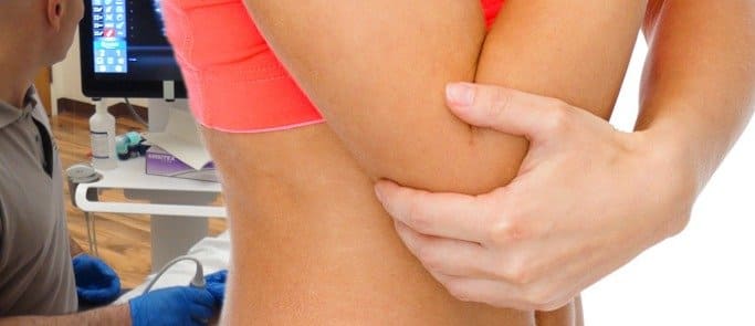
Lateral epicondylitis (LE) can be treated with the help of various techniques. Sometimes people suffering from lateral epicondylitis have to try more than one procedure to find relief from pain. Conventional treatment modalities available for lateral elbow pain are:
However, it has been observed that patients who have had steroid injections already still tend to visit the physiotherapy clinics regularly. It is highly advisable to extensively explore both manual therapy and exercise before opting for injection therapy. No particular protocol is going to work with every patient when it comes to the rehabilitation of LE; therefore, different combinations must be used on different patients to find out which works best to help relieve elbow pain.
Researchers in Sweden conducted a six-week randomized controlled trial. This trial observed the effects that forearm resistance exercise had on people who have LE. Forearm resistance exercises are essential in developing a firm, strong grip. These exercises engage not only the forearm but are also useful in strengthening all the other muscles in your arm including the shoulder.
Researchers divided the people with LE into two groups to compare the difference between the two different exercises. Each group had 42 participants who had been allocated randomly to each group. A bucket filled with water was used for the forearm resistance exercise. One group had to perform forearm resistance exercises with weights maintained at a pain-free range while the second group was asked to perform forearm resistance band exercises.
During the forearm resistance exercise, the patient was asked to hold the bucket with the non-affected. The purpose of this step was to prevent the concentric phase on the affected arm. He was then asked to place his arm on a table or the thigh griping the bucket with your affected arm. He was then asked to flex his wrist several times.
During the first week, participants were encouraged to perform two complete sets of 8 to 12 repetitions. These repetitions were increased to two full cycles in the second week and then three cycles in the third week thus increasing them as the week’s progress.
After the intervention of point marking three weeks, researchers observed that there was no difference in the wrist extensor strength or in the strength of the grip between both the groups, however, there was a commendable difference when both the groups were compared after six weeks.
Results showed that the group that performed forearm resistance exercises for six weeks experienced a significant decrease in symptoms of Lateral Epicondylitis with almost 56% of the participants testifying to being pain-free.
However, the group that performed resistance band exercises still suffered from elbow pain with practically 79% of the participants complained that they felt no change in their symptoms. This study indicates that doing forearm exercises daily is much better than sports medicine as they can help get rid of elbow pain permanently.

Verified Expert Profiles
Dr. Lev Kalika is a world-recognized expert in musculoskeletal medicine. with 20+ years of clinical experience in diagnostic musculoskeletal ultrasonography, rehabilitative sports medicine and conservative orthopedics. In addition to operating his clinical practice in Manhattan, he regularly publishes peer-reviewed research on ultrasound-guided therapies and procedures. He serves as a peer reviewer for Springer Nature.
Dr. Kalika is an esteemed member of multiple professional organizations, including:
Below is a prime example of how ultrasound can take the guesswork out of diagnosis.

A bad physical therapy experience is one of the primary causes of unnecessary surgery
In this instance, an athlete was originally diagnosed with minor quadriceps muscle strain and was treated for four weeks, with unsatisfactory results. When he came to our clinic, the muscle was not healing, and the patients’ muscle tissue had already begun to atrophy.
Upon examination using MSUS, we discovered that he had a full muscle thickness tear that had been overlooked by his previous provider. To mitigate damage and promote healing, surgery should have been performed immediately after the injury occurred. Because of misdiagnosis and inappropriate treatment, the patient now has permanent damage that cannot be corrected.
The most important advantage of Ultrasound over MRI imaging is its ability to zero in on the symptomatic region and obtain imaging, with active participation and feedback from the patient. Using dynamic MSUS, we can see what happens when patients contract their muscles, something that cannot be done with MRI. From a diagnostic perspective, this interaction is invaluable.
Dynamic ultrasonography examination demonstrating
the full thickness tear and already occurring muscle atrophy
due to misdiagnosis and not referring the patient
to proper diagnostic workup
Demonstration of how very small muscle defect is made and revealed
to be a complete tear with muscle contraction
under diagnostic sonography (not possible with MRI)

Complete tear of rectus femoris
with large hematoma (blood)

Separation of muscle ends due to tear elicited
on dynamic sonography examination


