Lateral epicondylitis – commonly known as tennis elbow – is an overuse injury affecting the tendons that attach your forearm muscles to your elbow. While it gets its popular name from tennis injuries, lateral epicondylitis can arise from a variety of activities that exert repetitive force on the forearm and outer elbow. If left untreated, tennis elbow can be painful and slow to heal, often persisting for months on end. Today, advanced regenerative technologies, orthobiologics and innovative therapies are revolutionizing the way tennis elbow is diagnosed and treated, reducing recovery time from months to just a handful of treatment sessions.
NYDNRehab stays on the cutting edge of rehabilitative medicine by embracing the latest evidence-based methodologies and making them available to our patients. Our integrative patient-centric approach combines highly skilled diagnostics with advanced technologies, for a speedy restoration of pain-free elbow function without drugs or surgery.
Clinical director & DC RMSK
Verified Expert Profiles
Dr. Lev Kalika, DC clinical director of NYDNRehab, is an internationally recognized expert in diagnostic and musculoskeletal ultrasonography, with multiple research papers to his credit. Dr. Kalika has studied with some of the world’s most prestigious experts in diagnostic, fascia, and nerve ultrasonography, and has presented his research at multiple international conferences.
Dr. Kalika is an active member of the American Institute of Ultrasound in Medicine (AIUM), and has developed his own unique approach to Dynamic Functional and Fascial Ultrasonography.

Orthobiologic, Sports Medicine and Regenerative Medicine Specialist
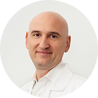
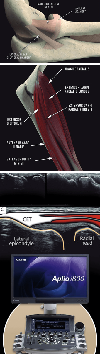
The lateral epicondyle is the bony bump on the outside of the elbow where the tendons of the wrist and forearm muscles attach to the humerus bone. Tendons are tough collagenous structures that are continuous with their associated muscles. The extensor carpi radialis brevis (ECRB), the forearm muscle that extends the wrist, is most frequently involved in tennis elbow.
The condition is caused by repetitive wrist extension and supination (palm-up rotation) of the forearm that leads to microtears. The radial nerve and its branches are often affected, especially the posterior interosseous nerve (PIN) and the superficial radial nerve. Fascial restrictions in the forearm can increase pressure on the tendons and nerves, intensifying pain.
Multiple factors slow down the tendon healing process:
Medical doctors often treat tennis elbow with corticosteroid injections, but patients should beware of steroid toxicity in the common extensor tendons that can lead to tendon degeneration and reduced performance.
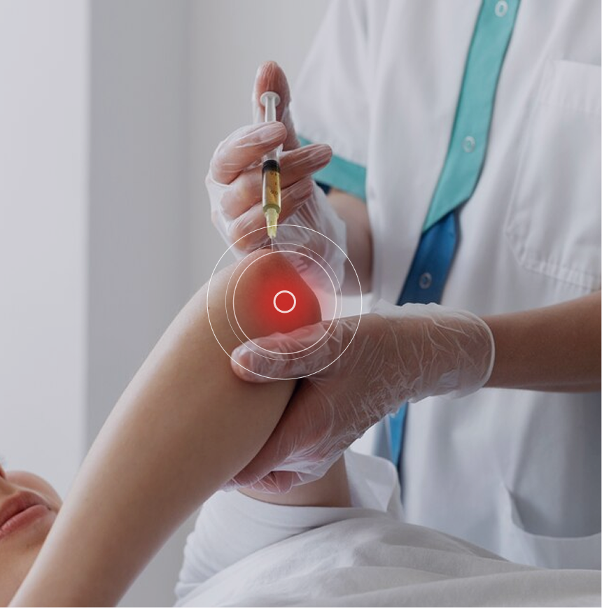
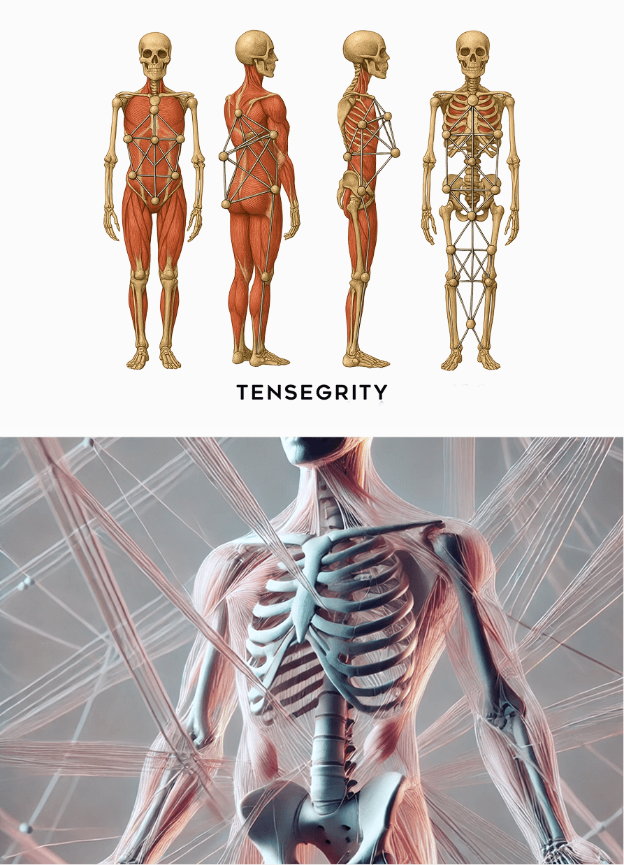
Biotensegrity refers to a physiological state where a system of individual components is held together under continuous elastic tension. In the human body, biotensegrity is created by the myofascial system, a complex network of muscles and fascia that exert elastic tension to control and guide movement, and to mediate outside forces. Biotensegrity is especially important in sports, where precise and efficient movement plays a key role in athletic performance.
Tennis elbow is a relatively common condition, and symptoms-based diagnosis can steer your elbow treatment in the wrong direction. Many other conditions mimic tennis elbow symptoms, and require different types of treatment. To get fast and effective results, it is important to work with a doctor who is able to differentiate lateral epicondylitis from its copycats.
Tennis elbow imposters include:
Identifying and treating underlying issues prior to beginning physical therapy is key to getting fast and effective results. Failure to pre-treat your tissues can completely undermine your treatment protocol, and in some cases, your condition may even worsen.
Obstacles to physical therapy success include:
At NYDNRehab, we use a combination of regenerative technologies, orthobiologics, and integrative therapies to restore tissue integrity before starting physical therapy. Our holistic treatment protocols are personalized, tailoring your recovery journey to your unique diagnostic results. Once we pre-treat your damaged tissues and eliminate compensation patterns, your body will be ready to begin physical therapy.
Advanced treatment approaches are changing the game in injury rehab, and NYDNRehab is on the cutting edge. Our clinic features the latest evidence-based technologies and innovative methodologies that are only now emerging in the injury rehab space.
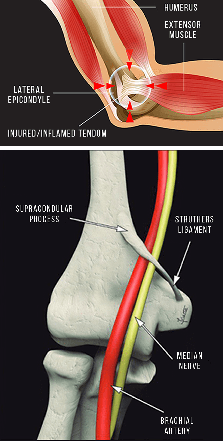
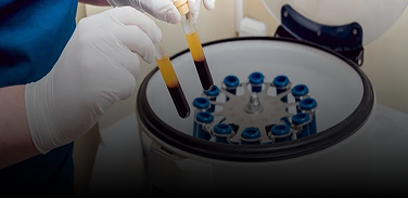
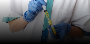
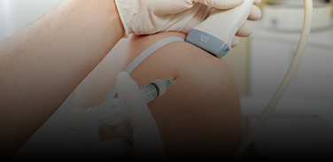
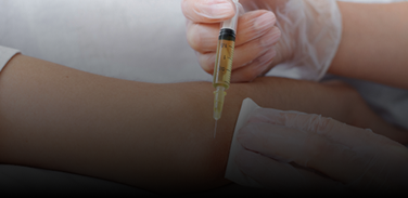
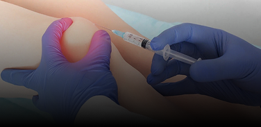
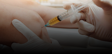
Dr. Kalika and Dr. Brosgol are pioneers in holistic rehabilitative medicine, leveraging the most current methodologies to tap into the body’s own healing mechanisms, to renew and repair tissues without drugs or surgery. Orthobiologic injection therapies use natural/neutral solutions, injected with precision thanks to ultrasound guidance. The injected solutions stimulate cellular repair by either nourishing or irritating the targeted cells.
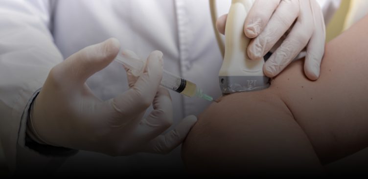
For needling procedures, Dr. Kalika and Dr. Brosgol work together as a team, with Dr. Kalika providing ultrasound guidance to ensure that the needles hit their mark. Treatment results are dramatically enhanced when tissues are pre-treated with focused extracorporeal shockwave therapy (fESWT) and myofascial release techniques.
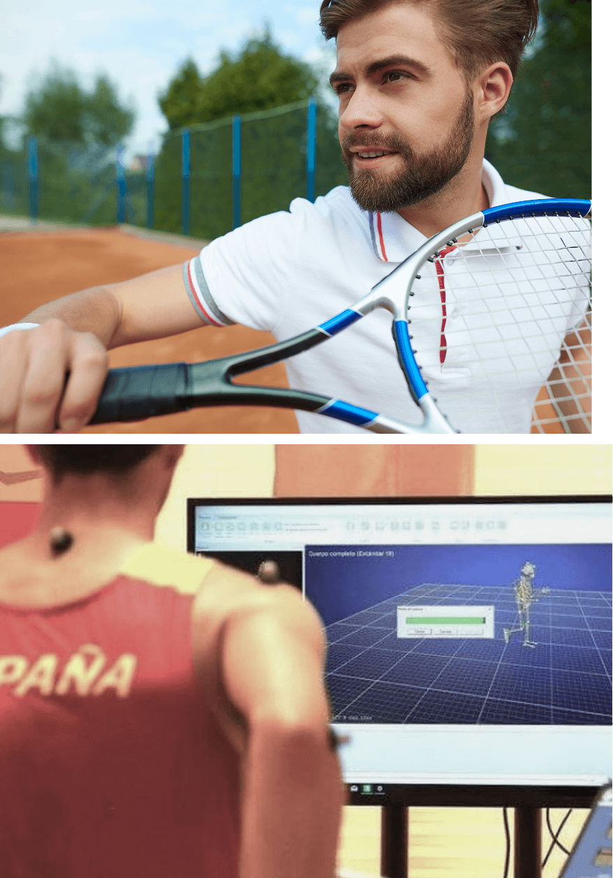
Tennis elbow is mostly a repetitive overuse injury, and symptoms often arise without warning. In addition to racquet sports, people in many occupations are prone to lateral epicondylitis, such as construction workers, cooks and chefs, mechanics, hairdressers, dentists, musicians, and many more.
Follow these guidelines to avoid lateral epicondylitis:
Modern medicine’s reductionist approach to tennis elbow focuses on treating the locus of pain, without considering other structures that impact elbow function. While pain management may provide temporary relief, it does not resolve the mechanical issues that contribute to tendon pathology. At NYDNRehab, our holistic approach ensures that all contributing factors are addressed, so you can return to your sport or occupation with confidence.
Your high-resolution ultrasound exam takes place on your first visit, enabling us to differentiate lateral epicondylitis from other issues that cause outer elbow pain. Our personalized one-on-one treatment approach is based on your unique diagnostic results. Our advanced regenerative technologies, orthobiologics and cutting-edge therapies accelerate healing, so you can quickly get back to your favorite activities without pain or dysfunction.


These case studies reflect real clinical conditions evaluated at NYDNRehab using advanced diagnostic methods and individualized rehabilitation strategies. All cases are evaluated and managed by Dr. Lev Kalika and the NYDNRehab clinical team.


Independent peer-reviewed research relevant to this treatment approach.
Research authored or co-authored by the clinic’s medical director. The following research publications inform the clinical approach used in this treatment program.
Conference abstract
2023
 Lev Kalika
  Rostyslav Bubnov
Below is a prime example of how ultrasound can take the guesswork out of diagnosis.

A bad physical therapy experience is one of the primary causes of unnecessary surgery
In this instance, an athlete was originally diagnosed with minor quadriceps muscle strain and was treated for four weeks, with unsatisfactory results. When he came to our clinic, the muscle was not healing, and the patients’ muscle tissue had already begun to atrophy.
Upon examination using MSUS, we discovered that he had a full muscle thickness tear that had been overlooked by his previous provider. To mitigate damage and promote healing, surgery should have been performed immediately after the injury occurred. Because of misdiagnosis and inappropriate treatment, the patient now has permanent damage that cannot be corrected.
The most important advantage of Ultrasound over MRI imaging is its ability to zero in on the symptomatic region and obtain imaging, with active participation and feedback from the patient. Using dynamic MSUS, we can see what happens when patients contract their muscles, something that cannot be done with MRI. From a diagnostic perspective, this interaction is invaluable.
Dynamic ultrasonography examination demonstrating
the full thickness tear and already occurring muscle atrophy
due to misdiagnosis and not referring the patient
to proper diagnostic workup
Demonstration of how very small muscle defect is made and revealed
to be a complete tear with muscle contraction
under diagnostic sonography (not possible with MRI)

Complete tear of rectus femoris
with large hematoma (blood)

Separation of muscle ends due to tear elicited
on dynamic sonography examination


