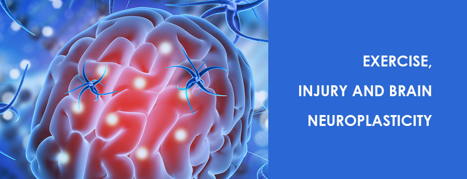
Despite the important role of the brain in regulating all metabolic and physiologic processes throughout the lifespan of the human body, the brain and central nervous system are given minimal attention from the medical community. In many cases, it is only when cognitive issues arise from trauma or disease that the brain takes center stage.
For ages, scientists and medical professionals have treated the brain and the body as distinct entities, ignoring the interrelated and interdependent relationship between the two. Only recently, thanks in part to advances in neuroimaging, scientific research has begun to unlock the important codependencies that exist between the brain, the central nervous system and human movement.
Despite our similarities, human beings have unique movement patterns and distinct anatomical characteristics. Treatment is often generalized, with exercise prescriptions based on what works in a research setting. IMA lets us look at the unique muscle activation patterns of each individual patient, so we can provide the most effective personalized treatment. Integrative motion analysis (IMA) provides tools for quantifying various movement parameters, to assess dysfunction and track recovery. In human movement, many factors come into play that define our quality of movement:
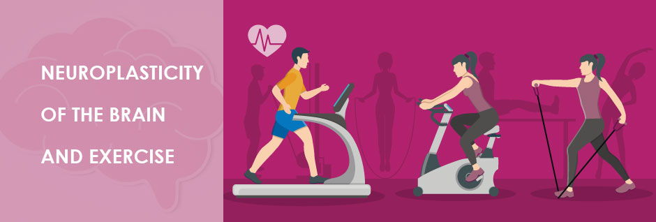
One important recent discovery is that neuroplastic changes take place in the brain in response to aerobic exercise. Aerobic exercise is defined as sustained rhythmic repetitive motion exercise that elevates the heart and respiratory rates, like running, swimming, cycling and other similar types of sustained exercise. Neuroplasticity describes how chemical changes cause the brain to form new connections between neurons, and to change the neurons themselves.
Regular exercise that increases blood flow to the brain sparks the regeneration of brain cells through a variety of mechanisms. Physical exercise actually increases the volume of white matter in the brain that represents connections to neurons, and also gray matter, which represents the neurons themselves. In addition, aerobic exercise stimulates the formation of new blood cells and elevates nerve growth factor.
Motor skills are acquired through practice and experience. They entail the fine-tuning of muscles to perform specific tasks in specific ways. For athletes, motor skills are essential to peak performance. From serving a tennis ball to the far corner of your opponent’s court, to throwing a football into the end zone with exacting precision, superior motor skills define elite athletes.
Aerobic exercise like running has been shown to enhance motor skills learning by increasing neuroplasticity in multiple regions of the brain. Plasticity of neurons and the glial cells that surround them is essential for adopting new skills. Neurotransmitter switching is a type of neuroplasticity that enables neurons to change their transmitter identity when subjected to ongoing stimuli.
In one new study, Li and Spitzer (2020) provided a running wheel to an experimental group of mice, which was used on a voluntary basis for just one week. Afterward, the runners had a greater ability to acquire new skills on the balance beam and rotarod, compared to controls who were not given a running wheel. The research team attributed the enhanced skill learning to chemical neurotransmitter switching, from ACh (acetylcholine) to GABA (gamma-Aminobutyric acid) in the caudal pedunculopontine nucleus (cPPN), a collection of neurons found in the upper region of the brain stem.
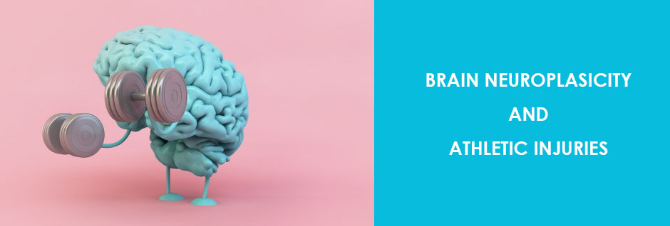
These findings have huge implications for the way we approach sports training and rehabilitation. By recognizing the role of brain neuroplasticity in skills learning and execution, and by adopting methods for retraining the brain in rehab, we can help both athletes and non-athletic patients avoid and recover from musculoskeletal injury. In the process, we can expect to achieve new heights in human performance.
Exercise benefits brains of any age. This is good news for anyone over 40, since the brain naturally begins to atrophy during the fifth decade of human life. Not only can exercise stimulate the neurogenesis of new brain cells, but it may be able to delay or even prevent neurodegenerative disease by improving the structure and function of the hippocampus, responsible for learning and memory, and the prefrontal cortex, responsible for executive function.
A popular theory about why the body ages zeroes in on oxidative stress. It is thought that free radicals naturally produced within cells can cause biochemical damage to cellular molecules, unless antioxidant defenses are in place to counteract free radicals. In particular, oxidative stress can cause damage to mitochondria, responsible for energy production and metabolism within each cell.
Exercise has been shown to enhance antioxidant defenses by ramping up the production of antioxidant enzymes. This in turn is thought to slow the aging process by combating oxidative stress, not only in the muscles and vital organs, but in the brain and nervous system. As a result, brain cells are able to adapt, repair and resist degenerative forces.
If you have suffered an injury or are experiencing pain and mobility issues, the team at NYDNRehab can help. We use cutting-edge technologies and innovative treatment methods to restore your brain and body to pain-free functional movement.
Some of our treatment tools include:
Resources:
Khan, Farid. “Run Regular, Age Slower, Be Neuroplastic.” Archives of Neuroscience 4.2 (2017).
Li, Hui-quan, and Nicholas C. Spitzer. “Exercise enhances motor skill learning by neurotransmitter switching in the adult midbrain.” Nature communications 11.1 (2020): 1-13.
Vivar, Carmen, and Henriette van Praag. “Running changes the brain: the long and the short of it.” Physiology 32.6 (2017): 410-424

Verified Expert Profiles
Dr. Lev Kalika is a world-recognized expert in musculoskeletal medicine. with 20+ years of clinical experience in diagnostic musculoskeletal ultrasonography, rehabilitative sports medicine and conservative orthopedics. In addition to operating his clinical practice in Manhattan, he regularly publishes peer-reviewed research on ultrasound-guided therapies and procedures. He serves as a peer reviewer for Springer Nature.
Dr. Kalika is an esteemed member of multiple professional organizations, including:
Below is a prime example of how ultrasound can take the guesswork out of diagnosis.

A bad physical therapy experience is one of the primary causes of unnecessary surgery
In this instance, an athlete was originally diagnosed with minor quadriceps muscle strain and was treated for four weeks, with unsatisfactory results. When he came to our clinic, the muscle was not healing, and the patients’ muscle tissue had already begun to atrophy.
Upon examination using MSUS, we discovered that he had a full muscle thickness tear that had been overlooked by his previous provider. To mitigate damage and promote healing, surgery should have been performed immediately after the injury occurred. Because of misdiagnosis and inappropriate treatment, the patient now has permanent damage that cannot be corrected.
The most important advantage of Ultrasound over MRI imaging is its ability to zero in on the symptomatic region and obtain imaging, with active participation and feedback from the patient. Using dynamic MSUS, we can see what happens when patients contract their muscles, something that cannot be done with MRI. From a diagnostic perspective, this interaction is invaluable.
Dynamic ultrasonography examination demonstrating
the full thickness tear and already occurring muscle atrophy
due to misdiagnosis and not referring the patient
to proper diagnostic workup
Demonstration of how very small muscle defect is made and revealed
to be a complete tear with muscle contraction
under diagnostic sonography (not possible with MRI)

Complete tear of rectus femoris
with large hematoma (blood)

Separation of muscle ends due to tear elicited
on dynamic sonography examination


