Osteoarthritis (OA) is a pro-inflammatory condition historically linked to aging that affects synovial joints throughout the body. In the 21st Century, knee osteoarthritis is on the rise due to growing trends toward sedentary lifestyles and obesity in the general population.
While OA can affect multiple joints, weight-bearing joints like the knees are especially susceptible. Recent research affirms that shockwave therapy is an effective treatment for knee OA that helps to regenerate knee cartilage, reduce pain and improve mobility in OA patients.
Osteoarthritis (OA) is a term used to describe degenerative joint disease resulting from progressive loss of cartilage, the protective tissue that covers the ends of bones and allows them to glide smoothly without friction. In the later stages of knee OA, reduced lubrication from synovial fluid contributes to further degeneration of the joint.
While the risk of OA increases with age, it can occur in younger people. Knee OA is more prevalent in women, and obesity and past injuries can contribute to the progression of knee OA.
Because OA is a pro-inflammatory condition, chronic systemic inflammation related to metabolic disease can be a significant factor. Lifestyle modifications can potentially slow or even halt OA progression.
or
Clinical director & DC RMSK
Verified Expert Profiles
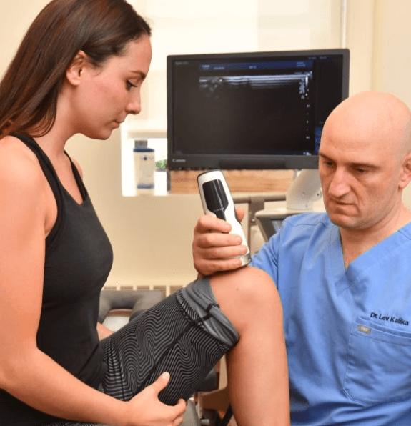
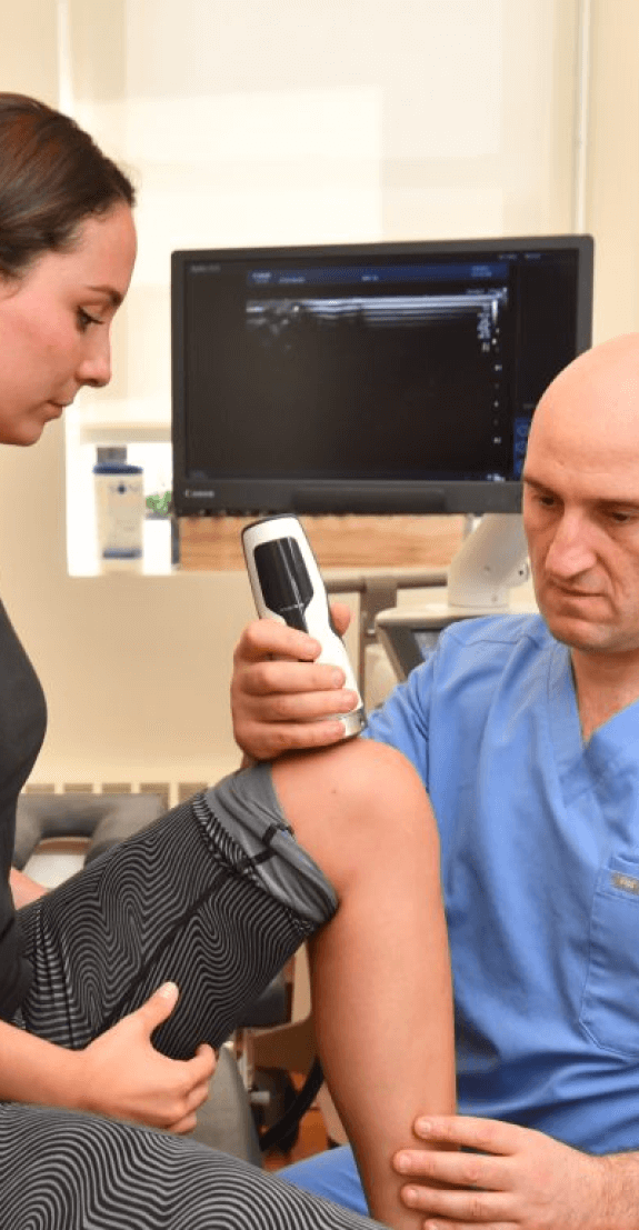
NYDNRehab clinical director Dr. Lev Kalika has revolutionized knee pain treatment by introducing high resolution diagnostic ultrasonography for structural diagnosis, combined with gait and motion analysis technology to visualize and objectify the functional movement of the lower extremities. Dr. Kalika’s use of shockwave therapy and other regenerative technologies is transforming the way degenerative knee osteoarthritis is treated and managed.
For over 15 years, Dr. Kalika has used extracorporeal shockwave therapy (ESWT) to treat knee OA and other amusculoskeletal disorders. His extensive training and hands-on experience with ESWT and other regenerative technologies make NYDNRehab the clinic of choice for knee OA treatment in NYC.
Listen to Dr. Kalika’s podcast interview with AOR.US to learn more about knee osteoarthritis
While joint osteoarthritis has long been associated with aging, multiple recent studies show that joint degeneration has significant links to sedentary lifestyles and obesity. Yet despite mounting evidence that lifestyle is a key factor in knee OA, medical doctors continue to treat the condition by managing symptoms rather than taking measures to reverse joint damage.
At NYDNRehab, we treat every patient as a unique case, paying full attention to the patient’s lifestyle and health profile. We never use a one-size-fits-all approach to patient care. Our personalized approach goes beyond treating knee OA symptoms to addressing their underlying cause, with the goal of restoring normal joint function and pain-free mobility, without drugs or surgery.
According to recent research, knee OA appears to be a modern condition associated with key lifestyle factors. One analysis reveals that the prevalence of knee osteoarthritis has more than doubled since the mid-20th Century, and another peer-reviewed article digs into prehistoric archaeology to debunk the notion that “wear and tear” is the primary cause of joint OA.
As knee OA progresses, it not only causes the degeneration of cartilage, but also affects the meniscus, ligaments, and muscles surrounding the knee. Knee OA is strongly associated with metabolic disease and premature death.
Rather than addressing lifestyle factors, common medical treatments involve prescription painkillers, NSAIDs, corticosteroid injections, knee braces, and avoiding physical activity — a factor known to cause the condition in the first place. When all else fails, surgery may be prescribed.
Knee osteotomy is a surgical procedure that seeks to offset cartilage loss by removing or reshaping bone tissue in either the femur or the tibia, to redistribute body weight and take pressure off the affected knee. As you can imagine, shifting weight to other parts of the body creates new problems, with little hope of restoring normal mobility.


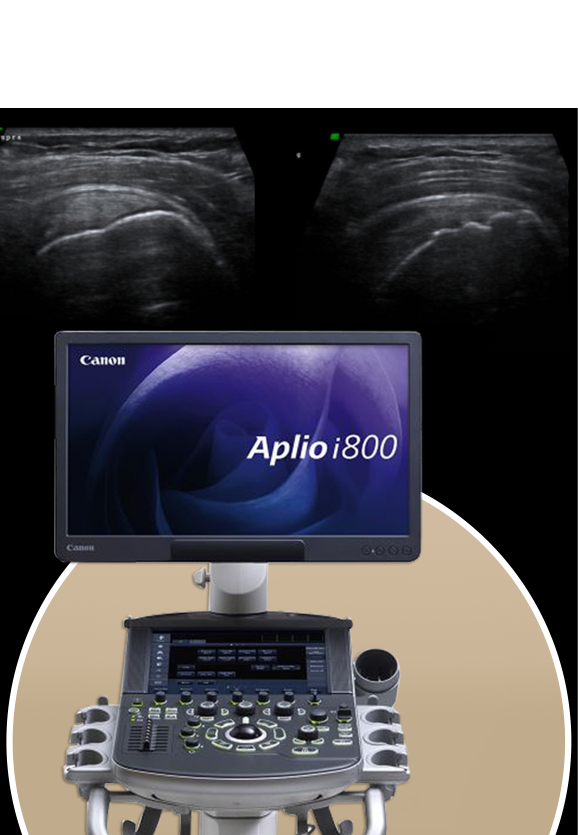
Dr. Kalika is a world-renowned expert in diagnostic musculoskeletal ultrasonography, with multiple research publications to his credit. Ultrasound imaging lets us view the knee joint in real time, to identify the exact location where cartilage has eroded.
We believe that every case of knee osteoarthritis should be treated based not only on the patient’s symptoms and imaging results, but also on each patient’s unique health profile and functional abilities. Ultrasound lets us assess how OA affects other knee structures, and how it impacts overall joint function.
A 3D gait analysis and other evidence-based assessments help us to uncover additional factors contributing to knee OA pain. Armed with this information, we then create a personalized treatment plan for you, to relieve OA pain and restore functional movement.
Degeneration of knee cartilage is a key characteristic of knee OA, but it is not the only factor to consider. At NYDNRehab, we have over 15 years of experience using a combination of treatment approaches for arthritic joint and bone conditions.
We not only treat the cartilage in arthritic knees, but we also address the entire lower kinetic chain and its associated soft tissues, to balance muscle tension, realign joints, rehabilitate joint function and restore pain-free movement.
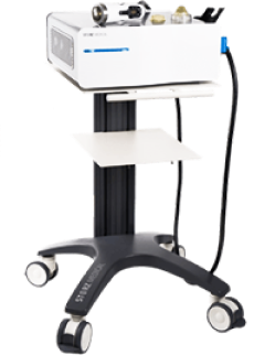
Focused Shockwave Therapy (ESWT)
Extracorporeal shock wave therapy (ESWT) delivers focused pulses of high-energy sound waves to affected tissues, to promote blood vessel formation and increase blood flow for greater oxygen and nutrient delivery. At the same time, ESWT reduces pain and inflammation, and improves joint function. ESWT is a recognized evidence-based approach for rebuilding cartilage in cases of degenerative knee OA.
Electromagnetic Transduction Therapy (EMTT) uses electromagnetic fields to reduce pain and stimulate cellular progenesis in cartilage and meniscus tissues. It is often used in conjunction with ESWT to promote the growth of healthy knee cartilage.
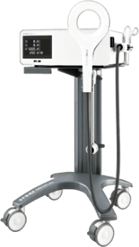
Electromagnetic Transduction Therapy (EMTT)
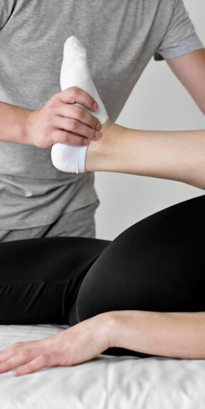
Physical therapy exercises work to strengthen and rebalance the muscles and fascia that support the knee joint, taking pressure off of nerves and connective tissues, and realigning the joints for optimal function.
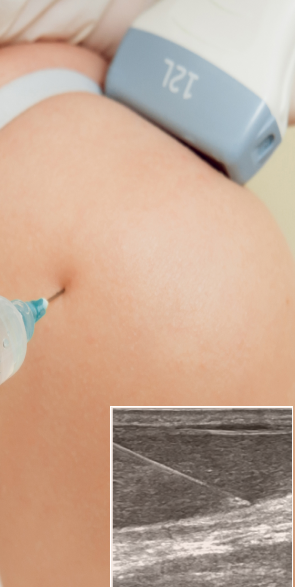
We sometimes use injection therapies for knee OA, to reduce pain and inflammation, and promote the regeneration of new collagen cells. Corticosteroids, platelet rich plasma, hyaluronic acid and other injections may be used when deemed appropriate. Ultrasound guidance ensures that the injected substance reaches its target with precision, for superior results and minimal patient discomfort.
Articular cartilage degeneration is a key feature of knee osteoarthritis, marked by the erosion and thinning of the cartilage layer that covers the ends of bones. It was once believed that degenerated cartilage could not be replaced or repaired, and that the only solution was eventual knee replacement surgery.
Thanks to new technologies and advanced research, we now know that, given the right environment, knee cartilage can regenerate itself. Multiple studies now demonstrate that shockwave therapy helps to create an environment in the knee capsule that promotes the regeneration and maintenance of knee cartilage.
Cartilage is a flexible and elastic connective tissue that enables the articular ends of bones to glide smoothly and prevents them from rubbing against each other. The low vascularity of cartilage tissue helps to maintain its integrity by maintaining a low-turnover state of equilibrium.
Chondrocytes are a unique type of protective cell embedded in the cartilage matrix that are responsible for maintaining its integrity. However, when cartilage is damaged by injury or overuse and begins to degenerate, the same chondrocytes that protect it now become resistant to the change necessary for building new tissue.
Knee osteoarthritis is essentially an abnormal wound healing response that creates an imbalance between the synthesis and degradation of the knee’s collagen matrix. The regeneration and maintenance of knee cartilage depends on chondrocyte synthesis of certain key mediators, including hyaluronic acid (HA) and platelet-rich plasma (PRP).
In one recent study, untreated chondrocyte cell cultures were compared with cultures exposed to ESWT, HA and PRP. The study concluded that ESWT enhances the ability of chondrocytes to utilize HA, thus encouraging cartilage regeneration. Data from the study also suggested that treatment with HA and ESWT can increase anti-inflammatory cytokine production in OA patients, helping to mitigate joint pain and inflammation.
Many clinics advertise shockwave therapy as a service, but the vast majority do not have the equipment or expertise to provide effective shockwave therapy for knee osteoarthritis.
At NYDNRehab, we use specialized equipment for focused, defocused, linear and radial shockwave therapy, depending on the type of tissues being targeted, the required depth of penetration, and the need to focus or zoom out on larger areas.
Once inflammation is reduced and the complex issue of cartilage degeneration is dealt with, we can proceed to address ancillary issues affecting the meniscus, ligaments, and muscles surrounding the knee. Physical therapy can help to retrain the muscles of the lower extremities in ways that optimize joint alignment, to redistribute body weight and reduce overall load on the knees.


Independent peer-reviewed research relevant to this treatment approach.

Below is a prime example of how ultrasound can take the guesswork out of diagnosis.

A bad physical therapy experience is one of the primary causes of unnecessary surgery
In this instance, an athlete was originally diagnosed with minor quadriceps muscle strain and was treated for four weeks, with unsatisfactory results. When he came to our clinic, the muscle was not healing, and the patients’ muscle tissue had already begun to atrophy.
Upon examination using MSUS, we discovered that he had a full muscle thickness tear that had been overlooked by his previous provider. To mitigate damage and promote healing, surgery should have been performed immediately after the injury occurred. Because of misdiagnosis and inappropriate treatment, the patient now has permanent damage that cannot be corrected.
The most important advantage of Ultrasound over MRI imaging is its ability to zero in on the symptomatic region and obtain imaging, with active participation and feedback from the patient. Using dynamic MSUS, we can see what happens when patients contract their muscles, something that cannot be done with MRI. From a diagnostic perspective, this interaction is invaluable.
Dynamic ultrasonography examination demonstrating
the full thickness tear and already occurring muscle atrophy
due to misdiagnosis and not referring the patient
to proper diagnostic workup
Demonstration of how very small muscle defect is made and revealed
to be a complete tear with muscle contraction
under diagnostic sonography (not possible with MRI)

Complete tear of rectus femoris
with large hematoma (blood)

Separation of muscle ends due to tear elicited
on dynamic sonography examination


