HomeBlogEye Brain Activity To Speed Up Healing Process Following ACL Surgery
Eye Brain Activity To Speed Up Healing Process Following ACL Surgery
Injuries often take longer to heal than we’d like, and new studies show this lag time might have more to do with our brains than previously thought. The brain seems to process movement in injured areas differently by putting more emphasis on visual cues than learned muscle memory. The sensation is a familiar one to anyone who’s been injured; movement becomes more hesitant and less instinctive, and the body is hyper-aware of any movement in the injured area.
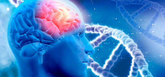
Recent research delves further into this possible brain-body connection. The research focuses on tears to the anterior cruciate ligament (ACL) and uses magnetic resonance imaging (MRI) to examine brain activity in athletes with and without injuries. The subjects were asked to move their legs while lying the MRI, and researchers looked for differences in how their brains responded.
The injured athletes showed major differences when asked to perform a simple knee movement involving their ACL. The visual centers of their brains were stimulated much more than the visual centers of non-injured athletes performing the same task, indicating that their brains were trying to rely more heavily on visual cues than normal to perform the motion.
Researchers described this difference as similar to walking down an unfamiliar hallway in the dark. Most people can navigate their own homes without too much trouble in the dark, but those same people will struggle to walk through a darkened hotel room without bumping into something. The brain is working overtime in this situation to find visual cues about the environment and relying very little on instinctive or remembered motion. According to recent studies, brains in injured bodies are doing much the same thing.
Relying more than usual on vision is distracting for the brain and can lead to further injuries. To counteract this distraction, researchers have come up with an ingenious way to keep the brain’s visual cortex occupied and force it to navigate familiar motions more naturally. The researchers, working with trainers, are having injured athletes wear special glasses with a built-in strobe effect while doing physical therapy for their injuries. The strobe keeps the brain’s vision centers busy and prompts the use of non-visual sensory systems the body usually uses to perform simple and familiar motions.
The goal here is to reduce the risk of injury after athletes go back to playing sports and to improve rehabilitation overall. Rebuilding range of motion, strength, and stability is the cornerstone of successful physical therapy after an injury. Adding the neuro-muscular re-education provided by the strobe could help athletes get back on the field faster, play to their full potential, and avoid being reinjured.
Further research is required to see just how effective strobe therapy is in speeding healing time after ACL injury. The strobe will also need to be tested to see how effective it is on rehabilitation from other types of injuries. However, the connection between the brain and the muscular system is an exciting area of research, and this new development has the potential to help many injured athletes with the recovery process.
In this instance, an athlete was originally diagnosed with minor quadriceps muscle strain and was treated for four weeks, with unsatisfactory results. When he came to our clinic, the muscle was not healing, and the patients’ muscle tissue had already begun to atrophy.
Upon examination using MSUS, we discovered that he had a full muscle thickness tear that had been overlooked by his previous provider. To mitigate damage and promote healing, surgery should have been performed immediately after the injury occurred. Because of misdiagnosis and inappropriate treatment, the patient now has permanent damage that cannot be corrected.
The most important advantage of Ultrasound over MRI imaging is its ability to zero in on the symptomatic region and obtain imaging, with active participation and feedback from the patient. Using dynamic MSUS, we can see what happens when patients contract their muscles, something that cannot be done with MRI. From a diagnostic perspective, this interaction is invaluable.
Dynamic ultrasonography examination demonstrating the full thickness tear and already occurring muscle atrophy due to misdiagnosis and not referring the patient to proper diagnostic workup
Demonstration of how very small muscle defect is made and revealed to be a complete tear with muscle contraction under diagnostic sonography (not possible with MRI)
Complete tear of rectus femoris with large hematoma (blood)
Separation of muscle ends due to tear elicited on dynamic sonography examination