While the term Femoroacetabular is quite a mouthful, it is just the clinical name for your hip joint, where the neck of your femur (the long bone of your upper leg) meets the acetabulum of your pelvis. Put simply it is the ball-and-socket complex that makes up your hip joint.
In a healthy person, the hip is a very strong and stable joint, capable of movement in multiple directions, thanks to wear and tear from daily activities, the hip joint can cause a great deal of pain and discomfort when injured.
Femoroacetabular Impingement (FAI)
There are actually two distinct types of FAI, and each has unique characteristics:
CAM Type FAI
A deformity of the femoral head-neck junction
Produces consistent intermittent stress on hip joint cartilage
Seen mostly in younger males, with a mean age of 32
Male to female ratio of incidence is 14:1
PINCER Type FAI
An abnormality in the pelvic acetabulum
Can cause ossification of the acetabular rim, which may lead to deepening of the acetabulum and over-coverage of the femoral head
Seen mostly in females, with a mean age of 40
Male to female ratio of incidence is 1:3
Patients typically first become aware of FAI as limited hip mobility. The condition gradually progresses to hip or groin pain, with hip rotation in a sitting position or during sports.
Causes of FAI
Essentially FAI is a “wear and tear” issue stemming from a number of potential causes, including:
Deficient movement mechanics
Poor postural habits
Muscle imbalances
Loose ligaments
Faulty motor control in the hip and pelvic region
Weak muscles surrounding the hip and core
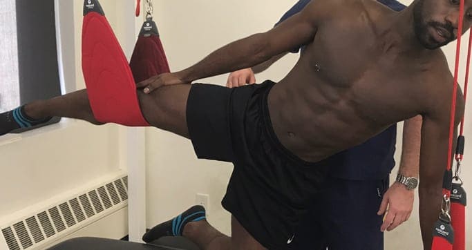
Treatment for FAI
Surgery is an option for FAI to correct those issues.
Conservative non-invasive physical therapy is often able to resolve FAI without surgical intervention. Therapy should focus on hip mobilization, range of motion and muscle strengthening.
A typical physical therapy protocol for FAI may include:
Aerobic exercise
Balance, coordination, and agility training
Corrective training to improve body mechanics and posture
Stretching exercises
Gait analysis and retraining
Relaxation therapy
Strength, power, and endurance training
Therapy strategies should be specifically tailored to the physical activity needs and preferences of the patient.
FAI Treatment in NYC
If you are suffering from limited hip mobility or chronic hip pain, the sports medicine specialists at NYDNRehab can help. We use diagnostic ultrasonography and other diagnostic to be the very best rehab clinic in NYC.
In this instance, an athlete was originally diagnosed with minor quadriceps muscle strain and was treated for four weeks, with unsatisfactory results. When he came to our clinic, the muscle was not healing, and the patients’ muscle tissue had already begun to atrophy.
Upon examination using MSUS, we discovered that he had a full muscle thickness tear that had been overlooked by his previous provider. To mitigate damage and promote healing, surgery should have been performed immediately after the injury occurred. Because of misdiagnosis and inappropriate treatment, the patient now has permanent damage that cannot be corrected.
The most important advantage of Ultrasound over MRI imaging is its ability to zero in on the symptomatic region and obtain imaging, with active participation and feedback from the patient. Using dynamic MSUS, we can see what happens when patients contract their muscles, something that cannot be done with MRI. From a diagnostic perspective, this interaction is invaluable.
Dynamic ultrasonography examination demonstrating the full thickness tear and already occurring muscle atrophy due to misdiagnosis and not referring the patient to proper diagnostic workup
Demonstration of how very small muscle defect is made and revealed to be a complete tear with muscle contraction under diagnostic sonography (not possible with MRI)
Complete tear of rectus femoris with large hematoma (blood)
Separation of muscle ends due to tear elicited on dynamic sonography examination