Five Common Causes of Pain in Your Hip

Five Common Causes

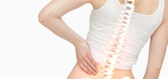
The structures surrounding your hip and pelvic region are a complex system of muscles, tendons, ligaments, nerves, joints and bones. They all interact together to transfer force loads between your upper and lower body, stabilize your spine, and support and protect your visceral and reproductive organs. As with any complex system, many moving parts means there are a number of things that can go wrong, and when something is out of whack, it often manifests as some type of hip pain.

Hip pain can present at the front, side, or back of the hip region, or in the groin area, and the exact cause can be difficult to diagnose. However, there are five common causes of hip pain that can be distinguished with a thorough diagnosis.

  1. Damage to the hip joint itself. Your hip joint is formed by the trochanter (ball) of the femur (or thigh bone), where it fits snugly into the socket of your pelvis. The structures are held in place by a strong network of ligaments, and facilitated by cartilage that allows the ball to glide freely within the socket. Damage to the hip joint itself is most often associated with wear and tear due to aging. Pain from hip joint damage is often felt at the front of the hip or groin area.
  2. Bursitis of the Hip. Bursae are fluid-filled sacs found in joints that provide a cushion between boney structures and soft tissues. An inflamed bursa at your hip joint can be quite debilitating, causing pain at the lateral or outside area of the hip that sometimes radiates down the side of the leg. Bursitis can be caused by overuse during sports or exercise, or can result from muscle weakness or obesity.
  3. Lumbar nerve compression or irritation. The nerves that govern your hip and leg movement descend from your lumbar spine, and they can become compressed or irritated as they exit the spinal canal. While the problem originates in your low back area, you may feel pain at the back of your hip or in your buttocks region.
  4. Deep gluteal syndrome. Previously called piriformis syndrome, deep gluteal syndrome occurs when the sciatic nerve becomes irritated or entrapped in the subgluteal space, where it passes beneath the piriformis muscle. The pain is deep beneath your gluteal muscles and can radiate down your leg.
  5. Hamstring insertional tendonitis. Your hamstring muscles originate at your pelvic region, beneath your buttocks. Pain in your lower buttock area that sometimes radiates down the back of your leg may indicate an inflamed hamstring tendon.

Hip Pain Diagnosis and Treatment in NYC

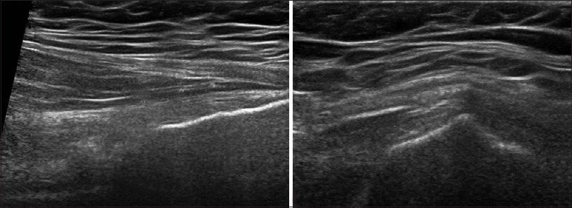
Hip pain can result from overuse, underuse or abuse of your muscles. Poor flexibility is often a contributing factor. The hip pain specialists at NYDNRehab use diagnostic ultrasonography, computerized gait analysis and other advanced diagnostic tools to identify the exact cause of hip pain. An individualized rehabilitation program is then designed to treat and correct the underlying cause of your pain, so you can return to an active lifestyle, pain-free.

Diagnosis

app

A clinical exam and diagnostic ultrasound imaging can help your therapist pinpoint the exact location and cause of your hip and groin pain.

Ultrasound enables you and your therapist to view the hip and groin region in real time, while in motion. In addition to ultrasound, video gait analysis can help us identify faulty movement mechanics that contribute to hip and groin pain. Once the exact cause is determined, an effective treatment plan can be initiated.

img

Explore more advanced diagnostic tools available only at NYDNRehab:

img

Hip dysfunction and pain can be a complex issue due to interactions of the trunk, pelvis, low back, groin and hip joint. Physical therapy and rehabilitation that is based only on subjective clinical analysis often addresses the symptoms without resolving the underlying cause.

icons

At NYDNRehab, our groundbreaking motion analysis technology and high resolution diagnostic ultrasonography have enabled us to develop a battery of tests that perfectly reveal the dynamic functional pathology of the hip joint and pelvis. Our tests are evidence-based protocols that are considered to be the gold standard in the world of research.

Our testing protocol includes:

1

Combined lumbopelvic hip stability test using DLEST methodology with C.A.R.E.N., our computer assisted rehab environment

2

Hip joint stability test using DLEST methodology with C.A.R.E.N.

3

3D star excursion banner test (SEBT) for assessing the involvement of the hip joint and muscles in postural stability

4

3D gait or running analysis

5

3D kinematic joint angle analysis during a squat, lunge, drop jump and pelvis on hip rotation

6

Rehabilitative ultrasonography for viewing intrinsic hip stabilizing muscle activation patterns

We also perform neuromotor testing with DD Robotech for:

  • Proprioception
  • Tracking ability
  • Force sense
  • Critical power
  • Reactive power
  • Flexibility

SEMG

Surface electromyography (SEMG) may be added to any of the above tests when needed.

Based on our experience and evidence-based information, we believe that physical therapy and rehabilitation should be based on objective quantifiable data.

bg

In this instance, an athlete was originally diagnosed with minor quadriceps muscle strain and was treated for four weeks, with unsatisfactory results. When he came to our clinic, the muscle was not healing, and the patients’ muscle tissue had already begun to atrophy.

Upon examination using MSUS, we discovered that he had a full muscle thickness tear that had been overlooked by his previous provider. To mitigate damage and promote healing, surgery should have been performed immediately after the injury occurred. Because of misdiagnosis and inappropriate treatment, the patient now has permanent damage that cannot be corrected.

The most important advantage of Ultrasound over MRI imaging is its ability to zero in on the symptomatic region and obtain imaging, with active participation and feedback from the patient. Using dynamic MSUS, we can see what happens when patients contract their muscles, something that cannot be done with MRI. From a diagnostic perspective, this interaction is invaluable.

Dynamic ultrasonography examination demonstrating
the full thickness tear and already occurring muscle atrophy
due to misdiagnosis and not referring the patient
to proper diagnostic workup

Demonstration of how very small muscle defect is made and revealed
to be a complete tear with muscle contraction
under diagnostic sonography (not possible with MRI)

image

Complete tear of rectus femoris
with large hematoma (blood)

image

Separation of muscle ends due to tear elicited
on dynamic sonography examination

bg
Buy now 3D Gait
Payment Success
Request Telehealth Request Telehealth Request in office visit Book now