June 14, 2024
The human body is uniquely designed to walk on two legs, and walking is our primary form of locomotion. In fact, when you walk, every joint in your body is activated, making walking an ideal form of exercise for anyone. But when you suffer from chronic low back pain, every step can be a challenge, sending pain signals along your central nervous system to your brain.
Many people subconsciously compensate for back pain by altering their gait to reduce force loads on the structures and nerves that cause the most pain. While this strategy may work in the short run, it can become a vicious cycle over time, creating gait abnormalities that cause more back pain.
Learn about gait fundamentals, the connection between low back pain and walking gait, and the benefits of 3D gait analysis.
While your body is much more than a machine, it adheres to certain mechanical principles governed by physics. In the same way that your car runs best when all parts are fine-tuned and in working order, your body moves best when joints are aligned, muscle and fascia tension are balanced, structures and systems are well-lubricated, and you give your cells the best fuel for peak performance.
Walking gait consists of a repetitive sequence of movements that make up the gait cycle. It involves proprioception from peripheral nerves, visual, aural and sensory input, coordinated muscle firing patterns, force generation and absorption, and tensegrity – the balanced tension provided by muscles and fascia that keep things in their place as you move. In a nutshell, gait mechanics is governed by the interplay of central nervous system commands and peripheral nervous system feedback.
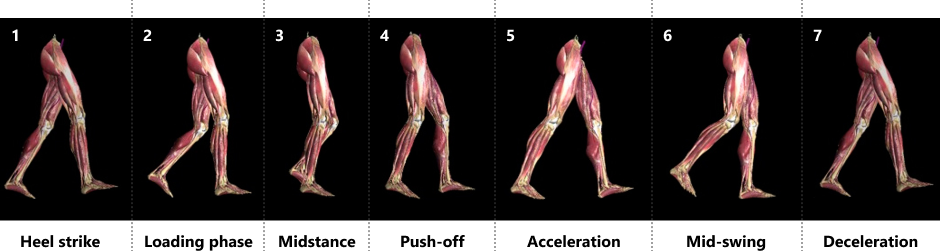
The gait cycle can be broken down into 7 distinct phases:
An optimal gait cycle depends on coordinated and synchronized action of the left and right sides of the body, which in turn depends on factors like muscle activation patterns, joint range of motion, and a plethora of other factors that regulate mobility and stability.
Unlike other mammals, human infants are not born fully developed. The size of the human brain necessitates premature childbirth, with the most profound developmental milestones occurring in the first year postpartum as the infant learns to defy gravity and becomes bipedal.
Early childhood development is driven by innate internal software that triggers progressive stages of development throughout the first year or so postpartum. In most infants, developing from a helpless neonate to an active toddler occurs naturally, and optimal gait patterns are established as the child progresses from walking to running. But as humans age, environmental and behavioral factors can undermine optimal motor patterns, causing gait abnormalities.
 Factors that alter gait include:
According to some studies, 85% of 60-year-olds have a “normal” gait, but by the age of 85, only 20% are able to maintain it. Staying physically active throughout your life and paying attention to other lifestyle factors like nutrition, sleep habits and stress management can reduce your risk of gait dysfunction as you age.
Low back pain often arises from compression of the nerve roots as they exit the lumbar spine – in some cases due to a herniated disc. However, disc herniation is itself a symptom, and does not fully explain low back pain. Optimal alignment of the lumbar vertebrae depends on a number of factors, including support from surrounding muscles and fascia, static and dynamic posture, pelvic alignment, and alignment of the lower kinetic chain from the feet up. When the spine is misaligned, discs can become compressed, placing pressure on the nerve roots.
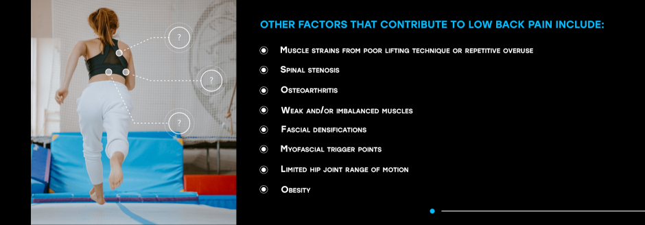
Other factors that contribute to low back pain include:
A Study on Central Sensitization, Gait and Low Back Pain
When your back hurts, you may subconsciously alter your gait in ways that affect your mobility. One recent study used artificial intelligence to analyze participants’ gait characteristics, based on data collected from a wearable tri-axial accelerometer device for one week.
Prior to the analysis, the 42 participants with chronic low back pain completed a central sensitization survey – central sensitization (CS) denotes a higher responsiveness of nociceptive neurons, manifesting as mechanical hypersensitivity. Participants were assigned to either a high or low sensitization group.
Upon analysis, the researchers found that the two groups had different motor control strategies, with differences in gait regularity, variability, predictability, smoothness, and stability. The high sensitization group exhibited tighter control of their gait, while the low sensitization group had smoother gait and greater stability.
Your lumbopelvic region plays a key role during gait. Throughout the gait cycle your pelvis tilts at varying angles to accommodate hip motion. Specifically, after initial contact the pelvis briefly tilts posteriorly (backward), then begins to tilt anteriorly (forward) again until about halfway through the gait cycle, where the pattern repeats itself.
Strong ligaments and fascia provide tension that stabilizes the pelvis to control movement. As your pelvis tilts, the vertebrae of the lower back move with it, compressing and expanding the intervertebral spaces. Inconsistencies between right and left hip action can cause uneven movement in the pelvis and lower spine, placing pressure on nerves and causing pain in the lower back.
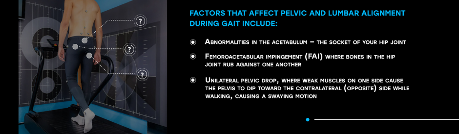
Factors that affect pelvic and lumbar alignment during gait include:
In addition, pelvic action during gait differs between males and females due to differences in pelvic architecture. A wider pelvis in females positions the hips farther apart, accounting for a wider stance and a lower center of gravity. Females tend to have a slight anterior pelvic tilt compared to males, whose pelvic tilt is closer to neutral. Females also have more motion in the transverse plane compared to males.
A full analysis of your walking gait mechanics can lend insight into the causes of your low back pain. However, beware of clinics that base their analysis on subjective observation – there are many nuances to gait mechanics that cannot be detected by the human eye. For example, observational gait analysis can only assess motion in the sagittal and frontal planes, overlooking critical gait deficits in the transverse plane.
To get an accurate and measurable picture of your gait, look for a clinic that uses advanced technologies to precisely measure your mechanical gait characteristics in all three planes of motion. By collecting precise data about joint angles, muscle firing patterns, stride length and frequency, foot mechanics, load distribution and more, your gait analysis provides an objective picture of your gait deficits.
From there, your therapist can devise a treatment plan to help you eliminate gait abnormalities that contribute to low back pain.
NYDNRehab is one of the few private physical therapy clinics to provide data-driven 3D gait analysis. Our proprietary software teams up with motion analysis technologies to create a comprehensive report, giving us key information needed to help you retrain your gait. At the same time, we use high resolution diagnostic ultrasound to look for other factors that contribute to low back pain, and create a personalized treatment protocol.
If walking makes your back hurt, or if back pain alters your walking gait, contact NYDNRehab today, and get to the source of your back pain so you can get back to pain-free movement.

Verified Expert Profiles
Dr. Lev Kalika is a world-recognized expert in musculoskeletal medicine. with 20+ years of clinical experience in diagnostic musculoskeletal ultrasonography, rehabilitative sports medicine and conservative orthopedics. In addition to operating his clinical practice in Manhattan, he regularly publishes peer-reviewed research on ultrasound-guided therapies and procedures. He serves as a peer reviewer for Springer Nature.
Dr. Kalika is an esteemed member of multiple professional organizations, including:
Below is a prime example of how ultrasound can take the guesswork out of diagnosis.

A bad physical therapy experience is one of the primary causes of unnecessary surgery
In this instance, an athlete was originally diagnosed with minor quadriceps muscle strain and was treated for four weeks, with unsatisfactory results. When he came to our clinic, the muscle was not healing, and the patients’ muscle tissue had already begun to atrophy.
Upon examination using MSUS, we discovered that he had a full muscle thickness tear that had been overlooked by his previous provider. To mitigate damage and promote healing, surgery should have been performed immediately after the injury occurred. Because of misdiagnosis and inappropriate treatment, the patient now has permanent damage that cannot be corrected.
The most important advantage of Ultrasound over MRI imaging is its ability to zero in on the symptomatic region and obtain imaging, with active participation and feedback from the patient. Using dynamic MSUS, we can see what happens when patients contract their muscles, something that cannot be done with MRI. From a diagnostic perspective, this interaction is invaluable.
Dynamic ultrasonography examination demonstrating
the full thickness tear and already occurring muscle atrophy
due to misdiagnosis and not referring the patient
to proper diagnostic workup
Demonstration of how very small muscle defect is made and revealed
to be a complete tear with muscle contraction
under diagnostic sonography (not possible with MRI)

Complete tear of rectus femoris
with large hematoma (blood)

Separation of muscle ends due to tear elicited
on dynamic sonography examination


