Hamstring tendon strain is one of the most common running injuries, affecting a set of muscles located in the back of the thigh. While mild hamstring strain may cause only a light feeling of discomfort, chronic hamstring strain can be severely crippling. What makes proper hamstring strain rehabilitation so crucial is that patients suffering from hamstring tendon strain have a 30 percent chance of re-injury. How to treat hamstring strain is the subject of this post.
The hamstrings are three muscles responsible for flexing and bending the knee. Each muscle begins as a tendon and runs from the pelvic bones through the femur, ultimately attaching itself to walk. This is when hamstring strain rehab becomes necessary.
Knowing how totally ruptured.
For those wondering how to the tissues, increasing blood circulation and degenerating damaged tendons.
Those at a loss for how to treat hamstring strain will find help at NYDRehab. We offer the first outpatient gait analysis in New York City. With over fifteen years’ experience treating orthopedic and sports injuries, we remain at the cutting edge of treatment for runners, sprinters, dancers, and other athletes.
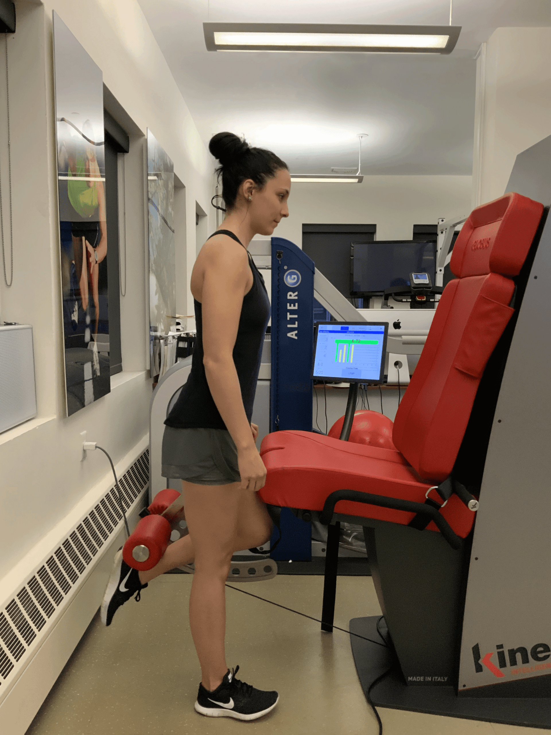
Reactive Neuromuscular Training on Kineo
Kineo – the most versatile muscle testing using artificial intelegence
In this instance, an athlete was originally diagnosed with minor quadriceps muscle strain and was treated for four weeks, with unsatisfactory results. When he came to our clinic, the muscle was not healing, and the patients’ muscle tissue had already begun to atrophy.
Upon examination using MSUS, we discovered that he had a full muscle thickness tear that had been overlooked by his previous provider. To mitigate damage and promote healing, surgery should have been performed immediately after the injury occurred. Because of misdiagnosis and inappropriate treatment, the patient now has permanent damage that cannot be corrected.
The most important advantage of Ultrasound over MRI imaging is its ability to zero in on the symptomatic region and obtain imaging, with active participation and feedback from the patient. Using dynamic MSUS, we can see what happens when patients contract their muscles, something that cannot be done with MRI. From a diagnostic perspective, this interaction is invaluable.
Dynamic ultrasonography examination demonstrating the full thickness tear and already occurring muscle atrophy due to misdiagnosis and not referring the patient to proper diagnostic workup
Demonstration of how very small muscle defect is made and revealed to be a complete tear with muscle contraction under diagnostic sonography (not possible with MRI)
Complete tear of rectus femoris with large hematoma (blood)
Separation of muscle ends due to tear elicited on dynamic sonography examination