An X-ray showing a heel spur can be alarming, but the bony spur is usually not the real source of heel pain. This guide explains what actually causes the pain, what genuinely helps, and how to get rid of it, in most cases without surgery.

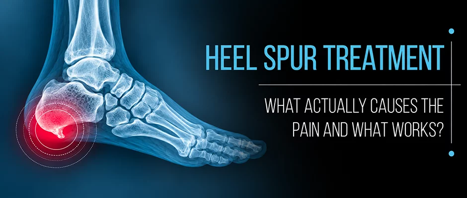
Your heel bone (calcaneus) is the largest of the bones in the foot, and is the main point of attachment for the plantar fascia underneath and the Achilles tendon behind. These attachment sites can develop a bony prominence called a calcaneal enthesophyte, or heel spur, as a result of chronic mechanical stress.
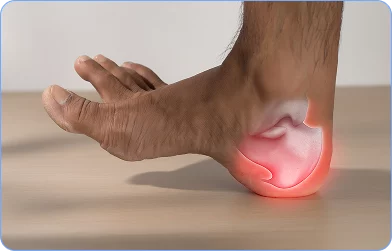
Plantar spurs develop at the point of origin of the plantar fascia at the inferior calcaneus. Posterior (also called dorsal) spurs develop at the insertion of the Achilles tendon. These two types behave differently, respond to different interventions, and should not be managed identically.
It is known that the presence of a spur on an X-ray is not a predictor of pain. Heel spurs are also common in adults who have no heel pain at all
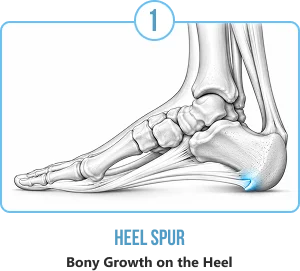
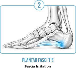
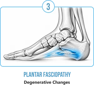
Although the term “plantar fasciitis” is the most common clinical term, it is becoming more apparent that the term is not precise. Histopathological examination has always shown collagen disorganization, angiofibroblastic hyperplasia and degenerative remodeling, but not classical acute inflammatory infiltrate.
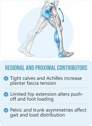
Regional and proximal contributors
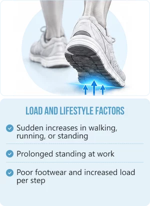
Load and lifestyle factors
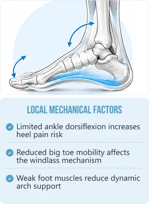
The plantar fascia is more than a passive band. It is a load-transmitting connective tissue structure with a viscoelastic (viscosity and elasticity) biomechanical complexity that has been significantly reinterpreted by contemporary fascia research.
The Windlass Mechanism
When your toes extend during push off, the plantar fascia tightens around the metatarsal heads, elevating the arch, stiffening the foot for propulsion and storing elastic energy.⁶ Disruption, whether caused by fascial degeneration, lack of extension of your great toes, or weakness of the intrinsic muscles of the foot, directly affects the way load is transferred through the foot.
Load Tolerance and Viscoelasticity
The plantar fascia stretches slightly under load and then recoils. If the volume is increased too quickly or there is a lack of rest, or tissue is degenerating, the tissue’s ability to recover from micro-damage is exceeded, and more micro-damage occurs than can be repaired. This is the mechanical mechanism of most presentations of plantar fasciopathy.
Mechanotransduction
Connective tissue fibroblasts are sensitive to mechanical signals. Progressive loading programs, and not complete rest, are the basis of modern fasciopathy rehabilitation, as they stimulate collagen synthesis and tissue remodeling.
The Achilles–Plantar Fascia Connection
The deep crural fascia of the calf, Achilles tendon and plantar fascia are anatomically contiguous as shown in cadaveric studies.⁷ Therefore, calf tightness does not just restrict ankle range, it also transmits tensile forces directly into the plantar fascia.
Walking and running are whole body rotational movements. As you step, your pelvis rotates, your trunk counter rotates and your arms swing opposite. Typical protective adaptations that occur when plantar heel pain develops are: decreased push-off, decreased step length, increased stance time, decreased ankle dorsiflexion, compensatory hip hiking, decreased stride length, and altered trunk and arm-swing mechanics.
These adaptations can lead to secondary restrictions over weeks and months, which may occur at the ankle, hip and thoracic spine, but not at the original site of pain.
Thoracic rotation is a part of normal arm swing and trunk counter-rotation during gait. Push-off may be painful, resulting in decreased contralateral arm swing, decreased trunk rotation to equal the shortened stride, and a possible restriction of the thoracic spine as a protective compensation. The question of whether thoracic restriction is an upstream contributor or a downstream compensation or whether it is an unrelated finding can only be answered by clinical examination, not by assuming it from the presence of both findings together.
Several other conditions can mimic heel spur pain.
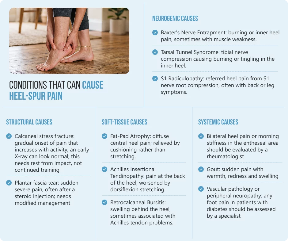
Structural causes
Soft-tissue causes
Systemic causes
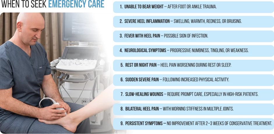
Conditions that require urgent medical attention.
If you have any of the following symptoms, you should get immediate assessment:
The presence of a calcaneal enthesophyte is confirmed on plain radiographs but has significant limitations as it cannot identify the pain generating tissue, a prominent spur may not be symptomatic, soft tissue pathology cannot be seen on plain film and a normal X-ray does not exclude early stress fracture. This is also why what heel spurs look like on an X-ray does not predict how much, or whether, they hurt.
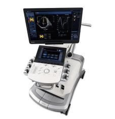
Diagnostic musculoskeletal ultrasound can measure the thickness of the plantar fascia and its echotexture, identify degenerative changes and bursae, and evaluate nerve involvement in the hands of an experienced practitioner, and direct injections precisely. It is dynamic, radiation-free and gives real-time information not available in radiographs.⁴
MRI is suitable for complex presentations, suspected stress fracture, or when diagnosis is still not certain after clinical and ultrasound evaluation.
History and physical examination are always required to be correlated with imaging. Treatment should focus on the clinically identified pain generator and not the radiographic finding.
The following measures are low risk of harm when applied as described and are suitable until a professional assessment is made. They should not be used in place of evaluation if symptoms are worsening, increasing, or if there are any red-flag features mentioned above.
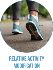
Decrease, but not eliminate, high impact activities. Complete rest is not usually helpful in the long term because the tissue needs a mechanical stimulus to remodel.
Gastrocnemius and soleus stretching is well supported in plantar heel pain management.⁹ Stretch gastrocnemius and soleus before the first steps in the morning. Repeat 2-3 times, hold for 20-30 seconds. The stretch should feel like gentle tension, not pain.
Heel cushioning and arch support decrease the plantar fascial load. Do not walk on hard surfaces without shoes on during the symptomatic period, especially first thing in the morning.
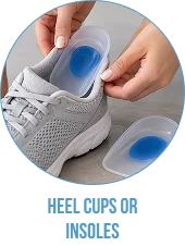
Cushioned heel cups can help decrease impact load during the acute phase and can give many patients meaningful short-term relief. In trials, prefabricated insoles have performed similarly to custom orthoses in the short term.
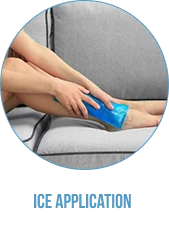
After activity, apply cold to heel for 10-15 minutes, using a cloth barrier. Avoid applying ice to skin that is cracked, broken or has reduced circulation or sensation.
Ibuprofen can be used at the lowest effective dose for the shortest period of time to relieve symptoms. Not recommended in patients with renal impairment, gastrointestinal disease, cardiovascular risk or drug interactions. Use with the advice of your pharmacist or doctor.

Acute heel pain with a degenerated/inflamed fascial insertion can be aggravated by forceful massage or ball-rolling. It is more suitable to work away from the area of maximum tenderness.
It is not appropriate and can be harmful to stretch into sharp pain. If there is a stress fracture, fascia tear or nerve entrapment, aggressive stretching can exacerbate the problem.
Excessive icing (longer than 20 minutes) or on skin can cause cold injury. People with neuropathy or vascular disease should avoid ice unless a clinician advises it.
There is no evidence to support any attempt to dissolve the spur with supplements or home remedies or self-applied devices, and this diverts treatment away from the true site of pain.
Self-care in the absence of a diagnosis is a real risk. Tenderness on palpation also occurs with stress fractures, nerve entrapments and inflammatory conditions. They will not respond to and may be exacerbated by conventional plantar fasciopathy self-care.
Returning to full training too quickly after a symptomatic period often triggers a flare-up. Unloaded tissue needs a “ramp-up” program.
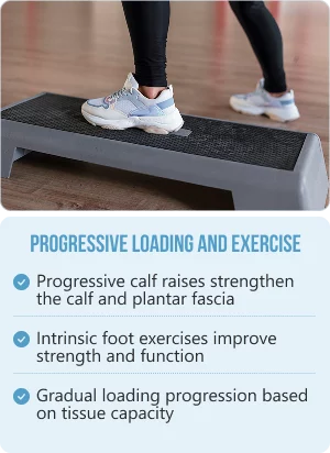
The most well supported intervention for plantar fasciopathy is progressive tendon and fascial loading programs. Calf-raise progressions plus intrinsic foot strengthening have been shown to produce clinically significant reductions in pain and improvements in function in clinical trials, and should be progressed systematically, rather than on arbitrary timelines, based on tissue capacity. In practice, the heel spur exercises that help most are calf and plantar-fascia loading, not stretching aimed at the spur itself.
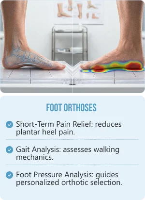
Prefabricated and custom orthoses often reduce plantar heel pain in the short term. Gait and foot pressure analysis can help guide orthotic choices better than a generic prescription.
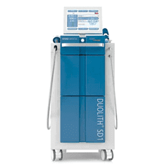
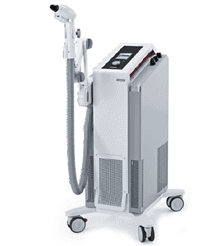
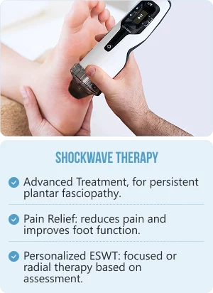
One of the more supported advanced treatments for chronic plantar fasciopathy that has not been helped by conservative treatment is extracorporeal shockwave therapy (ESWT). Multiple randomized controlled trials and systematic reviews show significant pain reduction and functional improvement, especially in presentations that last more than 3-6 months. It is not a cure-all; patient selection and dosing parameters are important. The focused and radial ESWT protocols are offered at NYDNRehab and are determined by the individual clinical assessment.
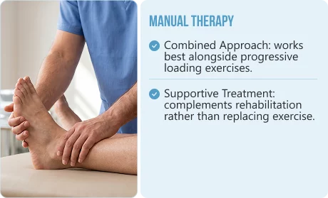
Manual therapy is best used in combination with loading programs, rather than as a standalone treatment for plantar heel pain.
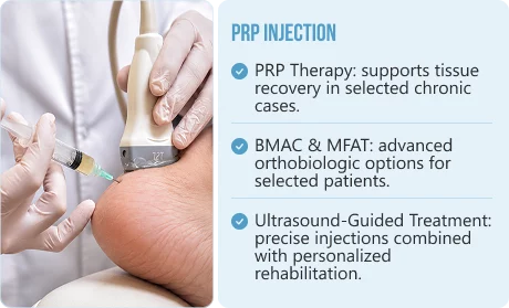
PRP injection has shown promising results in plantar fasciopathy, especially in chronic cases where ultrasound has demonstrated tissue degeneration. Quality of the evidence is improving and is still variable; PRP is not a first-line treatment and should be used in selected patients after conservative treatment. Ultrasound guidance helps ensure that it is delivered to the target tissue.
BMAC (Bone Marrow Aspirate Concentrate): an injection prepared from your own bone marrow. BMAC concentrates cells and other natural components from a small sample of your bone marrow. We may consider it for selected, more advanced joint or tendon problems when PRP alone may not be sufficient. Treatment combines precise ultrasound-guided injection with personalized rehabilitation.
MFAT (Micro-Fragmented Adipose Tissue): an injection prepared from your own fat tissue. MFAT uses a small amount of your fat, gently processed to preserve its natural cells and supporting tissue. We may consider it for selected, more advanced joint or soft-tissue problems when PRP alone may not be sufficient. Treatment combines precise ultrasound-guided injection with personalized rehabilitation.
Surgical interventions include partial plantar fascia release and, less frequently, spur resection if the spur has been particularly identified as a cause of pain. Surgery does not always resolve the pain and can have complications such as nerve damage, arch collapse and long recovery. It should be a last resort after all conservative measures have been tried.
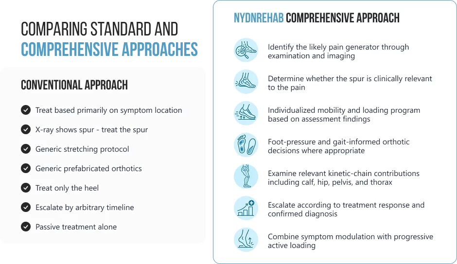
Rather than applying a uniform protocol to every presentation of heel pain, NYDNRehab combines diagnostic precision with individualized rehabilitation for heel pain to identify the primary pain generator and meaningful movement impairments contributing to each patient’s condition.
A comprehensive assessment at NYDNRehab may include:
This comparison should not be interpreted as a criticism of the quality of care provided by conventional clinicians. Comprehensive evidence-based management is already in practice by many practitioners. The difference is that the diagnostic assessment is more in-depth and treatment planning is more individualized.
Most patients with plantar heel pain respond well to the right conservative treatment, although recovery times are not uniform among patients and are influenced by length of symptoms, tissue pathology, loading and adherence to the rehabilitation program.
Patients with acute pain for less than 3 months and minimal degenerative tissue change on ultrasound tend to respond more quickly to loading programs, stretching and footwear modification. Patients with chronic presentations, proven degeneration in the fascia, and substantial biomechanical factors tend to need more time for rehabilitation and may need more advanced treatments like ESWT or PRP.
What is not conducive to recovery:
In most cases, no. The spur is an X-ray finding that often occurs along with but is not responsible for plantar heel pain. Treatment will focus on the tissue that is causing the pain, usually the plantar fascia, the surrounding soft tissues or an entrapped nerve.
Established bony projections do not dissolve with conservative treatment or soft-tissue interventions. But pain can go away completely even though the spur is seen on imaging, since the spur is typically not the cause of pain.
Stretching should give a slight tension but not sharp or reproducing pain. If stretching causes a lot of pain, it might not be simple plantar fasciopathy and further evaluation may be necessary before continuing with stretching.
ESWT may cause discomfort during treatment. Focused and radial protocols feel different, and the intensity is adjusted to your tolerance. Treatment is generally well tolerated, and discomfort usually goes away quickly following each treatment. Clinical assessment should guide selection of the appropriate protocol and dosing, rather than patient preference.
PRP is best used after conservative treatment (usually a structured loading program, manual therapy and shockwave therapy as indicated) has not resulted in adequate improvement, and ultrasound has identified degenerative change in the fascial tissue. It is not a first-line treatment. In selected, more advanced cases, BMAC or MFAT may also be considered.
This will largely depend on the duration of symptoms, the extent of tissue damage, the accuracy of diagnosis and the consistency of rehabilitation. Acute presentations where there is no significant tissue degeneration may resolve within weeks. If the chronic fasciopathy is accompanied by a clear demonstration of degenerative change, then a period of structured rehabilitation of several months is generally needed. Clinical evaluation is necessary to create an individual prognosis.
There is no overnight fix. The fastest realistic route is to reduce aggravating load, use supportive footwear or an orthotic, and begin a progressive calf and plantar-fascia loading program. Most people improve over a few weeks to a few months.
On an X-ray a heel spur looks like a small bony hook or shelf projecting from the heel bone, either under the heel (plantar) or at the back where the Achilles attaches (posterior). A spur under the heel usually cannot be seen or felt through the skin, while a bump at the back of the heel sometimes can.
In most cases you do not need to remove the spur at all. Treating the surrounding tissue with load management, exercise, orthoses and, when needed, shockwave therapy resolves the pain in the large majority of people, leaving the spur in place but no longer painful.
If you have been managing heel pain without a clear diagnosis, or if conservative self-care has not produced meaningful improvement after several weeks, a precise clinical assessment is your most important next step.
At NYDNRehab, we use high-resolution diagnostic ultrasound, gait and pressure analysis, and individualized rehabilitation to identify what is actually generating your pain and to build a treatment plan around that finding rather than around the most common label.
Schedule an assessment for heel spur treatment in NYC at NYDNRehab:
Request an appointment»

Our location: 11 West 25th Street 5th floor, New York, NY 10010
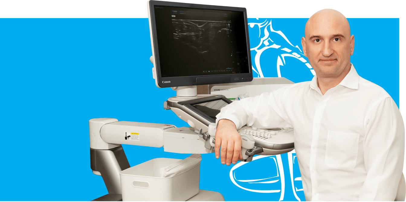
Dr. Lev Kalika is a world-recognized expert in musculoskeletal medicine. with 20+ years of clinical experience in diagnostic musculoskeletal ultrasonography, rehabilitative sports medicine and conservative orthopedics. In addition to operating his clinical practice in Manhattan, he regularly publishes peer-reviewed research on ultrasound-guided therapies and procedures. He serves as a peer reviewer for Springer Nature.
Dr. Kalika is an esteemed member of multiple professional organizations, including:

Below is a prime example of how ultrasound can take the guesswork out of diagnosis.

A bad physical therapy experience is one of the primary causes of unnecessary surgery

In this instance, an athlete was originally diagnosed with minor quadriceps muscle strain and was treated for four weeks, with unsatisfactory results. When he came to our clinic, the muscle was not healing, and the patients’ muscle tissue had already begun to atrophy.
Upon examination using MSUS, we discovered that he had a full muscle thickness tear that had been overlooked by his previous provider. To mitigate damage and promote healing, surgery should have been performed immediately after the injury occurred. Because of misdiagnosis and inappropriate treatment, the patient now has permanent damage that cannot be corrected.
The most important advantage of Ultrasound over MRI imaging is its ability to zero in on the symptomatic region and obtain imaging, with active participation and feedback from the patient. Using dynamic MSUS, we can see what happens when patients contract their muscles, something that cannot be done with MRI. From a diagnostic perspective, this interaction is invaluable.
Dynamic ultrasonography examination demonstrating
the full thickness tear and already occurring muscle atrophy
due to misdiagnosis and not referring the patient
to proper diagnostic workup
Demonstration of how very small muscle defect is made and revealed
to be a complete tear with muscle contraction
under diagnostic sonography (not possible with MRI)
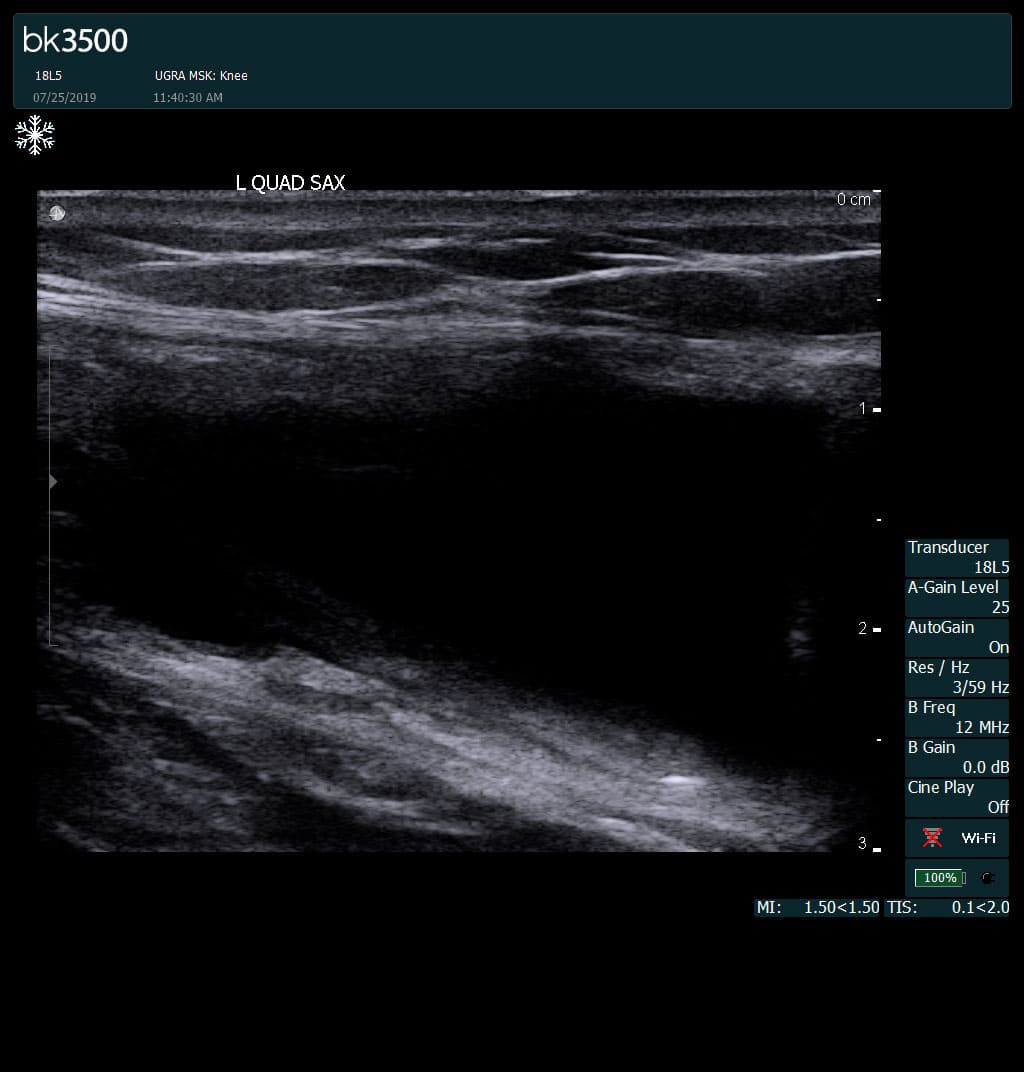
Complete tear of rectus femoris
with large hematoma (blood)
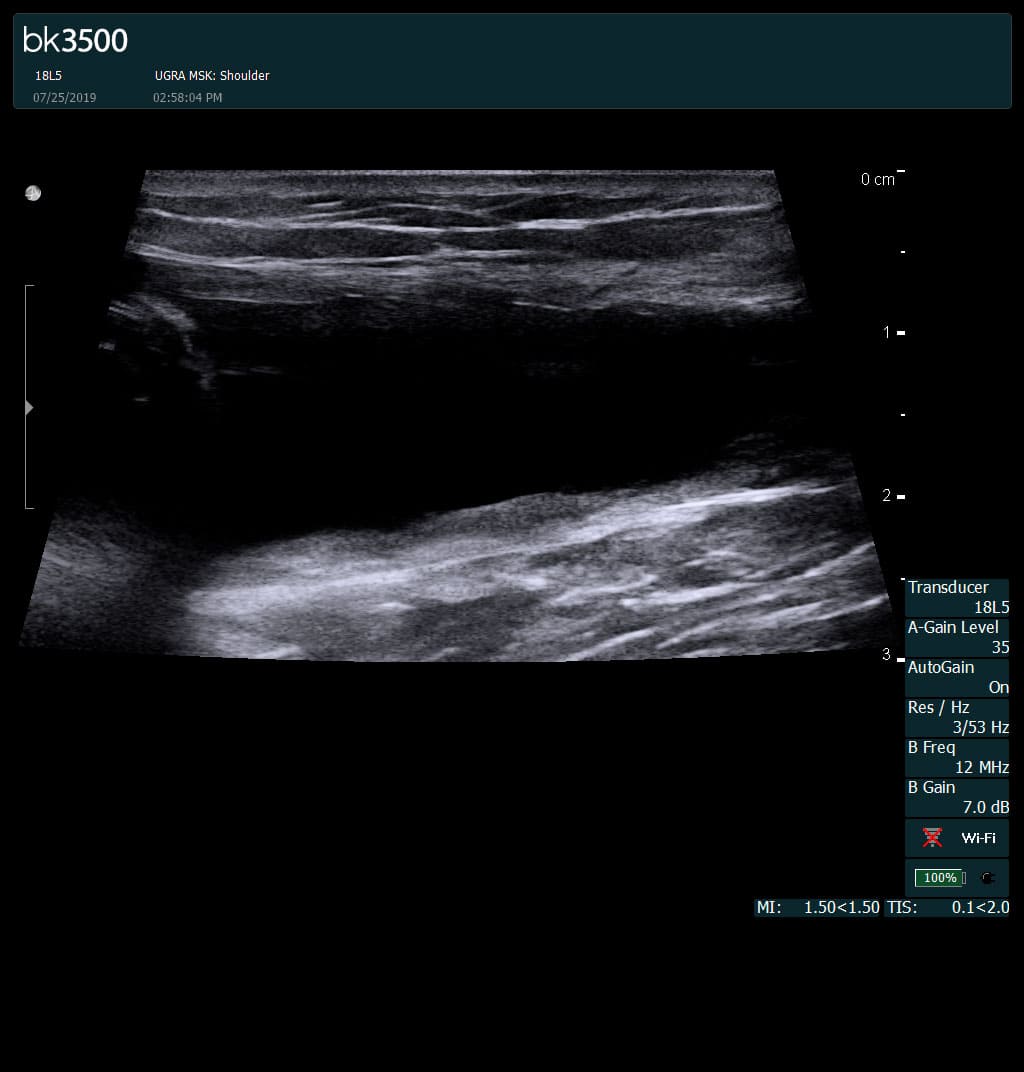
Separation of muscle ends due to tear elicited
on dynamic sonography examination

 Request Telehealth