Syndrome of impinged hip also called FAI is a medical condition implying a great deal of pain, discomfort and debilitation. Femoroacetabular Impingement hits adults in middle age. It heavily limits the way a person moved before the condition manifested itself. Hip joint lays at heart of the problem. Femoral head excessive rubbing causes inflammation and pain. The symptoms include:
- range of motion limitations
- groin related painful sensations
- lower back aches
- CT scan and MRI usually show signs of osteoarthritis
Acetabulofemoral joint is the joint in the pelvic area that supports the body weight in static and dynamic positions. The way a hip joint is built the way so the head of the greater femur rotates. This way the range of motion of the hip is almost unlimited. If an inflammation occurs, the range of motion becomes largely limited. The parts of this joint fit tight together when the joint is in healthy condition. It normally allows for unobstructed motion.
The smooth slippery glossy cartilaginous layer almost fourth of an inch thick covers the head of the femur. Thanks to this layer a joint moves freely.
In the case of Femoroacetabular impingement boney needley structures start to form on top of the femur and start rubbing the socket of this joint, prompting trauma and inflammation. The needles are called bone spurs, they are the cause of irregular excessive friction within the joint, inhibiting normal movement. Glenoidal labrum of the hip joint gets tears over time, causing osteoarthritis (swelling and inflammation within the joint) as well as slow cartilage breakdown.
Femoroacetabular impingement is usually treated using NSAIDs to manage pain, activity modification,PT complex. Rare cases are that surgery becomes an option to remove bone spurs.
Attempting to alleviate the pain and the obvious epicentre of it, the major picture may go out of focus. Human movement as we know it is not isolated to one joint or body part. Other factors may as well impact the situation. Faulty movement mechanics, poor postural habits, neglected injuries, muscle tightness, weakness could all contribute to all the wrong things going on in hip joint function. When addressing FAI, clinicians should pay attention and assess holistically.
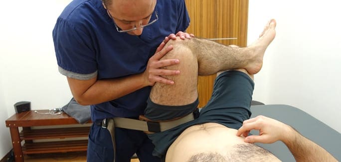
Sports medicine team at NYDNRehab in NYC understands this common integrated nature of human motion. We use the latest therapeutic methods and cutting edge technologies to figure out correct deficiencies in functional movement, eliminating pain at its source.
Contact NYDN Rehab today. Let our highly-trained hip pain specialists diagnose your condition properly. Your hip pain is about to be treated. Leave hip related aches behind.
Professional sports players whose lives revolve around their sport, having an injury that takes them off the playing field can be torture. In eagerness to get back in the game, athletes often play down their symptoms and exaggerate their degree of recovery.