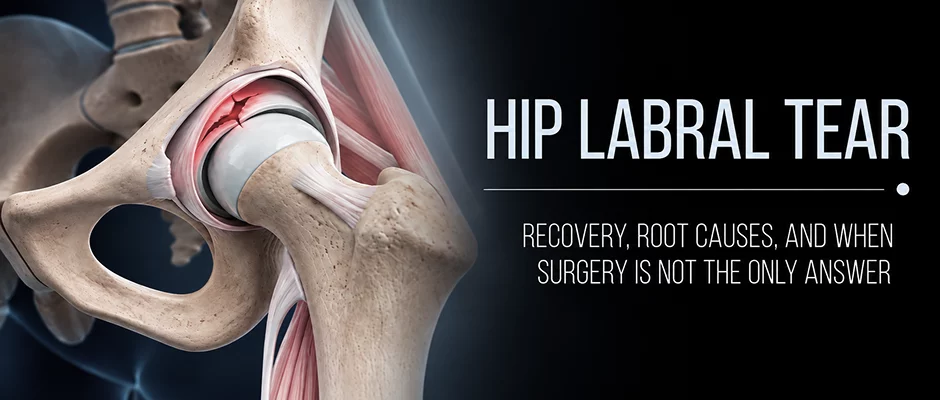
A hip labral tear on an MRI can feel like a definitive answer, but it rarely tells the whole story. Many people with a torn labrum have no pain, and many people with hip pain have more going on than the tear alone. This guide explains what the labrum does, why a tear is often a consequence rather than the sole cause, when conservative care is worth trying before surgery, and what realistic recovery looks like.

A hip labral tear is a true structural abnormality. However, an MRI scan does not always provide the full clinical picture.
Many people have imaging that shows a clear labral tear but no symptoms at all: a systematic review and meta-analysis found labral tears on MRI in about half of people without hip pain. Others suffer from substantial and debilitating pain, even though the tears on scan seem rather small. This disconnect is important because if the tear is just a component of the issue, then treating the tear alone may only help partially resolve the issue.
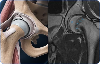
The first question asked in any hip evaluation at NYDNRehab is: Does this dysfunction contribute to the problem, is it a result of it or both?
A sore hip can affect the way the body functions. The pelvis can move. The lower back may adjust. The movement of the mid-back may become increasingly restricted. These adaptations over time can become abnormal movement patterns and can persist for an extended period of time after the original tissue has recovered.
It can be the other way around, too. Hip joint loading patterns can also be abnormal due to poor pelvic control, limited thoracic rotation, or other stresses on other parts of the movement system, which can make the hip joint labrum structurally vulnerable.
Knowing the direction of the problem or knowing that both directions are at play at the same time, can be a meaningful change in the treatment approach.
There is no single clinical history for hip labral tear. The diagnosis might be made at different stages in the patient’s process, and the things that need to be understood will vary by stage of the process.
This article is intended for three different types of patients.
You Have an MRI Finding and Are Considering Your Options
You’ve heard that you have a labral tear. Surgery called arthroscopy may have been suggested. You don’t necessarily have to be against surgery, but you want to know everything about it before deciding.
That instinct is clinically sound.
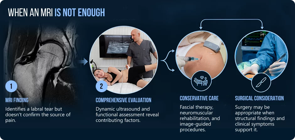
An MRI is used to confirm that there is a structural finding. It does not prove that the tear is the cause of your pain. It does not show how your hip actually moves under load, how your pelvis controls the forces during walking, or whether limited mobility in other parts of your body could have been contributing to hip stress for months or years prior to the onset of symptoms.
At NYDNRehab, patients come in with imaging results and a surgical plan, and when they are evaluated in detail, with a mechanical evaluation, the symptoms they have do not seem to be significantly related to the joint itself. Some of these patients undergo surgery in the end. Others benefit from complete conservative treatment. The distinction may be in the nature of what the assessment is really assessing.
You Are Preparing for Surgery or Recently Had Hip Arthroscopy
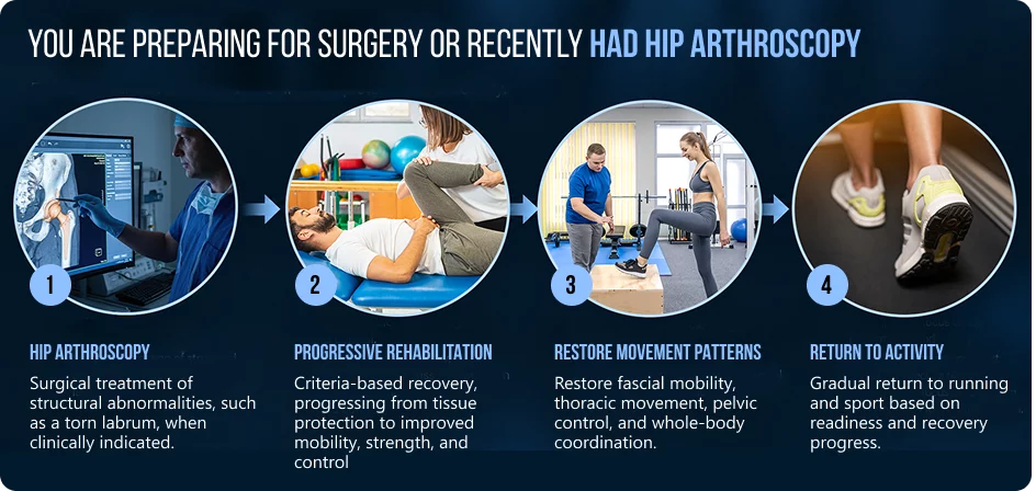
You have to undergo surgery or have already undergone surgery. You want the maximum recovery you can get. You want to get back to what you enjoy doing and you don’t want to get back to it re-injured or stuck in a plateau.
Many post-surgical patients come to NYDNRehab not because they doubt their surgeon’s work, but because typical post-surgical rehab that focuses on weekly goals and isolated hip exercises may not meet the needs of the movement system the hip operates in.
Surgery corrects the anatomical structure. It does not automatically retrain compensatory movement patterns that might have been established prior to the procedure. It cannot correct fascial restrictions that can occur over the course of months of pain-altered movement. Deliberate, progressive rehabilitation is necessary to restore thoracic rotation, lumbopelvic control and coordinated whole-body mechanics.
You Had Surgery and Your Hip Still Hurts
This can be the toughest role to have. You had the surgery. You have finished the rehabilitation program. And months or years after the procedure, you are still having deep groin pain, clicking, or persistent tightness, or your hip just doesn’t feel normal.
Your surgeon has looked at the imaging and informed you that the joint is structurally sound. Occasionally, symptoms persist even though the repair appears to be intact, and sometimes, several factors contribute to the symptoms.
Sometimes, continued pain may be a result of mechanical factors that existed preoperatively and were not diagnosed or treated during the rehab process. Compensatory movement patterns, fascial restrictions, involvement of adjacent structures, and/or limited thoracic mobility can all contribute, but to different extents in different people.
This doesn’t necessarily mean the surgery was a failure. It could be that the surgery solved one component of a problem that involved more than just the joint and a more comprehensive evaluation is necessary.
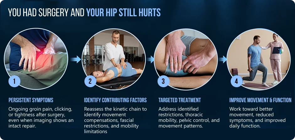
The clinical approach at NYDNRehab starts where you’re at in this process: a comprehensive evaluation of the entire movement system, rather than just the structure seen on imaging. The treatment pathway is based on the results of that assessment. One piece of information is important, and that is imaging. It’s not often the whole solution.
The labrum is a fibrocartilaginous ring around the rim of the hip socket. It creates a deeper socket, spreads the load evenly over the surface of the joint, helps to keep the fluid seal that lubricates the hip joint, and adds to the stability of the joint. These functions may be impaired to various extents when they are torn, due to trauma, repetitive loading, structural abnormality, or gradual degeneration.
The most common structural causes of labral tears are:
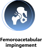
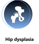


There is no change in bone morphology during rehabilitation. The amount of functional stress this morphology produces and if it is symptomatic or not may be significantly affected by the movement system around it.
The Fascial Network
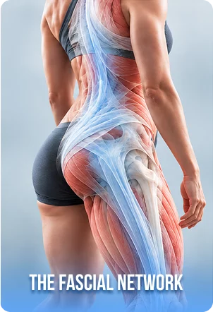
The hip isn’t a standalone joint. It is part of a continuous band of connective tissue, fascia, which mechanically connects the trunk, pelvis and lower limb.
The thoracolumbar fascia (lower back and pelvis), the abdominal wall and pelvic fascia (transmitting load between spine and hip), the iliotibial tract (from pelvis to knee), the hip capsule and deep rotator fascia (surrounding the hip joint), and the capsular ligaments, iliofemoral, pubofemoral and ischiofemoral (passive joint stability) are key structures in this network.
These structures are involved in the transmission of force from the trunk, pelvis, hip and leg during movement. As the hip pain develops, the body may start to recruit protective tension in portions of this network. Some of the normal gliding properties of the fascial planes may be lost. Movement patterns may become guarded and asymmetrical.
It is not always the case that these changes contributing to continued hip loading; they may simply be a result of it, and this will need to be assessed on a case-by-case basis.

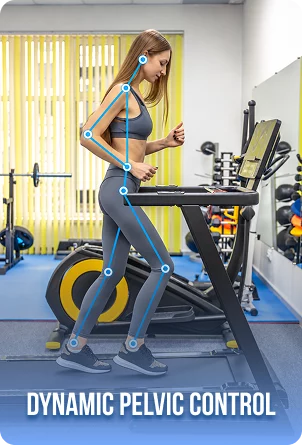
Thoracic Rotation and the Hip: A Two-Way Relationship
Good walking and running requires co-ordinated counter-rotation of thorax and pelvis. The mid-back rotates in one direction and the pelvis rotates in the other direction, which aids in efficient arm swing, hip extension and lower limb propulsion.
If the hip hurts, the body will decrease the amount of rotation required from the hip by limiting thoracic mobility. As time goes on, a decrease in thoracic rotation can lead to:
A higher load on the rotation of the spine in the lower back. Hip extension may be limited during walking, leading to increased loading on the anterior hip during walking. Gradual compensation further up the kinetic chain
The connection can be the other way around as well. If the thoracic mobility is restricted, either by stiffness or by previous injury or by habit, this could change the way the load is carried through the pelvis and hip. This doesn’t imply that thoracic restriction is directly responsible for labral tears. However, it could affect how the hips are loaded and if structural weaknesses are symptomatic.
Thoracic mobility is evaluated early and systematically incorporated into hip rehabilitation as the patient’s capacity increases at NYDNRehab, because restoring it may support hip mechanics during recovery.
The Dynamic Function of the Pelvic Control is More Important than its Static Position
The static position of the pelvis, either anterior or posterior tilt, is not clinically significant when compared with the ability to control the pelvis during actual movement. The clinical evaluation includes the ability to control pelvic orientation during walking, running, squatting, loading on one leg and rotational tasks.
The bony morphology of FAI is not caused by pelvic mechanics. The position of the pelvis during motion may affect the amount of functional impingement in vivo and if the prevailing bone shape is asymptomatic or may lead to anterior hip pain.
If the patient has mild cam and good pelvic control, they may not have any symptoms. In a patient with decreased lumbopelvic stability, the same morphology can create a sufficient, consistent anterior hip load leading to a painful hip. The difference can be a significant factor in treatment decisions.
MRI is an important diagnostic modality for the detection of labral pathology, joint morphology, and cartilage integrity. A thorough review of MRI is a part of each hip evaluation at NYDNRehab.
The one thing MRI does have as its limitation, however, is that it is performed while lying still. The movements that cause hip symptoms are in standing, walking, running and rotation.
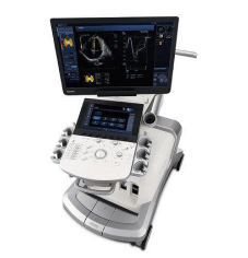
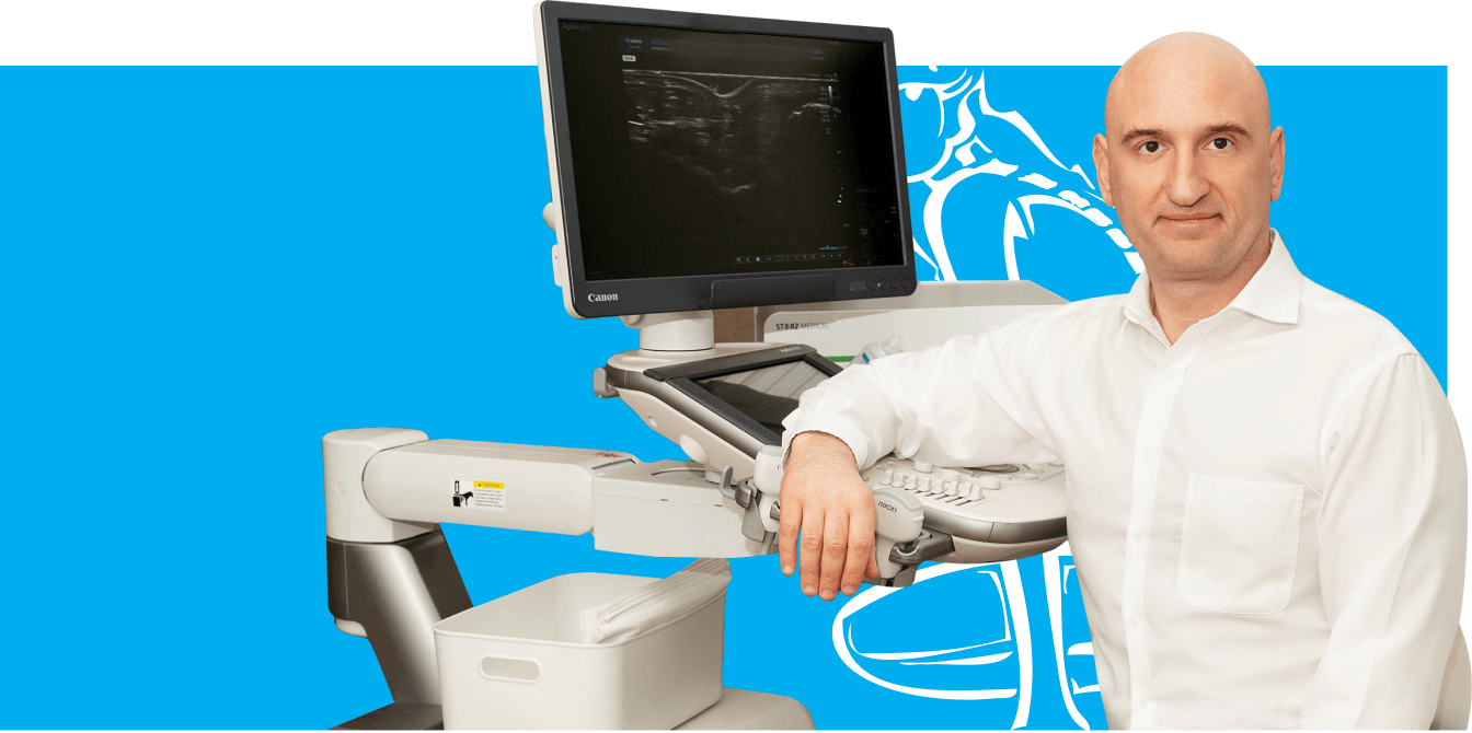
In addition to MRI, musculoskeletal ultrasound imaging is used to assess soft tissue structures, such as the hip capsule, periarticular tendons, bursae and fascial planes, in real time and during movement. Unlike static imaging, dynamic ultrasound can show how tissues react when they are moving during the precise motions that produce a patient’s symptoms.
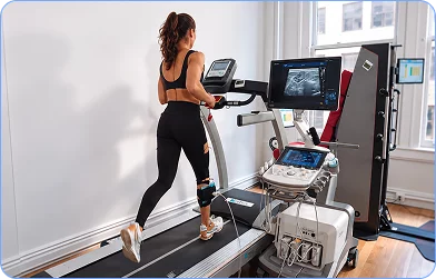
The real-time imaging accuracy of ultrasound also plays a key role in guiding injection procedures with precision to the correct anatomical target, which is a factor that impacts the clinical value of image-guided treatments.
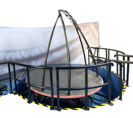
Computerized gait analysis and movement assessment provides an objective component to this assessment, quantifying compensatory patterns, loading asymmetries and movement strategies along the kinetic chain that may not be obvious on a routine clinical exam.
These tools will give a more comprehensive view of the hip’s actual function, rather than the view it shows on a scan when it is at rest.
The Tear as Possible Consequence, Not Certain Cause
One of the most important clinical questions is whether the labral tear is a major contributor to symptoms or whether mechanical contributors are present but not identified and may have contributed, at least in part, to the development of the labral tear.
If any of those contributors are not corrected, and/or the pelvic control is inadequate, the mobility of the fascia is restricted, the capsule is not sufficient, or the thoracic mechanics are altered, then surgical repair of the labrum may not fully resolve the mechanical environment that caused the original stress.
This is NOT an anti-surgery statement. It’s a case for a comprehensive evaluation prior to surgery.
Structural imaging may show no change, and the patient may be significantly less symptomatic and much more functional. The purpose of treatment is not to normalize the MRI; it is to restore movement, reduce pain and support return to meaningful activity.
Addressing the Fascial System
When clinical assessment suggests that fascial restrictions may be contributing to altered hip mechanics or reduced movement tolerance, several approaches may be considered as part of a broader rehabilitation strategy:
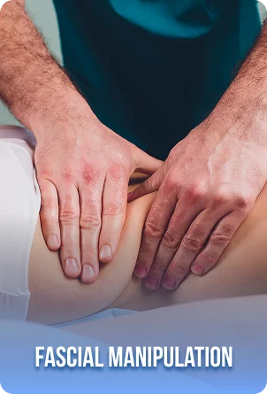
Manual fascial therapy is a targeted manual therapy technique that addresses clinically identified areas of altered fascial mobility. The intended goal is to improve the gliding capacity between fascial planes, potentially reducing restrictions that may be limiting movement quality or the effectiveness of exercise-based rehabilitation.
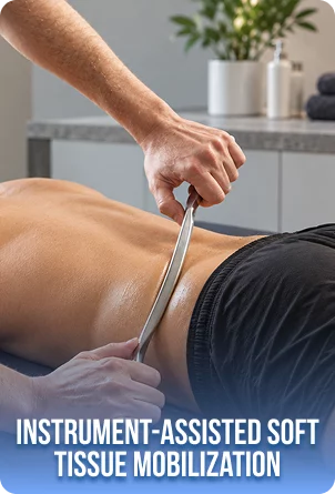
Instrument-assisted soft tissue mobilization uses specialized tools to address superficial and deeper fascial layers in a targeted way.
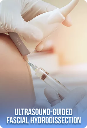
Ultrasound-guided fascial hydrodissection involves introducing fluid around a restricted fascial interface under real-time imaging guidance, targeting a specific tissue plane that clinical assessment has identified as potentially contributing to movement limitation or localized discomfort. This is not a permanent structural correction. Its proposed role is to improve the local tissue environment sufficiently to allow progressive rehabilitation to proceed more effectively.
None of these approaches replaces neuromuscular rehabilitation. Their role is to address tissue restrictions that may be reducing the effectiveness of exercise not to independently correct biomechanical dysfunction.
Prolotherapy for Capsular and Ligamentous Insufficiency
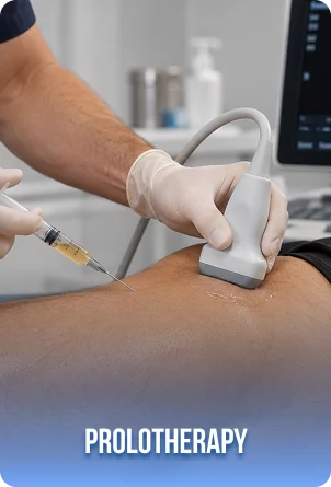
In selected patients where clinical assessment raises the possibility that capsular laxity or ligamentous insufficiency may be contributing to hip instability and labral stress, ultrasound-guided prolotherapy may be considered as one component of a broader treatment strategy.
Prolotherapy involves injecting a concentrated dextrose solution into or around a targeted ligamentous or capsular structure. The proposed mechanism involves a localized biological response that may support tissue remodeling in the treated area over time.
The hip capsule is reinforced by three primary ligaments, the iliofemoral, pubofemoral, and ischiofemoral ligaments, along with clinically relevant pelvic ligament structures. When these structures provide insufficient passive support, the labrum may be required to absorb stability demands that exceed what it is ideally designed to handle.
Prolotherapy does not mechanically repair a labral tear. It does not regrow tissue or reconstruct the joint. The evidence base for its use in hip instability is still developing, and it is most appropriately considered for specific clinical presentations, not as a universal treatment for hip pain.
PRP: A Possible Biological Window for Rehabilitation
In selected patients where clinical assessment raises the possibility that capsular laxity or ligamentous insufficiency may be contributing to hip instability and labral stress, ultrasound-guided prolotherapy may be considered as one component of a broader treatment strategy.
Prolotherapy involves injecting a concentrated dextrose solution into or around a targeted ligamentous or capsular structure. The proposed mechanism involves a localized biological response that may support tissue remodeling in the treated area over time.
The hip capsule is reinforced by three primary ligaments, the iliofemoral, pubofemoral, and ischiofemoral ligaments, along with clinically relevant pelvic ligament structures. When these structures provide insufficient passive support, the labrum may be required to absorb stability demands that exceed what it is ideally designed to handle.
Prolotherapy does not mechanically repair a labral tear. It does not regrow tissue or reconstruct the joint. The evidence base for its use in hip instability is still developing, and it is most appropriately considered for specific clinical presentations, not as a universal treatment for hip pain.
Orthobiologic Preparations
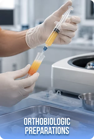
BMAC (Bone Marrow Aspirate Concentrate): an injection prepared from your own bone marrow. BMAC concentrates cells and other natural components from a small sample of your bone marrow. We may consider it for selected, more advanced joint or tendon problems when PRP alone may not be sufficient. Treatment combines precise ultrasound-guided injection with personalized rehabilitation.
MFAT (Micro-Fragmented Adipose Tissue): an injection prepared from your own fat tissue. MFAT uses a small amount of your fat, gently processed to preserve its natural cells and supporting tissue. We may consider it for selected, more advanced joint or soft-tissue problems when PRP alone may not be sufficient. Treatment combines precise ultrasound-guided injection with personalized rehabilitation.
These are not stem cell injections that regenerate a new labrum. They should not be described or understood in those terms. Their proposed role where clinically appropriate and in properly selected patients, is to potentially support the biological conditions in which rehabilitation and natural repair processes occur. The evidence base is still developing, realistic expectations are essential, and their use requires careful patient selection and individualized clinical judgment.
Surgery treats the structural anatomy. Rehabilitation is the process of restoring the movement system the hip has to operate in. Surgery followed by limited rehabilitation is likely to be less successful than surgery followed by comprehensive, individualized rehabilitation of the whole kinetic chain.
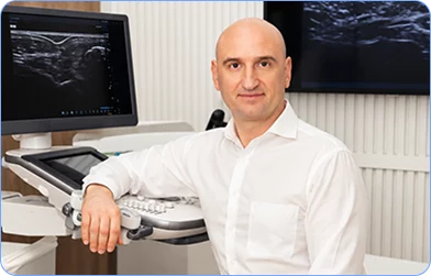
Thinking about surgery but looking to find out all the details beforehand?
A comprehensive biomechanical evaluation at NYDNRehab utilizing dynamic ultrasound and computer motion analysis can assist in determining if the factors causing your symptoms are modifiable and have been thoroughly identified and treated. Call us to schedule a thorough hip examination.
Post-surgical protocols are designed to allow tissues to heal over time and gradually load the tissue as healing progresses. This is necessary. It is also not enough by itself.
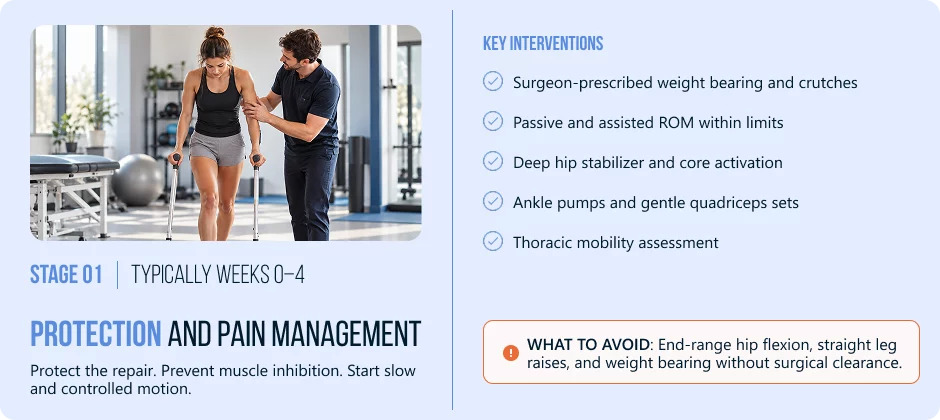
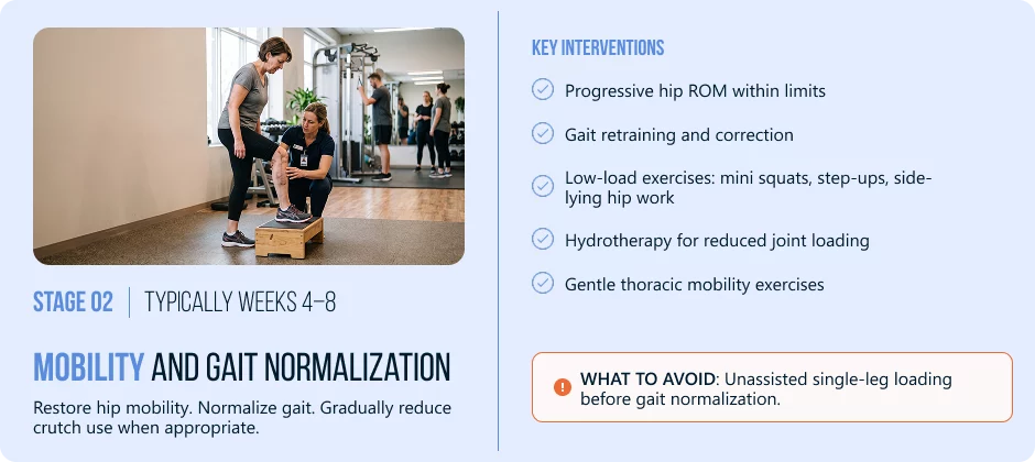
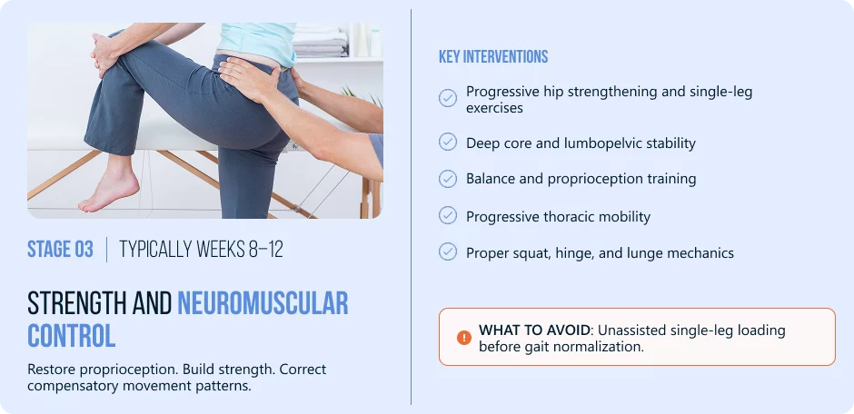
The following week ranges are only representative of the normal periods. The decision for an individual’s progression should always be based on objective factors and clinician guidance and not calendar date.
Stage 1 Typically Weeks 0–4 | Protection and Pain Management
Stage 2 Typically Weeks 4–8 | Mobility and Gait Normalization
Stage 3 Typically Weeks 8–12 | Strength and Neuromuscular Control
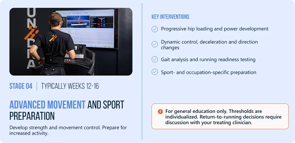
Stage 4 Typically Weeks 12–16 | Advanced Movement and Sport Preparation
There are several factors that are considered prior to returning to running. The thresholds listed are individual and should be verified with your treating clinician.
This checklist is for general education only. All return to running decisions should be discussed with your treating clinician.
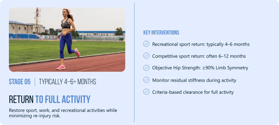
Stage 5 Typically 4–6+ Months | Return to Full Activity
Objective: Restore sport, work and recreational activity without the risk of re-injury.
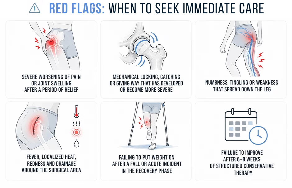
It is not uncommon for there to be incomplete relief after hip arthroscopy and this may not be attributed to any single cause.
Persistent symptoms may be due to mechanical factors that existed prior to surgery which were not detected or corrected during rehabilitation. These may include:
labral repair tissue may take months to mature after surgery, or due to factors outside of the mechanical contributors of the original injury.
A thorough reevaluation of the entire kinetic chain can be useful to determine what factors might be playing a role in a particular case. This doesn’t always give a straightforward answer but often reveals specific factors to address that would not have been looked at during routine post-surgical rehab.
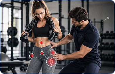
Still experiencing hip pain months after surgery?
If you don’t feel you are improving as you would expect following standard rehabilitation, a thorough kinetic chain assessment at NYDNRehab may help determine factors that have not been addressed. If you are considering a post-surgical second opinion assessment, please contact us to discuss the situation.
Request an appointment»

Our location: 11 West 25th Street 5th floor, New York, NY 10010
In many cases, yes. If the labral tear is not related to other significant structural deformities like large FAI morphology or advanced hip dysplasia, it can often be managed with non-surgical hip labral tear treatment. This usually includes physical therapy to improve movement mechanics, neuromuscular control and fascial mobility, activity modification and, if necessary, image-guided procedures. The suitability of conservative treatment will depend on the individual findings and a detailed clinical evaluation. It is possible to see the fibrocartilage tear on imaging even though symptoms have improved substantially.
Hip arthroscopy recovery time is typically 4-6 months after surgery, but may vary depending on the type of procedure performed, any other hip problems, and the quality and adherence to recovery. It takes 6-12 months for competitive athletes in sports that involve a lot of rotation to return to full sports participation. Objective measures of progress and strength, not calendar weeks, are the most meaningful measures of progress
It is not uncommon to have symptoms that last after a hip arthroscopy, and it is not a sign of a failure of surgery. In some cases, chronic pain can be a result of pre-existing movement compensations that were not diagnosed or treated (pelvic instability, fascial restrictions, lumbar and thoracic stiffness), and may be contributing to chronic hip loading. In others, it may represent a normal healing and remodeling process of tissues, or the involvement of neighbouring structures. The most useful next step is a full re-evaluation.
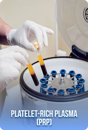
Platelet-rich plasma (PRP) is created from the patient’s own blood and concentrated to boost the number of platelets, which contain signaling molecules that are important in tissue repair. It’s not just an anti-inflammatory injection, it is likely to have a mild local response at first that may also affect the later remodeling stages. When used in the right patients, ultrasound-guided PRP may help alleviate symptoms enough to allow progressive rehabilitation. It does not ensure labral repair and cannot correct structural damage. The evidence base for the use of PRP in labral pathology in particular is still evolving.
Prolotherapy is a procedure that uses a highly concentrated dextrose solution to be injected into or around the specific ligamentous or capsular structure. For certain patients, clinical evaluation suggests that capsular laxity or ligamentous insufficiency may be playing a role in hip instability and labral stress, and ultrasound-guided prolotherapy can be a part of a comprehensive treatment plan. It is not a mechanical repair for a labral tear and there is still an ongoing body of evidence to support its use in hip instability. It is best used in certain clinical situations, and should be discussed with realistic expectations.
Fascial hydrodissection is the process of placing fluid into the fascial interface around a restricted area, under real-time ultrasound guidance. It may be recommended in cases where clinical evaluation indicates that a particular tissue plane is thought to be involved in limiting motion or causing symptoms in a specific area. It does not permanently correct the structural defect and does not by itself correct biomechanical dysfunction. It is proposed that it will create a better local tissue environment to enable rehabilitation to take place more effectively.
Surgery is most clearly indicated if there is significant structural instability that is not addressable with conservative treatment, if there is mechanical pathology (true locking or severe giving way), if the FAI morphology is too severe to allow for meaningful functional improvement despite well-designed movement mechanics, or if there has been no meaningful functional improvement with a well-designed conservative treatment program over an appropriate period of time. Surgical consultation is appropriate when all conservative options have been fully considered and are not proving to be of benefit.
Typical signs of recovery are a progressive decrease in pain over weeks, improving walking and activity tolerance, and working toward rehabilitation milestones within expected ranges and progressive improvements in ability to perform daily activities. Recovery is not always smooth, some days will be better than others, and it’s not uncommon to have some mild fluctuations. If the progress has not been made consistently, the pain has increased or new symptoms have developed, this is a reason to directly discuss the matter with your surgeon or physical therapist, not to continue the same program without re-evaluating.
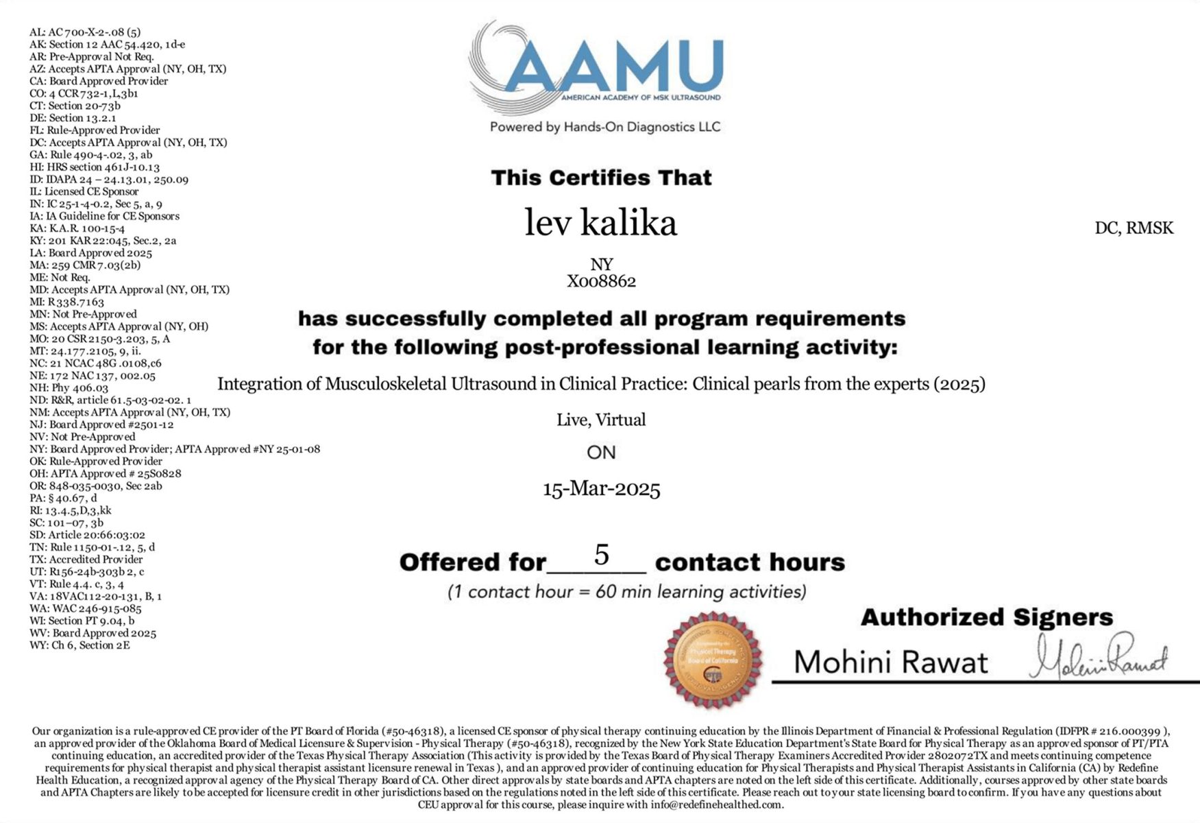
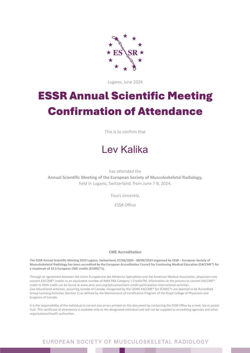
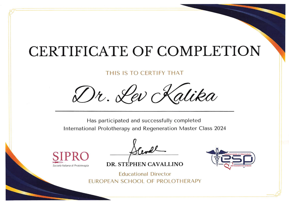
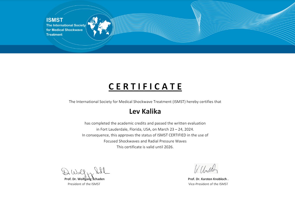
Dr. Lev Kalika is a world-recognized expert in musculoskeletal medicine. with 20+ years of clinical experience in diagnostic musculoskeletal ultrasonography, rehabilitative sports medicine and conservative orthopedics. In addition to operating his clinical practice in Manhattan, he regularly publishes peer-reviewed research on ultrasound-guided therapies and procedures. He serves as a peer reviewer for Springer Nature.
Dr. Kalika is an esteemed member of multiple professional organizations, including:

Below is a prime example of how ultrasound can take the guesswork out of diagnosis.

A bad physical therapy experience is one of the primary causes of unnecessary surgery

In this instance, an athlete was originally diagnosed with minor quadriceps muscle strain and was treated for four weeks, with unsatisfactory results. When he came to our clinic, the muscle was not healing, and the patients’ muscle tissue had already begun to atrophy.
Upon examination using MSUS, we discovered that he had a full muscle thickness tear that had been overlooked by his previous provider. To mitigate damage and promote healing, surgery should have been performed immediately after the injury occurred. Because of misdiagnosis and inappropriate treatment, the patient now has permanent damage that cannot be corrected.
The most important advantage of Ultrasound over MRI imaging is its ability to zero in on the symptomatic region and obtain imaging, with active participation and feedback from the patient. Using dynamic MSUS, we can see what happens when patients contract their muscles, something that cannot be done with MRI. From a diagnostic perspective, this interaction is invaluable.
Dynamic ultrasonography examination demonstrating
the full thickness tear and already occurring muscle atrophy
due to misdiagnosis and not referring the patient
to proper diagnostic workup
Demonstration of how very small muscle defect is made and revealed
to be a complete tear with muscle contraction
under diagnostic sonography (not possible with MRI)
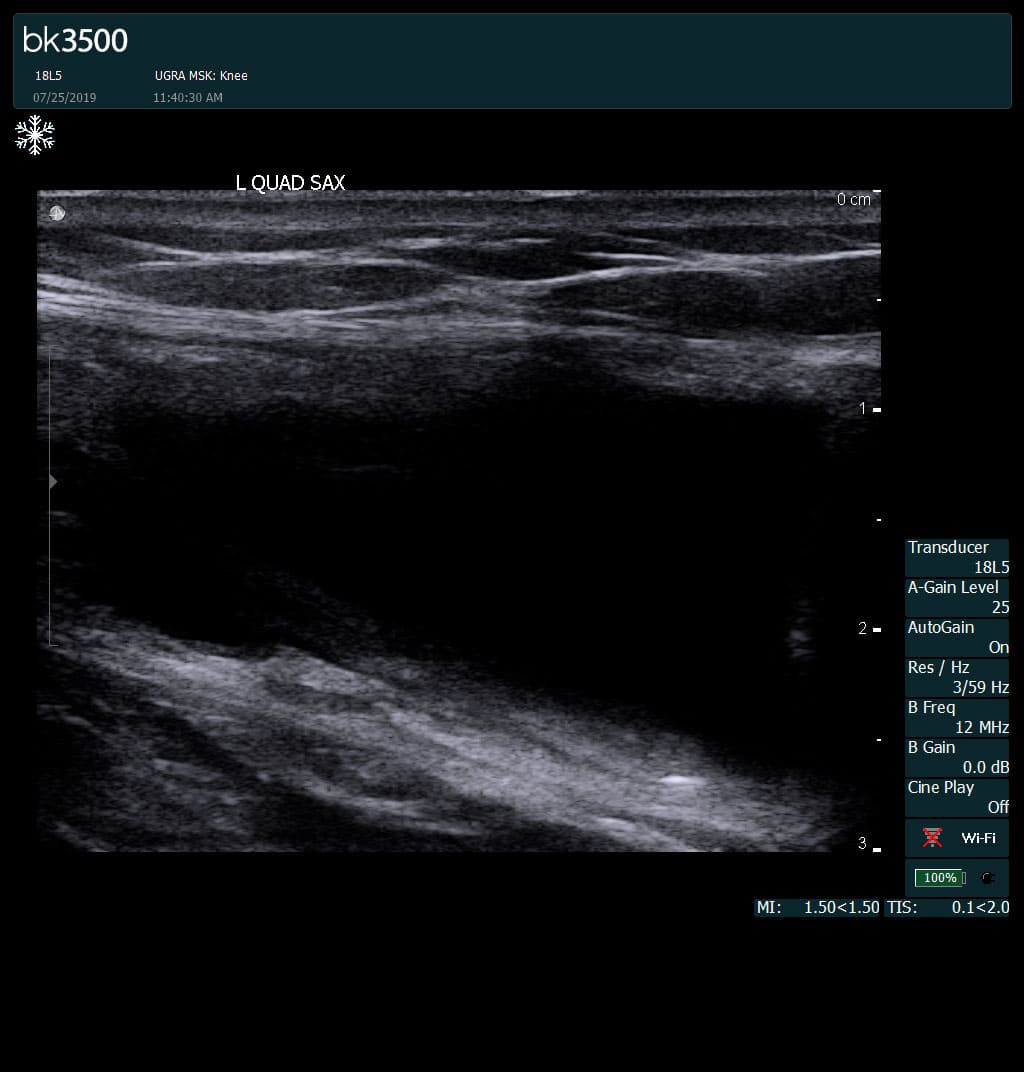
Complete tear of rectus femoris
with large hematoma (blood)
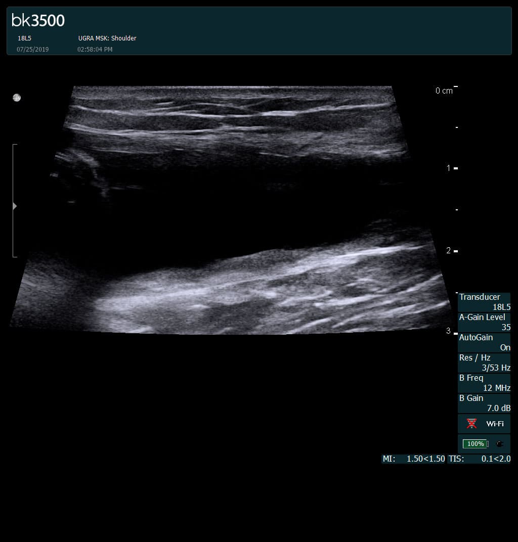
Separation of muscle ends due to tear elicited
on dynamic sonography examination

 Request Telehealth