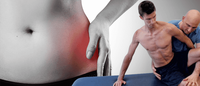
Injury, illness or congenital defect can cause hip pain. Pain in this joint can be debilitating and interfere with activities of daily living essential to quality of life. Here is an overview of the joint, causes of problems and types of hip pain treatment.
The hip joint is a major joint. It is classified as a ball-and-socket joint. The top of the femur (thigh bone) is rounded and fits into a socket in the pelvis. The proper name for the pelvic bone in question is “acetabulum”. The size and shape of the acetabulum make this joint one of the hardest joints to dislocate.
This joint is capable of a wide variety of complex motions. These motions are aided by a large group of muscles. In addition to the well known gluteal and hamstring muscles, there are lesser well known muscles such as the iliopsoas, rectus femoris, Sartorius, pectineus, and the gracilis. They typically work in groups of two or three to create each distinct motion, such as flexion, rotation, and abduction.
There is a layer or cartilage cushioning the joint and reducing the friction that occurs when it moves. The cartilage is further assisted by bursae, which are fluid filled sacs that lubricate the joint.
The pelvis is a central area. Unsurprisingly, multiple nerves pass through it, if only to connect other parts back to the brain. When the hip is injured, the two most commonly nerved are the sciatic nerve and the femoral nerve.
Osteoarthritis is one of the most common causes of joint pain and is a major cause of hip pain. It involves degeneration of the joint cartilage. Pain is usually worse after sustained periods of walking. Typically, symptoms begin as sporadic or intermittent problems, including pain, stiffness, decreased range of motion, and popping sounds while moving. If left untreated, these issues get steadily worse and can lead to sleep difficulties. Treatment includes gentle exercises and hot and cold treatments. Doctors sometimes prescribe non-steroidal anti-inflammatory drugs to reduce pain and inflammation. In extreme cases, steroidal injections can be part of the treatment plan.
Hip flexor strain is a tear in the muscles involved in flexion, such as the iliopsoas muscle. Minor tears can cause minimal problems. Severe tears can create extreme pain and impairment. You are most likely to notice a problem when taking stairs or engaging in other activities that cause the hip to flex in that fashion. People suffering from this condition frequently wake up with pain and stiffness. They may also notice bruising. It is typically treated with rest and physical therapy.
Sciatica is a commonly used term for pain involving compression of the sciatic nerve in the lower back. It is often caused by a ruptured disc or a bone spur. It can be due to either a sudden trauma or slow degeneration. It is particularly debilitating because the pain may not remain confined to the lower back. It can spread to the lower body.
In most cases, misalignment or dysplasia is congenital. This condition can lead to an increased risk of dislocation. Generally speaking, the acetabulum is misshapen in some way, and this reduces the stability of the joint. It may not need any particular treatment, unless it does lead to dislocation.
When the bursae become inflamed, the condition is called bursitis. Trochanteric bursitis is the name of the condition when the inflamed bursae surround the greater trochanter, a bony prominence that anchors several gluteal muscles. This can be a repetitive stress injury, incurred from excess jumping, lunging, or running. It can also be due to trauma. It typically causes pain in the outer hip and along the outside of the thigh, running down to the knee. Treatment typically starts by cutting back on the activities that created the condition to begin with. It may include other exercises aimed at strengthening or stretching the joint, plus hot and cold treatment. Occasionally, cortisone injections are used.
Snapping hip is the term for a condition that involves a loud, often painful, snapping sound when the joint is flexed or extended. It is very frequently caused by tendinitis of the iliopsoas tendon. As with most hip conditions, it can be caused by either overuse or injury. This tendon is stretched over the bone of the socket. If something goes wrong, it can make a clicking or snapping sound as it rubs back and forth. The most common treatments include rest, reducing certain problematic activities, taking NSAIDs, and cortisone injections. The worst cases are sometimes treated surgically.
Hip pain can impede the simplest and most basic activities of daily life, such as sitting down, getting up again or getting into or out of a car. It should not be ignored.