Hip Pain? Visit Your Doctor Right Away!

As soon as we experience pains and aches, we try stalling going to the doctor as much as we can and try all the home remedies that come to mind. However, there always comes the point where we realize that none of the treatments are working and we inevitably, have to visit the doctor. This article will outline how an orthopedic specialist will help treat your hip pain.

Biomechanics

What factors are responsible for your hip pain?

The first question that comes to mind, of course, is what the reason behind your hip pain is? Generally, there are plenty of factors responsible for hip pain. But the most frequent question which pops up is if the pain is because of arthritis. And if arthritis is not reason, then what is?

To figure out the cause, our orthopedic specialists in NYC begin the medical exam by reviewing the patient’s medical history along with a physical medical exam. However, the orthopedist specialist may need to conduct further tests if he is unable to diagnose the pain. The most commonly asked medical history questions include:

  • When and how did the pain start?
  • Do you perform any activities which increase the pain?
  • Have you experienced similar pain before? If so, what was done for its treatment?
  • Do you perform any activities which help ease the pain?
  • Did you have an accident or were you injured before the pain started?
  • Have you experienced pain in any other joint or part of your body?
  • Has the pain been preventing you from performing daily physical activities like walking?

You may also needto describe the pain like if it is a snapping sensation, a dull ache, a sharp stabbing pain, or a burning sensation.

What does a good medical exam include?

Make sure your doctor has conducted a thorough physical examination before he tells your diagnosis. The doctor will usually look for weakness in the muscles, pain near or around the hip, and tenderness to the touch of the hip.

For a thorough medical exam and an accurate diagnosis, you will have to lie down on the table while the orthopedic specialist will examine you in a supine or flat position. Your orthopedist specialist may also instruct you to change the position of your legsor walk to determine the exact location and how the hip pain occurs.

Is there any other approach to diagnose hip pain?

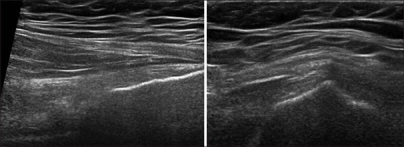
There are several other ways to diagnose hip pain, and X-rays are on the top of the list. Actually, X-rays are the most useful tools for diagnosing degenerative joint disease, particularly osteoarthritis in the hip. However, sometimes orthopedic specialists need to conduct blood tests as well, to rule out systemic inflammatory disease and inflammatory arthritis which can lead to joint disease in the hip. An increased ESR (erythrocyte sedimentation rate) along with an increased WBC count can also indicate hip infection.

CT scans are also occasionally used if your X-rays indicate any sign of lesions or raise any suspicion of other factors responsible for hip pain and pelvic pain. Sometimes, CT scans are considered to be more useful than standard X-rays in identifying certain hip problems because they show soft tissues, including muscles and ligaments more clearly than the X-rays. If the X-rays fail to diagnose the disease and give a clear picture, an MRI is particularly helpful in ruling out osteonecrosis, a degenerative disorder of the bone.

What to expect in a typical X-ray

It is very easy to see if your hip is in perfectly good health or if it needs treatment. In fact, you can figure it out yourself! If you have a good joint, there should be some clear space between the ball and socket of the femoral head. However, if you see no clear joint space, you need to seek immediate medical attention. You can further confirm a bad joint if you see a white and hardened bone where the hip has degenerated.

Diagnosis

app

A clinical exam and diagnostic ultrasound imaging can help your therapist pinpoint the exact location and cause of your hip and groin pain.

Ultrasound enables you and your therapist to view the hip and groin region in real time, while in motion. In addition to ultrasound, video gait analysis can help us identify faulty movement mechanics that contribute to hip and groin pain. Once the exact cause is determined, an effective treatment plan can be initiated.

img

Explore more advanced diagnostic tools available only at NYDNRehab:

img

Hip dysfunction and pain can be a complex issue due to interactions of the trunk, pelvis, low back, groin and hip joint. Physical therapy and rehabilitation that is based only on subjective clinical analysis often addresses the symptoms without resolving the underlying cause.

icons

At NYDNRehab, our groundbreaking motion analysis technology and high resolution diagnostic ultrasonography have enabled us to develop a battery of tests that perfectly reveal the dynamic functional pathology of the hip joint and pelvis. Our tests are evidence-based protocols that are considered to be the gold standard in the world of research.

Our testing protocol includes:

1

Combined lumbopelvic hip stability test using DLEST methodology with C.A.R.E.N., our computer assisted rehab environment

2

Hip joint stability test using DLEST methodology with C.A.R.E.N.

3

3D star excursion banner test (SEBT) for assessing the involvement of the hip joint and muscles in postural stability

4

3D gait or running analysis

5

3D kinematic joint angle analysis during a squat, lunge, drop jump and pelvis on hip rotation

6

Rehabilitative ultrasonography for viewing intrinsic hip stabilizing muscle activation patterns

We also perform neuromotor testing with DD Robotech for:

  • Proprioception
  • Tracking ability
  • Force sense
  • Critical power
  • Reactive power
  • Flexibility

SEMG

Surface electromyography (SEMG) may be added to any of the above tests when needed.

Based on our experience and evidence-based information, we believe that physical therapy and rehabilitation should be based on objective quantifiable data.

bg

In this instance, an athlete was originally diagnosed with minor quadriceps muscle strain and was treated for four weeks, with unsatisfactory results. When he came to our clinic, the muscle was not healing, and the patients’ muscle tissue had already begun to atrophy.

Upon examination using MSUS, we discovered that he had a full muscle thickness tear that had been overlooked by his previous provider. To mitigate damage and promote healing, surgery should have been performed immediately after the injury occurred. Because of misdiagnosis and inappropriate treatment, the patient now has permanent damage that cannot be corrected.

The most important advantage of Ultrasound over MRI imaging is its ability to zero in on the symptomatic region and obtain imaging, with active participation and feedback from the patient. Using dynamic MSUS, we can see what happens when patients contract their muscles, something that cannot be done with MRI. From a diagnostic perspective, this interaction is invaluable.

Dynamic ultrasonography examination demonstrating
the full thickness tear and already occurring muscle atrophy
due to misdiagnosis and not referring the patient
to proper diagnostic workup

Demonstration of how very small muscle defect is made and revealed
to be a complete tear with muscle contraction
under diagnostic sonography (not possible with MRI)

image

Complete tear of rectus femoris
with large hematoma (blood)

image

Separation of muscle ends due to tear elicited
on dynamic sonography examination

bg
Buy now 3D Gait
Payment Success
Request Telehealth Request Telehealth Request in office visit Book now